The Anemia Sorting Hat

Your patient is anemic. That's the easy part. Now figure out why.

Microcytic - Macrocytic / Normocytic - Hemolytic - IDA workup

A 25-year-old woman presents with fatigue. Labs show Hgb 9.2 and MCV 68. What's the FIRST thing you check?
B12 level
Iron studies
Reticulocyte count
Peripheral smear
MCV framework · micro · macro · board walkthrough
Attending Rounds
Attending 65-year-old man. Tired. Cold hands. MCV is 72. What's the first thing you do for him?
Student I'd... give him some ferrous sulfate?
Attending GIVE HIM IRON? You're gonna give him iron and let the cancer finish the job? In an adult, iron deficiency is a bleed until proven otherwise. I don't care if he says he's a vegetarian. I don't care if he's poor.
Student I should check for a bleed first?
Attending YES. He is bleeding from his colon until you prove otherwise. You get him a colonoscopy. You find that tumor before it spreads. THEN you can talk to me about iron. Answer like a doctor, not a student.
Adult iron deficiency without an obvious cause = investigate GI blood loss first. Treat the iron deficit, but do not stop there.

The MCV Framework

MCV is the one number that sorts everything. It tells you how big the red cells are, which tells you what kind of problem made them that way.
MCV < 80 = microcytic (cells too small)

MCV 80-100 = normocytic (cells normal size)

MCV > 100 = macrocytic (cells too big)

Drag Each Anemia Into Its MCV Bucket

If you get one wrong, it bounces back - try again.

Iron deficiency
B12 deficiency
Thalassemia
Anemia of chronic disease
Folate deficiency
Sideroblastic
Lead poisoning
Aplastic anemia
Hemolytic anemia
Liver disease
MCV < 80
Microcytic
MCV 80-100
Normocytic
MCV > 100
Macrocytic

Microcytic Anemias

Small cells = not enough hemoglobin packed inside. The cell wanted to be bigger, but the hemoglobin ran out before it could finish. 🔑 TAILSTAILS - Thalassemia, Anemia of chronic disease*, Iron deficiency, Lead poisoning, Sideroblastic. (*ACD can be micro or normo)

Iron Deficiency - The #1 Anemia on Earth

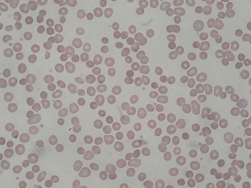
Most common anemia worldwide. The body doesn't have enough iron to make hemoglobin, so the cells come out small and pale.

  • Smear: microcytic, hypochromic (pencil cells = ovalocytes flattened by low hemoglobin) 🔑Pencil cells = pencil thin on iron - they're writing a letter asking for more
  • Iron studies:ferritinFerritin = storage form of iron. Low ferritin = empty warehouse. Most specific single test for iron deficiency., ↓ serum iron, ↑ TIBCTotal Iron Binding Capacity - how many empty seats are on the transferrin bus. High TIBC = lots of empty seats = body desperate for iron., ↓ % saturation
  • Causes: Blood loss (#1 in adults - menstruation, GI bleed), poor intake (infants, vegans), poor absorption (celiac, gastrectomy)
Bone Wizardry pencil cell version Low iron means low hemoglobin. A red cell that should be a full biconcave disc gets stretched into a pale, thin oval, like dough rolled too flat.
Pencil cell schematic for iron deficiency anemia normal disc less Hb inside pencil cell / elliptocyte
Peripheral blood smear showing hypochromic microcytic red cells in iron deficiency anemia
Iron deficiency smear. The cells are small and pale because there is not enough iron to load hemoglobin. Free-use image: Dr Graham Beards, CC BY-SA 3.0.

Thalassemia - Genes Made Less Hemoglobin

What's actually broken? The gene that codes for globin chains is defective. Iron is completely fine. The protein framework is the problem.
So what happens to hemoglobin? Hemoglobin needs both heme AND globin to assemble. No globin chains = the cell can't fill itself up normally → small, pale cell.
Why the bullseye on smear? One teaspoon of jam spread over a big piece of toast: edges have jam, middle looks thin. The membrane is too big for the hemoglobin filling it. That underfilled center folds and creates the pale ring with the dark rim. Target cell.
Iron studies are normal. That's the killer differentiator from iron deficiency. Same small cells, completely different iron labs.
Walk Beta-Thalassemia Like a Disease

Blur → reveal → next beat.

BEAT 1 OF 19
Smear finding
Microcytic + target cells
Why the bullseye? Less hemoglobin makes the membrane look oversized. Pale center = underfilled cytoplasm. Dark rim = membrane folding back on itself. Liver disease is the other lane.
Iron studies key differentiator
NORMAL
Iron stores are completely fine. This is what separates thalassemia from IDA - both are microcytic, but only IDA runs out of iron.
Diagnosis
Hb electrophoresis
Shows the hemoglobin fraction breakdown. Beta-thal raises HbA2. Alpha-thal trait may look normal - the pattern depends on which chain is affected.
  • What the test looks like Beta-thal shifts the bands: HbA falls because HbA needs beta chains, so HbA2 and HbF rise. Alpha-thal often looks normal on adult electrophoresis because HbA, HbA2, and HbF all need alpha chains, so there is no clean beta-to-delta/gamma reroute to expose.
    Normal
    HbA
    HbA2
    HbF
    Beta-thal trait
    HbA
    HbA2
    HbF
    Beta-thal major
    HbA
    HbA2
    HbF

    Diagnosis pearl: beta-thal trait = HbA2 > 3.5%. The broken part is the beta chain, so the lab cannot build normal adult HbA well. Delta chains still work, so some alpha chains pair with delta instead, making extra HbA2. That is why high HbA2 points to beta-thal trait. Alpha-thal is different: HbA, HbA2, and HbF all need alpha, so the fractions can stay proportionally normal and adult electrophoresis may look normal. Severe alpha disease can make HbH after birth or Hb Bart's in the newborn.

  • Genetic defect:tap to expand
    Alpha-thal
    • Deletion of HBA1/HBA2 on chr 16
    • 4 total alpha-gene copies. Severity tracks how many are lost.
    • 1 deleted = silent carrier. 2 = trait. 3 = HbH disease. 4 = Hb Bart hydrops.
    • Adult electrophoresis can look normal in trait (all fractions need alpha).
    Beta-thal
    • Production defect at HBB on chr 11.
    • beta+ = too little beta globin made. beta0 = none made.
    • Two severe hits = major. After HbF falls in infancy, HbA cannot take over.
    • Result: severe microcytic anemia, marrow expansion, extramedullary hematopoiesis, transfusion dependence.
    • Trait = small RBCs, normal iron, often high RBC count, HbA2 > 3.5%.
    Do NOT confuse with HbC disease Thalassemia = not enough globin made. HbC = structurally wrong globin. Hexagonal crystals or very high MCHC = HbC, not thal. Beta-thal trait makes mild microcytosis, not hemolytic anemia with strong reticulocytosis.
    HBA1/HBA2 -> chr 16: read HBA as HemogloBin Alpha. The labels are 1 and 2, and alpha has four total gene copies. Remember it as 41x2 = 16.
    HBB -> chr 11: the beta-globin gene is HBB on chromosome 11.
    HBB chromosome 11 mnemonic H B B = 11 Only look at the two colored backs of the B letters: those two vertical bars are the chromosome number.
    Optional Thalassemia chromosome memory
    CHROMOSOME BEATS1/1
  • How the patients present:tap to expand
    Tap a disease row, then open the bullet you need.
    Alpha silent / trait
    Molecule formed
    Mostly normal HbA with slight alpha chain deficit.
    ααββHbA α₂β₂
    How they show up
    Often asymptomatic or mild microcytosis with normal iron. Electrophoresis can look normal.
    Board clue
    African or Southeast Asian ancestry, small RBCs, no iron deficiency.
    Fatality risk
    Not fatal
    Carrier states matter for counseling, not acute mortality.
    HbH disease
    Molecule formed
    Almost no alpha chains left. Excess beta chains clump together forming HbH.
    α?ββββHbH β₄
    How they show up
    Moderate to severe hemolytic anemia, splenomegaly, jaundice. Those β₄ tetramers precipitate inside the RBC, punching holes in it.
    Board clue
    Microcytosis plus hemolysis, not just a quiet carrier state.
    Fatality risk
    Can decompensate
    Usually survivable, but infections or oxidant stress can trigger dangerous hemolytic crises.
    Hb Bart hydrops
    Molecule formed
    Zero alpha chains. Fetal gamma chains self-pair into Hb Bart's.
    α✗γγγγHb Bart's γ₄
    How they show up
    Fetus cannot make working HbF (which needs alpha). Gamma chains form γ₄ instead, which can't deliver oxygen. Develops hydrops fetalis.
    Board clue
    Alpha chain is needed before and after birth.
    Fatality risk
    Usually fatal
    Lethal in utero or soon after birth without fetal transfusion and intensive support.
    Beta trait / minor
    Molecule formed
    Mostly HbA, but extra alpha chains pair with delta chains instead (making extra HbA2).
    ααδδHbA2 ↑ α₂δ₂
    How they show up
    Mild anemia or incidental microcytosis. RBC count often high because marrow makes many small cells.
    Board clue
    Normal iron studies + HbA2 > 3.5%.
    Fatality risk
    Not fatal
    The board danger is misdiagnosing it as iron deficiency, not sudden death.
    Beta intermedia
    Molecule formed
    Some HbA made (partial beta output), but HbF persists because gamma production compensates.
    ααβγHbA↓ + HbF↑
    How they show up
    Variable anemia, sometimes transfusions during stress, growth delay if severe.
    Board clue
    Lives between trait and major.
    Fatality risk
    Severity varies
    Not automatically fatal, but severe cases can become transfusion-dependent.
    Beta major
    Molecule formed
    No HbA possible. After HbF falls, only broken HbF remnants and HbA2 remain.
    β✗ααγγHbA = 0 - HbF↑↑
    How they show up
    Infant becomes severely anemic after HbF naturally falls. Regular packed RBC transfusions needed to maintain oxygen delivery, growth, cardiac workload.
    Board clue
    Symptoms start after 6 months. Marrow expansion, frontal bossing, transfusion dependence.
    Fatality risk
    Fatal untreated
    Life-threatening in early childhood without chronic transfusions and iron-overload management.
  • Red cell count: Often ↑ (body compensates by making MORE small cells)
Peripheral smear showing target cells in thalassemia
Target cells. This collage includes the bullseye cell in the lower-right panel. Less hemoglobin inside the RBC means the membrane looks relatively oversized, so the smear gets that target look.
The Board's Favorite Trick: Iron Deficiency vs Thalassemia

Both are microcytic. Both have low MCV. The split:

Iron deficiency: ↓ ferritin, ↑ TIBC, ↑ RDW. RDW = red cell distribution width, which means how different the RBC sizes are from each other. In IDA, iron stores fall gradually, so older cells may be closer to normal while newer cells are progressively smaller. Mixed sizes = high RDW.

Thalassemia: Normal iron studies, normal RDW, ↑ RBC count. Here the marrow is making small cells from the start because the globin blueprint is faulty every time, so the cells are uniformly small instead of mixed-size. Think alpha-globin gene deletions or beta-globin mutations causing reduced chain production.
Board Trap
"Microcytic anemia that doesn't respond to iron supplementation" = think thalassemia. If they gave iron for weeks and the MCV didn't budge, the problem was never iron.

Sideroblastic Anemia - Iron Trapped in the Wrong Place

What's happening here? The body has plenty of iron. The heme factory is broken. Iron enters the developing red cell, hits a wall, and piles up inside the mitochondria around the nucleus instead of getting built into hemoglobin.
What does that look like on biopsy? The iron-loaded mitochondria form a ring around the nucleus. That's a ringed sideroblast. The name is literally what it looks like.
Iron stores are full (high ferritin, high serum iron), but the cells are still small and pale. The warehouse is stuffed. The factory can't use it.
Bone marrow smear showing a ringed sideroblast with iron granules encircling the nucleus
Ringed sideroblast. The blue/green granules are iron-loaded mitochondria that have formed a collar around the nucleus. They piled up because lead (or alcohol, B6 deficiency, isoniazid) broke the heme assembly line. The iron had nowhere to go. Free-use image: Makysm, CC0.
Causes: lead, alcohol, B6 deficiency, isoniazid, MDS Smear: microcytosis + basophilic stippling Iron labs: HIGH ferritin, HIGH serum iron

Lead Poisoning - Two Enzymes, One Disaster

Lead hits two specific points in the heme synthesis pipeline. Everything else in lead poisoning follows from that.
Succinyl-CoA + Glycine
ALA synthase
ALA (aminolevulinic acid)
LEAD BLOCKS ALA dehydratase → ALA builds up in urine
Porphobilinogen (PBG)
...several steps...
Protoporphyrin IX
LEAD BLOCKS Ferrochelatase (Fe²⁺ insertion) → FEP rises in red cells
HEME(never made)
What even is ALA in urine? ALA (aminolevulinic acid) is the precursor that would become heme. ALA dehydratase is supposed to push it forward. Lead blocks the enzyme. ALA has nowhere to go → it spills into urine. That's your screening marker for lead exposure.
What even is FEP? FEP = Free Erythrocyte Protoporphyrin. Ferrochelatase was supposed to stick iron into protoporphyrin to finish building heme. Lead blocks it. Naked protoporphyrin piles up inside the red cell with no iron attached. That's FEP. When lead hits, FEP skyrockets.
Peripheral smear showing basophilic stippling with blue dots in red cells from lead poisoning
Basophilic stippling. The blue dots you see inside the red cells are clumps of retained ribosomal RNA. Normally the maturing red cell destroys its RNA before it leaves the marrow. Lead disrupts this process, so RNA gets trapped. Under the microscope, it coagulates into visible blue specks. Classic for lead and also seen in thalassemia and sideroblastic anemia. Free-use image: Dr. Roshan Nasimudeen, CC BY-SA 4.0.

Lead's Other Damage (Beyond the Blood)

Burton's lines: blue-black band at the gumline of a lead-mill worker
Burton's Lines
Blue-black band at the gumline. Lead deposits in gingival tissue as lead sulfide. The dark stripe appears exactly where the teeth meet the gum. Lead also deposits at growth plate lines on X-ray (dense white bands at the ends of long bones in kids). Wellcome Collection, public domain.
Foot drop: inability to dorsiflex the foot due to peroneal nerve damage
Wrist / Foot Drop
Lead demyelinates the radial nerve (wrist drop, can't extend wrist) and peroneal nerve (foot drop, can't dorsiflex foot). Peripheral motor neuropathy, not central. Classic in adults with occupational lead exposure. Commons, public domain.
Abdominal Colic
Lead causes smooth muscle spasm in the GI tract. Severe crampy abdominal pain with constipation. Often misdiagnosed as appendicitis or obstruction. No peritoneal signs - the abdomen is soft.
Encephalopathy (Kids)
Lead crosses the blood-brain barrier easily in children. It swaps for calcium in neural signaling. Result: cerebral edema, seizures, vomiting, papilledema. Even low levels cause permanent IQ drop. No safe level exists in kids. Classic trigger: old paint chips.

The Anemia Villains

Each anemia type is a villain with a different weapon. Tap a card to flip it and see the lab signature that gives them away.

Villain #1
Iron Deficiency

The empty warehouse. Body is out of iron. Cells come out thin and pale.

Tap to see how to catch it →
How to catch IDA:
  • Ferritin: LOW <12 (empty stores)
  • TIBC: HIGH >400 (desperate for iron)
  • Serum iron: LOW
  • RDW: HIGH (mixed cell sizes)
  • Smear: pencil cells, hypochromic
  • Key clue: menstrual loss in women; GI bleed in men >40
Villain #2
Thalassemia

The genetic imposter. Looks like IDA but iron studies are completely normal.

Tap to expose the imposter →
How to catch thalassemia:
  • Iron studies: NORMAL
  • RBC count: HIGH (makes more small cells)
  • RDW: Normal. RDW = how much RBC size varies. Thal cells are usually uniformly small.
  • Smear: target cells (bullseye)
  • Confirm: hemoglobin electrophoresis for beta-thal. Alpha-thal may have normal electrophoresis.
  • Key clue: Mediterranean, African, Middle Eastern, or Southeast Asian ancestry depending on alpha vs beta type
Villain #3
Sideroblastic

The hoarder. Has iron but can't use it. Iron piles up in the mitochondria.

Tap to see the stash →
How to catch sideroblastic:
  • Ferritin: HIGH or normal (iron accumulates)
  • Serum iron: HIGH
  • TIBC: Normal or low
  • Smear: basophilic stippling
  • Confirm: ringed sideroblasts on bone marrow biopsy
  • Causes: lead, alcohol, INH, B6 deficiency
Villain #4
Anemia of Chronic Disease

The saboteur. Body hides iron on purpose via hepcidin. Has iron. Won't share it.

Tap to unmask ACD →
How to catch ACD:
  • Ferritin: HIGH (iron locked in macrophages)
  • TIBC: LOW (not even looking for more)
  • Serum iron: LOW (can't get to marrow)
  • MCV: usually normal, can be mildly low
  • Smear: normochromic or mildly hypochromic
  • Context: active RA, TB, malignancy, CKD
Villain #5
B12 Deficiency

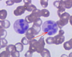
The neurologist's nightmare. Big cells AND neuro damage. The only one that hurts your spine.

Tap to see the damage →
How to catch B12 deficiency:
  • MCV: >100 (macrocytic)
  • Smear: hypersegmented neutrophils, macro-ovalocytes
  • MMA: HIGH (only in B12, not folate)
  • Homocysteine: HIGH
  • Neuro: subacute combined degeneration
  • Cause: pernicious anemia (no intrinsic factor)
Villain #6
Hemolytic Anemia

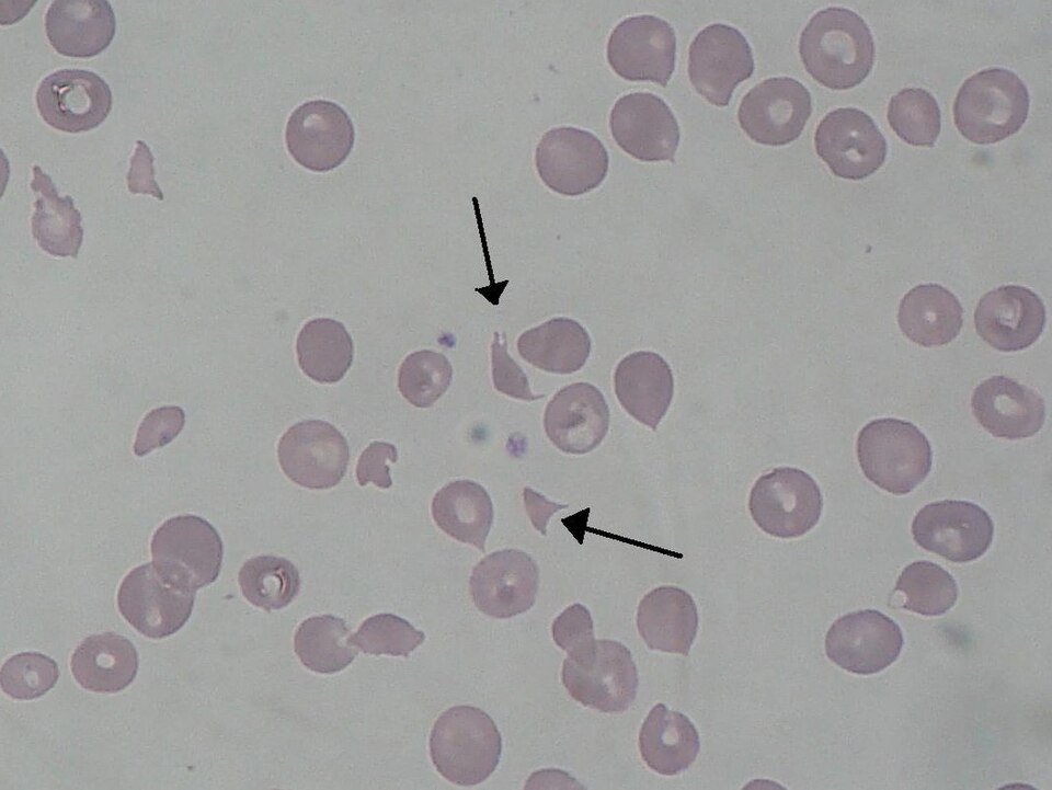
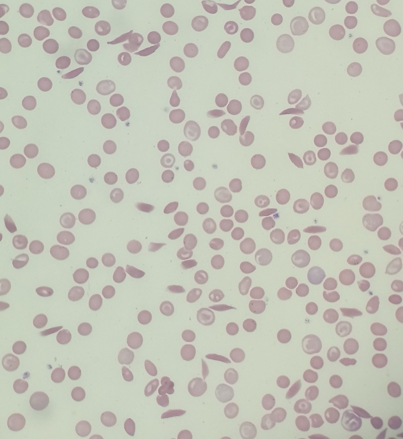
The destroyer. Cells are made fine but burst before their time. Marrow can't keep up.

Tap to see the wreckage →
How to catch hemolysis:
  • LDH: HIGH (cells spilling contents)
  • Indirect bili: HIGH (Hgb breakdown)
  • Haptoglobin: LOW (consumed binding free Hgb)
  • Reticulocytes: HIGH (marrow compensating)
  • Intravascular: schistocytes, hemoglobinuria
  • Extravascular: spherocytes, splenomegaly

Clinical Images

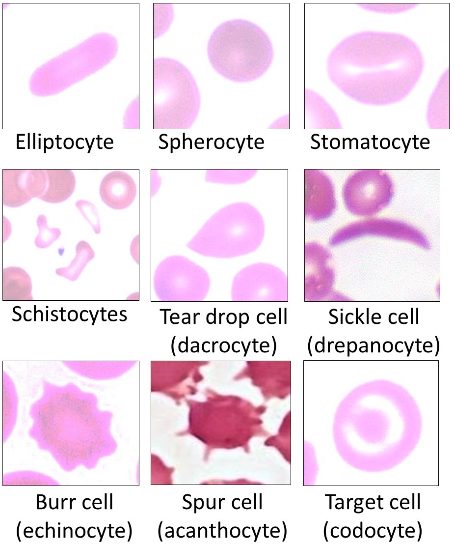
Free-use morphology anchors for the diseases above. Tap any image to expand.

Iron deficiency anemia smear with hypochromic microcytic red cells
Iron deficiency
Peripheral smear with target cells
Target cells
Ringed sideroblast on bone marrow smear
Ringed sideroblast
Basophilic stippling on peripheral smear
Lead stippling
Hypersegmented neutrophil in megaloblastic anemia
Hypersegmented PMN
Schistocytes on peripheral blood smear
Schistocytes
Spherocytes on peripheral blood smear
Spherocytes
Peripheral smear showing sickled red blood cells
Sickle cells

Why the Labs Look the Way They Do

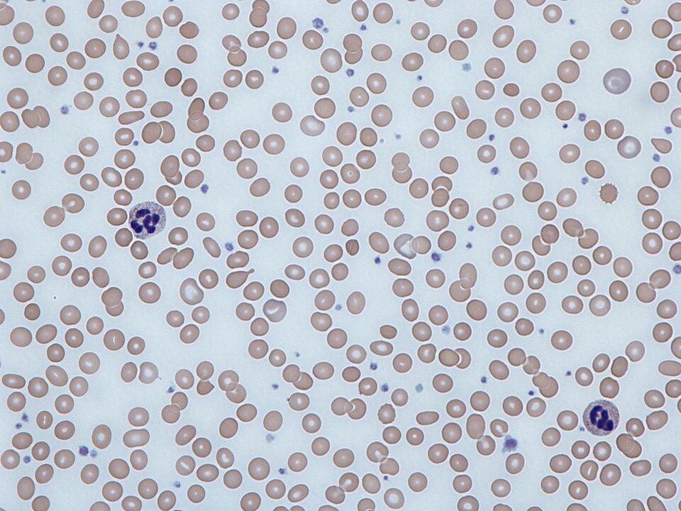
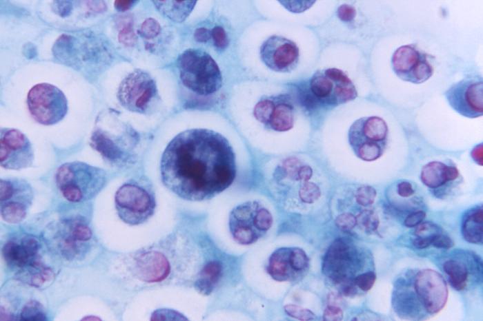
Hemolytic Anemia Lab Logic

The labs in hemolysis aren't random. Every single one is a direct consequence of red cells bursting. Once you understand the chain, you stop memorizing and start deriving. Tap each lab to reveal why it goes the way it does.
LDH ↑ why high?
Red blood cells are packed with enzymes. LDH (lactate dehydrogenase) is one of them - it manages energy inside the cell. When a cell bursts, its contents flood into the bloodstream, LDH included. High LDH = cells are leaking. Not specific - MI, liver injury, and rhabdomyolysis also raise it. But very sensitive: if LDH is normal, cells aren't bursting in significant numbers.
Optional - Biostats drill
A test is highly sensitive for disease X. A patient tests negative. What does that tell you?
Very unlikely to have disease X. High sensitivity = few false negatives. A negative result rules it out. SnNout: Sensitive test, Negative = rule Out.
LDH rises in MI, hepatitis, hemolysis, rhabdomyolysis. What does that tell you about its specificity?
Low specificity. Many conditions trigger it. A positive LDH can't confirm hemolysis - you need haptoglobin, indirect bili, and smear to pin it down.
You want to screen for hemolysis. You'd rather miss no one than have a few false alarms. Which property matters more?
Sensitivity. Screening = catch everyone. LDH's high sensitivity makes it a good screener. Use LDH to screen, haptoglobin to confirm.
tap to reveal
Indirect Bili ↑ why high?

Every red cell that dies gets taken apart. Hemoglobin breaks down into heme, and heme becomes bilirubin. The indirect form is the raw, freshly made version before the liver conjugates it. When cells die faster than the liver can process them, indirect bilirubin backs up in the blood. Enough backup and the patient turns yellow. The liver is not failing. It is buried under the load.

tap to reveal
Haptoglobin ↓ why low?

Haptoglobin is a blood protein whose entire job is to grab free hemoglobin that spills out when red cells burst. Think of it as the cleanup crew. When a lot of cells burst fast, free hemoglobin floods the plasma, and haptoglobin binds it until the supply runs out. Empty crew = it was all used up = active hemolysis. Low haptoglobin is one of the most specific signs of hemolysis.

tap to reveal
Reticulocytes ↑ why high?
Reticulocytes are young red cells that normally spend 1-2 days maturing in the bloodstream before becoming adult cells. The bone marrow releases them a little early when demand goes up. When cells are being destroyed faster than the marrow can fully replace them, it panic-ships immature cells early, still carrying residual RNA. High reticulocytes = the marrow is in overdrive. It's the bone marrow saying: we're losing them faster than we're making them.
tap to reveal

Intravascular vs Extravascular

Schistocytes what are those?
Helmet-shaped or triangular cell fragments. A red cell gets physically sheared apart while still inside a blood vessel - a fibrin strand, a damaged vessel wall, or a mechanical heart valve slices through it. Each fragment was once a whole cell. You see schistocytes in TTP, HUS, DIC, mechanical valves: anything that physically chops cells mid-stream.
tap to reveal
Hemoglobinuria dark urine?
When cells burst inside vessels, free hemoglobin floods the plasma. The kidneys filter it out into the urine. The haptoglobin cleanup crew got overwhelmed and couldn't catch it all - so it spills into the urine instead. Cola-colored urine in someone with no prior kidney disease = intravascular hemolysis until proven otherwise.
tap to reveal
Spherocytes why round?
Red cells going through the spleen get nibbled by macrophages. The macrophages chew off membrane fragments - but leave the hemoglobin inside. Less membrane, same contents = the cell rounds into a sphere. Spherocytes have no central pallor. The spleen destroys them on the next pass. Overworked spleen gets bigger = splenomegaly.
tap to reveal
The Rule which is which?
Intravascular = cells burst inside vessels. Schistocytes + hemoglobinuria + very low haptoglobin. Violent, dramatic, happens fast.

Extravascular = spleen destroys cells. Spherocytes + splenomegaly + less dramatic haptoglobin drop. Slower, chronic, macrophage-mediated.
tap to reveal

Macrocytic Anemias

Big cells = problem with DNA synthesis. The cell wants to divide but can't, so it just keeps growing. Split into two lanes: megaloblastic (B12/folate, you'll see hypersegmented neutrophils) and non-megaloblastic (alcohol, liver disease, hypothyroid, no hyperseg on smear).

B12 Deficiency - The Neuro One

Megaloblastic anemia + neurological symptoms. B12 is needed for DNA synthesis AND myelin maintenance.

  • Smear: macro-ovalocytes + hypersegmented neutrophils (≥5 lobes = pathognomonic for megaloblastic anemia)
  • Neuro: Subacute combined degeneration - loss of position sense, vibration sense, ataxia. Affects dorsal columnsPosterior columns of the spinal cord carry proprioception and vibration. B12 deficiency demyelinates these first. Patient can't tell where their feet are without looking. and lateral corticospinal tractsUpper motor neuron tracts. Damage here causes spasticity, hyperreflexia, positive Babinski. Combined with dorsal column loss = "subacute combined degeneration."
  • Causes: Pernicious anemia (#1 - autoimmune destruction of parietal cellsStomach cells in the gastric body and fundus. They make hydrochloric acid and intrinsic factor. If autoimmune disease destroys them, B12 cannot be absorbed well. → no intrinsic factorMade by parietal cells. Binds B12 in the stomach, carries it to the terminal ileum for absorption. No IF = no B12 absorption regardless of intake.), vegan diet, Crohn's/ileal resection, Diphyllobothrium latum (fish tapeworm)
  • Pernicious vs megaloblastic: Megaloblastic describes the blood picture from broken DNA synthesis. Pernicious names the autoimmune B12-malabsorption cause. Same smear pattern, but pernicious adds anti-intrinsic factor/parietal cell antibodies, lifelong B12 replacement, and gastric cancer risk.
Medical distinction: Parietal cells are stomach cells that make acid and intrinsic factor. Pernicious anemia is the autoimmune B12-malabsorption cause of megaloblastic anemia, not a separate smear pattern.
Peripheral blood smear showing a hypersegmented neutrophil with 6 nuclear lobes in megaloblastic anemia
Hypersegmented neutrophil (5+ lobes). Normally neutrophils have 2-4 lobes. In B12/folate deficiency, DNA synthesis slows and the nucleus can't divide properly, so it just keeps lobing. A neutrophil with 5 or more lobes = megaloblastic until proven otherwise. One of these on a smear is enough to flag the diagnosis. Public domain image.

Folate Deficiency - The Diet One

Same megaloblastic picture as B12 - hypersegmented neutrophils, macro-ovalocytes. But NO neuro symptoms.

  • Causes: Poor diet (#1 - alcoholics, elderly), pregnancy (↑ demand), methotrexate/phenytoin/TMP-SMX (folate antagonists)
  • Key difference from B12: Folate stores only last ~4 months. B12 stores last ~3-4 years. Folate deficiency develops FAST.
Tiny pop quiz: Why do folate antagonists cause macrocytosis?
Drug logic: Methotrexate intentionally blocks human folate recycling to slow rapidly dividing immune, tumor, or trophoblast cells. TMP-SMX blocks bacterial folate production because bacteria make folate from scratch. Phenytoin is the odd one: folate lowering is an adverse effect, not the therapeutic goal.
B12 vs Folate - The One Difference That Matters

Both: megaloblastic, hypersegmented neutrophils, macro-ovalocytes, ↑ homocysteine

Only B12: ↑ methylmalonic acid (MMA), neurological symptoms

Only folate: Normal MMA, no neuro 🔑MethylMalonic acid = only in B12 → Myelin problems. Both M's go together.
Board Trap
Never give folate alone to a B12-deficient patient. Folate fixes the anemia (hides the blood findings) but the neuro damage KEEPS PROGRESSING silently. Always check B12 before treating with folate.

Non-Megaloblastic Macrocytic

Big cells but NO hypersegmented neutrophils. DNA synthesis is fine - the cell membrane is just weird.

  • Alcohol: Direct toxic effect on RBC membrane + usually folate-deficient too
  • Liver disease: Excess cholesterol deposits in RBC membranes → cells swell
  • Hypothyroidism, reticulocytosis (reticulocytes are bigger than mature RBCs), myelodysplastic syndrome
The easy split: sometimes the RBC is big because it cannot divide DNA. Other times it is big because the marrow or membrane is acting strange. These three are the strange-but-testable ones.
Hypothyroidism why big?
Thyroid hormone is a marrow gas pedal. Low thyroid slows production and can make red cells mature sluggishly. The result is often mild macrocytosis with no hypersegmented neutrophils. Board move: unexplained macrocytosis + tired/cold/constipated → check TSH.
tap to reveal
Reticulocytosis why big?
Reticulocytes are baby RBCs, and baby RBCs are larger than mature RBCs. If the marrow is dumping them out after bleeding or hemolysis, the average cell size rises. This is macrocytosis from a good marrow response, not a B12 or folate problem.
tap to reveal
MDS what is it?
Myelodysplastic syndrome is a broken bone marrow factory. A clonal stem cell keeps making blood-cell precursors, but they mature badly and fail quality control. Think older patient, macrocytosis, low counts in more than one cell line, normal B12/folate, dysplastic marrow. The danger is transformation to AML.
tap to reveal

Normocytic Anemias

MCV is normal. The cells are the right size - there just aren't enough of them, or they're being destroyed. Split by reticulocyte count.

Reticulocyte count = is the bone marrow TRYING?

↑ Reticulocytes (>2%): Marrow is working overtime → cells are being DESTROYED or LOST (hemolysis, bleeding)

↓ Reticulocytes (<2%): Marrow is failing → cells aren't being MADE (aplastic, ACD, renal failure)

Hemolytic Anemias - Cells Dying Too Fast

Red cells are being destroyed before their 120-day lifespan. The marrow tries to compensate → ↑ reticulocytes.

  • General labs:LDHLactate dehydrogenase - released when cells burst. Elevated in any hemolysis. Not specific but very sensitive., ↑ indirect bilirubin, ↓ haptoglobinHaptoglobin binds free hemoglobin released from lysed cells. When lots of cells lyse, haptoglobin gets used up → levels drop. Low haptoglobin = hemolysis until proven otherwise., ↑ reticulocytes
  • Intravascular hemolysis (in the blood vessels): hemoglobinuria, hemoglobinemia, schistocytes on smear
  • Extravascular hemolysis (in the spleen): splenomegaly, spherocytes on smear
Peripheral smear showing schistocytes in microangiopathic hemolysis
Schistocytes. Intravascular hemolysis shears cells inside vessels, leaving helmet-like fragments.
Peripheral smear showing spherocytes in extravascular hemolysis
Spherocytes. Extravascular hemolysis removes membrane in the spleen, leaving round cells without central pallor.

Anemia of Chronic Disease - The Body's Sabotage

What's happening in ACD? Chronic inflammation tells the liver to pump out hepcidinHepcidin is the iron gatekeeper. It blocks ferroportin on gut cells and macrophages, trapping iron inside cells and macrophages. The body hides iron from bacteria during infection, but it accidentally hides it from your own red cells too.. Hepcidin locks iron inside macrophages and gut cells. The marrow can't access it.
Iron is present but locked. Ferritin is high (full warehouse). TIBC is low (not asking for more). Giving iron won't help. Treat the disease causing the inflammation.
ACD mechanism map. Inflammation makes the liver release hepcidin. Hepcidin locks ferroportin on gut cells and macrophages. Iron stays trapped in storage, transferrin cannot deliver enough to marrow, and RBC production stalls.
CDC public-domain macrophage activation image used as the macrophage-side anchor for iron recycling
Macrophage-side anchor. Macrophages recycle iron from old RBCs. In ACD, hepcidin blocks ferroportin, so recycled iron stays inside the macrophage pool. Public domain CDC PHIL #10613 image.
CC0 small-intestine mucosa histology showing crypt cells and Paneth cells
Gut-cell-side anchor. Intestinal epithelial cells normally pass absorbed iron into blood through ferroportin. Hepcidin closes that exit, so less dietary iron reaches plasma. CC0 Wikimedia Commons image.
  • Iron studies: ↑ ferritin (iron is there, just locked up), ↓ TIBC (body isn't looking for more iron), ↓ serum iron
  • Usually normocytic but can be mildly microcytic
  • Treatment: Treat the underlying disease. Iron supplements won't help - the iron is already there.
Iron Deficiency vs Anemia of Chronic Disease - Boards LOVE This
Iron DeficiencyChronic Disease
Ferritin↓↓↓ (empty)↑ (locked up)
TIBC↑ (hungry)↓ (not looking)
Serum iron
% Sat

Both have low serum iron. The split is ferritin (empty vs full warehouse) and TIBC (desperate vs indifferent).


The Anemia Algorithm

Patient is anemic. Walk through the decision tree. I'll quiz you at each branch.

Step 1: You get the CBC back. What's the FIRST number you look at?
Reticulocyte count
MCV
Ferritin
Step 2: MCV is 72 (microcytic). What do you order next?
Iron studies (ferritin, TIBC, serum iron)
B12 and folate levels
Bone marrow biopsy
Step 3: MCV is 110 (macrocytic). Smear shows hypersegmented neutrophils. Is this megaloblastic or non-megaloblastic?
Megaloblastic → check B12 and folate
Non-megaloblastic → check liver, thyroid, alcohol
Step 4: MCV is 88 (normocytic). What splits the differential?
Iron studies
Reticulocyte count
Direct Coombs test
You walked the tree.
MCV → bucket → specific workup. That's the algorithm. On test day, don't get fancy. Follow the tree.

Decision Tree: Anemia Workup by MCV

Use the CBC like a routing console: size the red cells first, then order the lab that actually separates the bucket.

CBC routing console
Pick the MCV lane. The next test changes immediately.
Checkpoint 1 Hgb is low The patient is anemic. Do not guess the cause yet.
Checkpoint 2 Read the MCV Small, normal, or large cells decide the first fork.
Checkpoint 3 Order one splitter Iron studies, retics, or B12/folate. Not everything at once.
The CBC is back. Which cell-size lane owns the workup?
Pick a lane. The page will keep the route visible so the algorithm stays in working memory.
Route<80

Microcytic route: iron studies before electrophoresis.

Small cells usually mean heme or globin production failed. Ferritin and TIBC decide whether iron is missing, trapped, or not the problem.

Choose the lab pattern. Ferritin is the hinge: empty, locked, normal, or overloaded.
Iron deficiency anemiaLow ferritin seals it. Next move: find the source, especially GI blood loss in older adults, then replete iron.
Source hunt
Thalassemia patternMicrocytosis with normal iron points to globin chain underproduction. Electrophoresis helps beta-thalassemia; alpha may need genetic context.
Hb panel
Anemia of chronic diseaseInflammation raises hepcidin, trapping iron in storage. Ferritin rises because the warehouse is full but locked.
Inflammation
Sideroblastic anemiaIron reaches the marrow but cannot enter heme efficiently. Think alcohol, B6 deficiency, lead, INH, or MDS.
Marrow clue
Route80-100

Normocytic route: reticulocytes tell you production vs loss.

The cells are normal-sized. The key is whether the marrow is trying hard enough to replace what is missing.

Use reticulocytes as the marrow tachometer. High means demand is being met; low means production is failing.
Blood loss or hemolysisHigh retics mean the marrow is trying. Add LDH, indirect bilirubin, haptoglobin, smear, and DAT when hemolysis is on the table.
Hemolysis labs
Production problemLow retics point to underproduction: CKD with low EPO, aplastic anemia, marrow infiltration, hypothyroidism, or chronic disease.
TSH/Cr/marrow
Route>100

Macrocytic route: smear decides megaloblastic vs not.

Large cells need a DNA-synthesis check first. Hypersegmented neutrophils push you toward B12 and folate.

Do not give folate alone if B12 is possible. Folate can fix the anemia while neurologic damage continues.
B12 deficiencyBoard giveaway: neurologic symptoms, elevated MMA, pernicious anemia, gastric surgery, ileal disease, or vegan diet.
B12 first
Folate deficiencyMegaloblastic anemia without neurologic deficits. Replete folate only after B12 is ruled out or treated too.
Folate
Non-megaloblastic macrocytosisNormal B12/folate or nonmegaloblastic smear should send you to alcohol/liver pattern, TSH, medication review, and marrow disease when older.
LFT/TSH

Board Walkthrough

Board-style vignettes. One at a time. Cross out wrong answers, highlight the clue. Reveal the chain.

Question 1
Board-Style Walkthrough

Board-Style Walkthrough

Original board-style vignettes. Shuffled, never-repeat, full explanations for every choice.