Anemias Deep Dive

Macrocytic, Normocytic & PNH

Iron Studies Decoder

Random iron panel pops up. Read the four arrows. Pick the anemia. The pattern is the test · memorize the table once and you own the bench question.

CORRECT 0
SEEN 0
Serum Fe
·
60-170
TIBC
·
240-450
Ferritin
·
15-200
% Sat
·
20-50
Tap Roll to pull a random patient's iron panel.
From the Attending
Four labs, four anemias, one table. IDA: Fe down, TIBC up, Ferritin down. ACD: Fe down, TIBC down, Ferritin up. Sideroblastic: Fe up, TIBC down, Ferritin up. Hemochromatosis: Fe up, TIBC down, Ferritin way up, sat > 60. The ferritin direction breaks every tie.

๐Ÿฅ MCV Triage Station

Read the scenario, classify by MCV, then click to drill down to the diagnosis.

๐Ÿ“ˆ Macrocytic (MCV >100)

Defective nuclear division

โ†’ Normocytic (MCV 80-100)

Normal RBC size, not enough of them

๐Ÿ“‰ Microcytic (MCV <80)

Iron issues or thalassemia

From the Attending

MCV is the first triage gate on every anemia stem. <80 = microcytic → iron deficiency, thalassemia, anemia of chronic disease (late), lead, sideroblastic. 80·100 = normocytic → ask: hemolysis (retics up) or underproduction (retics down, BM, kidney, ACD)? >100 = macrocytic → ask: megaloblastic (B12, folate, drugs) or non-megaloblastic (liver, alcohol, hypothyroid, MDS, reticulocytosis)? The single number splits the entire workup tree · never start anemia from "what's the Hgb"; start from MCV + retics.

Clinical Images

Hypersegmented neutrophil
Hypersegmented neutrophil ยท megaloblastic
Hypersegmented neutrophil in megaloblastic anemia
Macrocytes ยท pernicious anemia smear
Urine color spectrum including dark urine in hemoglobinuria
Hemoglobinuria ยท dark morning urine (PNH)
Megaloblastic marrow
Megaloblastic marrow ยท giant cells
Aplastic anemia bone marrow biopsy
Aplastic anemia ยท hypocellular marrow biopsy

The Villains: Key Diagnoses

Tap each card to reveal the crime file (key labs and clinical features).

๐Ÿ”ด
B12 Deficiency
Megaloblastic

Crime File

MCV: >100 (macrocytic)
B12: Low
Methylmalonate: Elevated (pathognomonic)
Homocysteine: Elevated
Smear: Hyperseg neutrophils
Neuro: Subacute combined degeneration
Cause: Pernicious anemia (anti-parietal cell Ab)
๐ŸŸก
Folate Deficiency
Megaloblastic

Crime File

MCV: >100 (macrocytic)
Folate: Low
Methylmalonate: Normal (key differentiator)
Homocysteine: Elevated
Neuro: NONE (never)
Cause: Overcooked vegetables, methotrexate
๐Ÿฉธ
PNH
GPI Anchor Defect

Crime File

MCV: Normocytic (hemolytic)
Retics: Elevated
CD55/CD59: Absent on flow cytometry
Urine: Hemoglobinuria (dark morning void)
Thrombosis: Budd-Chiari, portal, cerebral veins
Rx: Eculizumab (C5 blocker)
๐Ÿฆด
MDS
Myelodysplastic Syndrome

Crime File

MCV: Often macrocytic
CBC: Pancytopenia (all lines low)
Smear: Hyperseg neutrophils, ringed sideroblasts
Marrow: Hypercellular but ineffective
Risk: Transforms to AML (leukemia risk)
Age: Older patients (>60)
๐Ÿชฃ
Aplastic Anemia
Marrow Failure

Crime File

MCV: Normocytic
CBC: Pancytopenia
Retics: Very low (marrow not responding)
Marrow biopsy: Hypocellular, replaced by fat
Cause: Idiopathic (autoreactive T-cells) #1
Rx: ATG + cyclosporine, or BMT
๐Ÿซ˜
Renal Failure
EPO Deficiency

Crime File

MCV: Normocytic
Retics: Low (marrow not stimulated)
EPO level: Inappropriately low
Creatinine: Elevated (CKD >Stage 3)
Rx: Procrit / Epogen / Darbepoetin

Macrocytic Workup: Decision Tree

Follow the algorithm to reach the diagnosis.

MCV > 100 fL detected
โ†“ Check B12 and folate levels
Are B12 and/or folate LOW?
ย 
YES โ†’ B12 or Folate Low
Is methylmalonate ELEVATED?
ย 
YES โ†’
B12 Deficiency
Check for neuro sx, pernicious anemia Ab
NO โ†’
Folate Deficiency
No neuro sx. Check diet, methotrexate
NO โ†’ B12 and Folate Normal
Check smear + TSH + med list
ย 
Anticonvulsant use?
Drug-induced macrocytic anemia
Phenytoin blocks Ca + folate
Hypothyroid / Alcohol?
Hypothyroidism or Alcohol
TSH elevated or EtOH history
โ†“ Smear shows hypersegmented neutrophils in B12/folate deficiency
Still unclear? โ†’ Order bone marrow biopsy to rule out MDS

๐Ÿ“ˆ Macrocytic Anemias (MCV >100)

Defective nuclear division slows down, RBCs become bigger

From the Attending

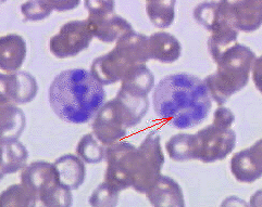
Macrocytic splits cleanly: megaloblastic (B12 or folate) vs non-megaloblastic. The smear tells you: hypersegmented neutrophils (>5 lobes) = megaloblastic. B12 vs folate · both give pancytopenia and macrocytosis, but only B12 deficiency causes subacute combined degeneration (posterior columns + corticospinal tracts → loss of vibration/proprioception + UMN signs). Folate gives no neuro findings. Pregnancy + macrocytic + no neuro = folate. Vegan or pernicious anemia + numb feet = B12. Give folate alone to a B12-deficient patient and you'll fix the anemia but accelerate the neuro damage.

The Core Problem: Nuclear division is broken or slowed. Cytoplasm keeps growing while the nucleus doesn't replicate โ†’ big, immature RBCs (megaloblasts) that don't work well.

B12 vs Folate: The Key Difference

Feature Vitamin B12 Deficiency Folate Deficiency
Anemia Type Megaloblastic Megaloblastic
Neurologic Symptoms YES - Subacute Combined Degeneration NO - Never
Paresthesias Dorsal columns affected Not present
Elevated Homocysteine YES YES
Elevated Methylmalonate YES (B12-specific) NO
Most Common Cause Type A Gastritis (Pernicious) Overcooked vegetables
Key triggers: Methylmalonate elevatedMethylmalonate is B12-specific. Folate deficiency does NOT raise methylmalonate. If both homocysteine AND methylmalonate are up, it is B12 deficiency no matter what the folate level looks like. Subacute combined degenerationB12 is needed to make myelin. Without it, dorsal columns (position sense, vibration) and lateral corticospinal tracts (weakness) both degenerate. Pattern: feet numb first, weakness climbing up. Folate never does this. Intrinsic factor linkIntrinsic factor (IF) is made by parietal cells in the stomach fundus. Pernicious anemia = autoimmune attack on parietal cells. No IF = no B12 absorption in terminal ileum. That is why gastrectomy patients also get B12 deficiency. Phenytoin blocks what?Phenytoin blocks two things: (1) Calcium channels needed for microtubule formation during cell division, and (2) Folate absorption. Result: macrocytic anemia. Boards love pairing "patient on phenytoin + MCV over 100."

๐Ÿ”ด Vitamin B12 Deficiency

Cofactors B12 Regulates:

  • Malonyl-CoA Mutase - needed for fatty acid synthesis (can't build myelin without this)
  • Homocysteine Methyl Transferase - converts homocysteine to methionine
๐Ÿ”‘
Memory Hook
Pernicious = Deadly Serious. Type A gastritis = immune attack on stomach parietal cells โ†’ kills intrinsic factor production. No IF = no B12 absorption in terminal ileum = neurologic catastrophe (weak legs, numbness in feet climbing up like a ladder).

Most Common Cause: Type A Gastritis (Pernicious Anemia)

  • Autoimmune attack on stomach parietal cells
  • Kills production of intrinsic factor (IF)
  • B12 cannot be absorbed in terminal ileum without IF
  • Results in megaloblastic anemia + neurologic damage

Neurologic Features (Subacute Combined Degeneration):

  • Paresthesias (numbness/tingling) in feet
  • Weakness progressing upward
  • Loss of position sense (dorsal column pathology)
  • Ataxia (uncoordinated movement)

Diagnosis Clue:

Elevated methylmalonate is pathognomonic for B12 deficiency (not seen in folate deficiency)

๐ŸŸก Folate Deficiency

๐Ÿ”‘
Memory Hook
FOLate = FOLiage = Leaves. Overcooked vegetables kill folate (heat-sensitive). First vitamin to run out when cells divide rapidly. No neurologic symptoms ever (unlike B12).

Why Folate First?

Folate is the first vitamin to become depleted during periods of rapid cellular division because the body has minimal stores. Unlike B12 (which can be stored for years), folate reserves last only months.

Most Common Cause: Overcooked Vegetables

  • Folate is destroyed by heat
  • Excessively cooked food = no folate
  • Causes megaloblastic anemia
  • NO neurologic symptoms

Other Causes:

  • Methotrexate (blocks folate metabolism)
  • Malabsorption: Crohn's disease, celiac sprue, cystic fibrosis
  • Rapid cell turnover (cancer, hemolysis)

Key Difference:

Folate deficiency causes anemia only. Never causes neurologic symptoms (no myelin damage).

Other Causes of Macrocytic Anemia

Hypothyroidism

Thyroid hormones are permissive for all body functions. Without them, everything slows down โ†’ including bone marrow. RBCs grow large but slowly.

Alcohol

Denatures all proteins. Impairs nuclear division directly. Also commonly causes folate deficiency (poor diet + malabsorption).

Chemotherapy

Drugs damage DNA and stop nuclear replication. Cytoplasm keeps growing โ†’ macrocytic cells.

โš ๏ธ Anticonvulsant Drugs & Macrocytic Anemia

All anticonvulsants that cause macrocytic anemia block calcium and/or folate metabolism. Calcium is needed for microtubules during cell division.

Phenytoin
Blocks Calcium
Blocks Folate
Ethosuximide
Blocks Calcium
Valproic Acid
Blocks Sodium
Blocks Calcium
Blocks Folate
Carbamazepine
Blocks Sodium
Blocks Calcium
Board Trap: Student sees "macrocytic anemia + seizure medication" and thinks epilepsy first. Actually think: Which drug blocks folate/calcium?

โ†’ Normocytic Normochromic Anemias

Normal MCV, Normal MCH โ†’ nothing wrong with the RBC itself, just not enough of them

From the Attending

Normocytic forks on reticulocyte count. Retics up (>2% corrected) = bone marrow is responding · you're losing or destroying RBCs (hemorrhage, hemolysis). Then ask: extravascular (spleen) vs intravascular (haptoglobin down, LDH up, hemoglobinuria). Retics down or inappropriately normal = marrow can't keep up · think kidney (low EPO), chronic disease, marrow failure (aplastic, MDS, infiltration), early iron deficiency. "Normocytic + retics 4% + indirect bili up + LDH 800 + haptoglobin undetectable" is intravascular hemolysis until proven otherwise. The retic count is the second triage gate after MCV.

The Core Problem: RBCs look normal, but the body isn't making enough of them or is destroying them too fast.

Timeline-Based Normocytic Causes

Acute Hemorrhage (<4 days)

When a patient bleeds out suddenly, the body loses RBCs faster than it can replace them. Reticulocytes (immature RBCs) don't show up until day 3-4. Before that: normal RBC size, low count.

Board Clue: Patient just had a car accident, lost blood. Labs show Hgb 8.5 with low reticulocyte count. That's acute โ†’ the marrow hasn't had time to respond yet.

Chronic Disease (Early Phase, First 60 Days)

Infections, cancer, rheumatologic diseases trigger mild anemia by damping down erythropoietin (EPO). Before iron stores fully deplete (which takes months), the anemia is normocytic.

Renal Failure

Kidneys make erythropoietin (EPO). No kidneys = no EPO = bone marrow doesn't make RBCs.

Treatment: Procrit, Epogen, or Darbepoetin โ†’ synthetic EPO

Hypothyroidism (Again)

Thyroid hormones are required for EPO to work. Even if kidneys make EPO, without T3/T4 it's useless.

Bone Marrow Production Problems

Aplastic Anemia

Most Common Cause: Idiopathic (Autoimmune T-Cell Mediated)

In most cases, autoreactive T-cells attack and destroy hematopoietic stem cells. Marrow becomes replaced with fat and cannot make any blood cells (pancytopenia). Parvovirus B19 causes aplastic crisis in patients with chronic hemolysis (e.g., sickle cell), and pure red cell aplasia in immunocompromised patients, but it is NOT the most common cause of acquired aplastic anemia.

Other Causes:

  • Chloramphenicol - antibiotic that's toxic to marrow
  • Benzene - industrial chemical
  • AZT - antiretroviral (older drugs, less used now)
  • Vinblastine - chemotherapy agent
๐Ÿ”‘
Memory Hook
Aplastic anemia = T-cells going rogue on stem cells. Most cases are idiopathic. Treatment: immunosuppression (anti-thymocyte globulin) or bone marrow transplant.

Myelofibrosis

Cancer of the bone marrow. Marrow becomes fibrotic (scarred, rigid) instead of making blood. RBCs can't be produced.

Fanconi Anemia

Genetic aplastic anemia (autosomal recessive DNA repair defect). ALL cell lines fail (pancytopenia, not just RBCs). Associated with physical anomalies including absent or hypoplastic thumbs and radius abnormalities. Increased cancer risk. Do NOT confuse with Diamond-Blackfan (pure RBC aplasia).

Diamond-Blackfan Syndrome

RBC aplasia + finger abnormality (classically triphalangeal thumb โ†’ thumb with 3 bones instead of 2).

Board Clue: Normocytic anemia + weird hand (extra thumb bone or webbing) = think Diamond-Blackfan before aplastic anemia.

๐Ÿ”ด Paroxysmal Nocturnal Hemoglobinuria (PNH)

Rare, acquired, life-threatening hemolytic anemia

The Core Problem: Mutation in gene for GPI anchor โ†’ RBCs can't protect themselves from complement โ†’ complement destroys them.
๐Ÿ”‘
Memory Hook
Paroxysmal = comes and goes (episodic attacks).
Nocturnal = worse at night (concentrated urine makes hemoglobinuria visible).
Hemoglobinuria = peeing hemoglobin (pink/dark red urine).

The Anchor Story: RBCs are boats. The GPI anchor is the boat's anchor that keeps complement sharks away. Gene mutation = anchor fell off. Now the boat (RBC) gets attacked by complement sharks and sinks. No anchor = no protection.

Complement Attack Simulator

Watch how the GPI anchor (yellow) protects RBCs from complement attack.

Normal RBC (Protected)
๐Ÿ”ด
GPI Anchors Present
CD55 (DAF) + CD59 (MAC inhibitor)
Complement can't attack
PNH RBC (Unprotected)
๐Ÿ”ด
C
C
C
GPI Anchors Missing
No CD55 or CD59
Complement destroys RBC

The Genetics

  • Gene mutation in PIGA gene (X-linked)
  • Loss of GPI anchor synthesis
  • Without GPI anchor: CD55 (DAF - Decay Accelerating Factor) and CD59 (MAC inhibitor) can't attach to RBC surface
  • Complement proteins (C3b, C5b-9) tag RBCs for destruction
  • RBCs hemolyzed (destroyed)

Clinical Presentation

Fatigue Out of Proportion

Hemolysis is chronic but episodic. Patients are constantly exhausted because they're losing RBCs constantly.

Jaundice

Hemolysis releases bilirubin. Yellowing of skin and sclera.

Pink/Red Urine (Hemoglobinuria)

Hemoglobin gets filtered into urine. Concentrated in the morning (hence "nocturnal"). Not always at night โ†’ can happen anytime, despite the name.

Board Trap: Student thinks PNH only happens at night. Actually, hemoglobinuria is worse at night because urine is more concentrated, making the color visible. It happens 24/7, just looks worse on the 3am void.

Thrombosis in Unusual Sites

PNH has a weird tendency to cause clots in abnormal places:

  • Hepatic vein thrombosis (Budd-Chiari syndrome)
  • Portal vein thrombosis
  • Cerebral vein thrombosis
  • Mesenteric vein thrombosis
  • Can cause MI or stroke despite young age
Red Flag: Young patient with unexplained thrombosis in the abdomen or brain = think PNH first.

Diagnosis

Historical: Ham's Test

RBCs placed in a petri dish + complement + acid (decreased pH) โ†’ RBCs lyse (hemoglobinuria visible). Positive = PNH. But outdated.

Modern: Flow Cytometry (Gold Standard)

Detects absence of CD55 and CD59 on RBC surface. Much more sensitive and specific.

Treatment

Block the C5 protein of the complement system to prevent RBC destruction.

C5 Blockers (complement inhibitors):

  • Eculizumab - IV infusion, first-line C5 blocker for PNH
  • Ravulizumab - longer-acting C5 blocker, less frequent dosing
Remember: Both are monoclonal antibodies that bind C5 and prevent formation of the membrane attack complex (MAC). No MAC = RBCs not destroyed. (Note: Mepolizumab, Reslizumab, and Benralizumab are IL-5 inhibitors used for eosinophilic asthma, NOT PNH.)

Decision Tree: Macrocytic Anemia Workup

Start with MCV. Click each branch to follow the workup algorithm.

What is the MCV?
MCV greater than 100
Check B12 and folate levels
Low B12
Neurologic symptoms? Subacute combined degeneration (posterior + lateral column) = B12 only, not folate. Check intrinsic factor antibodies: IF Ab positive = pernicious anemia (monthly B12 IM). Other causes: vegans, gastrectomy, terminal ileum disease.
Low folate
No neurologic symptoms. Check for alcoholism, methotrexate use, phenytoin, malabsorption. Supplement PO folate. Prevent with prenatal folate supplementation.
Both normal
Hypothyroidism
Hypothyroidism: macro + fatigue + cold intolerance. Check TSH. Treat with levothyroxine.
Liver disease
Liver disease: target cells + macrocytosis. Treat underlying liver disease.
Drugs / retics
Drugs: hydroxyurea, AZT, methotrexate cause macrocytosis. OR reticulocytosis: large reticulocytes inflate MCV. Find and treat underlying cause of hemolysis.
MCV 80-100 (normocytic with anemia)
Check reticulocyte count
High reticulocytes
Bleeding or hemolysis: marrow is compensating. Work up for bleeding source or see hemolytic anemia branch (DAT, peripheral smear).
Low reticulocytes
Bone marrow suppression: CKD (low EPO, check creatinine), aplastic anemia (pancytopenia, check bone marrow biopsy), hypothyroidism, or anemia of chronic disease.

Quiz: Test Your Knowledge

25 board-style vignettes covering macrocytic, normocytic, hemolytic, and PNH. Shuffled each session.

1 of 5