Master the MCV-based approach to anemia diagnosis and management
Anemia sorting hat · Macrocytic / Normocytic · Hemolytic · IDA workup
A 42-year-old man presents with fatigue, dyspnea on exertion, and angular cheilitis. Labs show Hgb 9 g/dL, MCV 62 fL, RBC 5.8. Serum iron 20 mcg/dL, ferritin 8 ng/mL, TIBC 480 mcg/dL. What's the diagnosis?

Measures the size of RBCs. <80 is microcytic (small cells). Directly reflects hemoglobin production capacity.
Amount of hemoglobin per RBC. Low in microcytic anemias (impaired Hgb production).
Calculated as MCH ÷ MCV. Usually low or normal in true microcytic production problems. A high MCHC points you away from this bucket toward spherocytosis or another hemolytic process.
Low in production problems (iron deficiency, chronic disease, lead). HIGH in hemolysis or acute bleeding.
Most common category overall. Low MCV with low MCH indicates impaired hemoglobin synthesis. RBCs are small and pale on smear (target cells). The bone marrow is struggling to make adequate hemoglobin.
Low serum iron, low ferritin🔑Ferritin under 12 = virtually diagnostic for IDA. Ferritin is an acute-phase reactant, so it can be falsely normal during inflammation. If in doubt, check both ferritin AND TIBC., HIGH TIBC🔑TIBC = "empty seats on the iron bus." High TIBC means tons of transferrin sitting empty, waiting for iron. Body is so desperate it made more buses. (transferrin elevated because body is desperately hunting for iron). Microcytic, hypochromic smear with target cells.
Iron stores depleted → iron-binding capacity exceeded → iron can't get incorporated into heme → RBCs produced are small and pale.
Ferritin <30 ng/mL is virtually diagnostic. Target cells on smear. Angular cheilitis and koilonychia (spoon nails) with chronic deficiency.
Ferritin can be falsely NORMAL in concurrent inflammation/malignancy. Check both ferritin AND iron/TIBC. Also: iron deficiency CAN cause reactive thrombocytosis. Platelets are not the discriminator. Ferritin and TIBC are.
Until age 21: Poor dietary intake. Middle age: IBD (especially Crohn's). After age 40: Mucosal bleeding (males: colon cancer; females: endometrial). Always colonoscope a middle-aged man with IDA.
Ferrous iron (ferrous sulfate, ferrous fumarate, ferrous gluconate) with vitamin C to enhance absorption. PO preferred; IV for intolerance. Expect Hgb rise 1-2 g/dL per month. Always investigate the cause (GI bleed, poor intake, malabsorption).
Low serum iron, ferritin NORMAL or INCREASED, LOW TIBC (opposite of IDA!). This distinction is critical on the board.
Any chronic disease >3 weeks triggers inflammation → ↑ hepcidin🔑Hepcidin = the iron gatekeeper. It blocks ferroportin on gut cells and macrophages, trapping iron inside. The body hides iron from bacteria during infection, but also from its own red cells. Chronic inflammation = permanent hepcidin spike. (IL-6 mediated) → hepcidin blocks ferroportin → iron trapped in macrophages → can't get to marrow even though stores are full → low serum iron with high ferritin.
RBCs die in 60-90 days (not from bleeding or hemolysis; just normal lifespan). Ferritin and TIBC direction OPPOSITE to iron deficiency. Mild reticulocytosis but inadequate.
Ferritin is normal/high (inflammation), not low. TIBC is low, not high. Students who only memorize "low iron, low ferritin" will miss this. The KEY DIFFERENTIATOR is ferritin direction.
Infections (TB, endocarditis), autoimmune (RA, SLE), malignancy, renal disease, heart failure. Any condition lasting weeks to months.
Treat the underlying disease. ESA (erythropoietin) for selected patients (especially CKD, cancer). Iron supplementation is usually ineffective (iron trapped, not deficient).
Click on a case to see labs light up. Then choose the diagnosis:
Case 1: 38-year-old woman with fatigue and GI bleeding history
Case 2: 55-year-old man with RA, anemia, normal GI workup
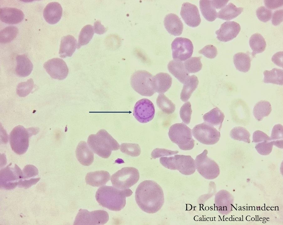
Case 3: 6-year-old with developmental delay, living in old house, basophilic stippling on smear
| Finding | Iron Deficiency | Chronic Disease | Lead Poisoning |
|---|---|---|---|
| Serum Iron | LOW | LOW | NORMAL |
| Ferritin | LOW (<30) | NORMAL/HIGH | NORMAL |
| TIBC | HIGH (>350) | LOW (<280) | NORMAL |
| FEP | NORMAL | NORMAL | HIGH |
| Smear | Target cells | Target cells, RBC indexes acceptable | Basophilic stippling |
Each microcytic villain has a lab signature. Tap to flip and expose the pattern.
Answer each step before the next branch reveals. This is how you work up a real patient.
Normal serum iron, normal or reduced TIBC, HIGH free erythrocyte protoporphyrin (FEP). Basophilic stippling on smear (RBCs with basophilic inclusions).
Lead blocks Delta-ALA Dehydratase (early step) and Ferrochetolase (late step in heme synthesis) → protoporphyrin accumulates → can't incorporate iron into heme → microcytic, hypochromic RBCs.
Basophilic stippling🔑Basophilic stippling = blue dots in the red cell from precipitated RNA. The ribosomal RNA that should have been cleared out is stuck because the heme synthesis enzymes are blocked. Lead = stuck ribosomes = blue polka dots. (RBCs packed with precipitated ribosomes/RNA). Elevated FEP is the confirmatory test. Normal serum iron (unlike IDA), so the lab pattern is distinct.
Easy to confuse with iron deficiency if you only look at MCV. But serum iron and TIBC are normal in lead poisoning. FEP differentiates. Also: lead causes BASOPHILIC STIPPLING, not target cells.
Children eating peeling paint in old buildings (lead paint). Also occupational exposure (battery workers, miners). Chronic toxicity affects CNS (developmental delay, encephalopathy), kidneys, GI tract.
Remove source of exposure. Chelation therapy (EDTA, dimercaprol, succimer) if blood lead >45 mcg/dL or symptomatic. Iron supplementation won't help (iron isn't the problem; heme synthesis is blocked).
4 alpha-globin genes total. Each deletion incrementally worsens disease.
Silent carrier. Normal labs, no symptoms. Only detectable if you look.
Alpha-thalassemia trait. Microcytic anemia, mild ± symptoms. RBC count often disproportionately high relative to Hgb (RBC/Hgb ratio useful for diagnosis).
Hemoglobin H disease. Moderate anemia, splenomegaly, symptoms. Forms Hb H (β4 tetramers). More microcytic. Hemolytic component.
Hydrops Fetalis (Hemoglobin Bart = γ4). Incompatible with life in utero. Fetal death or severe hemolytic disease at birth.
Loss of alpha chain → excess beta chains precipitate → hemolysis. No alpha → only beta available → Hb H or Bart forms. Microcytic because fewer normal globin chains.
Trait: observation, genetic counseling. Hb H disease: transfusion support PRN, folate supplementation, splenectomy if hypersplenism. Prenatal testing critical for at-risk couples.
2 beta-globin genes total. Mutations reduce or abolish beta production.
Beta-thalassemia trait (carrier). Microcytic anemia, asymptomatic or mild symptoms. Elevated Hb A2 and Hb F on electrophoresis (compensation). Normal or elevated bilirubin due to turnover.
Beta-thalassemia major (Cooley anemia). Severe anemia, jaundice at 6 months (when Hb F switches off and only abnormal beta available). Requires transfusions. Extramedullary hematopoiesis.
Beta-thalassemia trait: ↑ Hb A2 (>3.5%), ↑ Hb F.🔑Hb A2 > 3.5% = beta-thalassemia trait on electrophoresis. The body compensates for less beta by making more delta chains (which form Hb A2 = alpha2-delta2). Think: beta's down, delta's up. Beta-thalassemia major: Hb A2 + Hb F only, NO Hb A (normal). This electrophoresis pattern is diagnostic.
At birth, Hb F (fetal, made from gamma chains) protects. At 6 months, gamma → beta switch occurs. If beta is defective, suddenly only Hb A2 (α2δ2) and Hb F remain. Severe anemia and hypoxia result.
Trait: observation. Major: chronic transfusions, iron chelation (transfusions overload iron), folate. Splenectomy if hypersplenism. Bone marrow/gene therapy curative but high-risk.
Click gene buttons to delete them and see how phenotype changes:
Iron reaches the erythroblast, but the heme assembly line stalls. The marrow has iron, yet cannot load it into heme, so the red cell still comes out small and hypochromic.
Serum iron high or normal, ferritin high, and TIBC low or normal. This is the opposite of iron deficiency, where the tank is empty.
Prussian blue stain shows ringed sideroblasts: iron-laden mitochondria circling the nucleus like beads around the erythroblast.
Alcohol, isoniazid through B6 depletion, lead, copper deficiency, and myelodysplastic syndrome. The question usually hands you one trigger plus the ringed sideroblast clue.
Remove the trigger when possible. Give pyridoxine for B6-responsive or INH-related disease. If the cause is MDS, treat it as a clonal marrow disorder, not a vitamin problem.
Do not give iron just because the MCV is low. Sideroblastic anemia already has iron in the wrong place. More iron can worsen overload.
HbS polymerization causes vaso-occlusion and hemolysis. The anemia is usually normocytic, and trait often has normal hemoglobin. HbS belongs on the hemolysis page, not as a microcytic anemia cause.
Membrane protein defects make dense spherocytes with high MCHC. It is a normocytic, normochromic hemolytic anemia. High MCHC is a kick-out clue, not a microcytic exception.
Can show target cells and mild hemolysis, but the electrophoresis shows HbC. Target cells alone are not enough to call a microcytic production problem.
B12 and folate deficiency make big cells, not small ones. If the MCV is below 80, do not chase megaloblastic anemia first unless the smear is clearly dimorphic.
MCV below 80 starts with ferritin, serum iron, TIBC, RDW, RBC count, and the smear. Only move to electrophoresis after iron studies are normal or the patient fails iron.
Crohn disease, rheumatoid arthritis on NSAIDs, cancer, and chronic infection can drain iron while inflammation raises ferritin. A "normal" ferritin may hide real iron deficiency.
Thalassemia trait can coexist with iron deficiency. If iron corrects but the MCV stays very low, the genetic floor is still there. That is when electrophoresis or genetic testing matters.
Two cell populations can average into a misleading MCV. Iron deficiency pulls cells small; B12 or folate deficiency pulls cells large. The smear beats the average.
EPO will not work if the marrow has no iron. Dialysis patients often need IV iron plus an erythropoiesis-stimulating agent, not one or the other.
Do not let one abnormal number flatten the case. Ferritin, TIBC, RBC count, reticulocytes, and smear morphology tell you whether the anemia is empty stores, trapped iron, globin failure, or two things at once.
Treat the process that is actually limiting erythropoiesis: replace iron when stores are empty, calm inflammation when hepcidin is high, add B6 for responsive sideroblastic disease, and avoid unnecessary iron in thalassemia trait.
25 original vignettes. One at a time. Shuffle restarts fresh.

Original board-style vignettes. Shuffled, never-repeat, full explanations for every choice.
