
Hearing Loss: Weber, Rinne, and the CN VIII Wire
The trick is not memorizing the table. The trick is knowing whether the problem blocks sound before the cochlea, or damages the cochlea or nerve after sound arrives.
The ear is a relay race.
Tap each step. The diagnosis changes depending on where the relay breaks.
Tympanic membrane vibration
Sound waves hit the tympanic membrane and turn air pressure into movement. A blocked canal, fluid behind the drum, or a torn drum makes a conductive problem.
Why the table works: a conductive block does not stop the cochlea from sensing vibration through bone. It blocks outside sound, so internal skull vibration feels louder in that ear. Sensorineural loss damages the detector or wire, so vibration in that ear is weaker no matter how it arrives.

Outer to Inner Ear
The conductive system is the canal, tympanic membrane, and ossicles. The sensorineural system starts at cochlear hair cells.
Weber tells direction. Rinne tells the doorway.
Do not ask "what did the table say?" Ask: did the bad ear trap bone vibration, or did the bad ear lose nerve signal?
Conductive Loss
Something blocks air sound before the cochlea: wax, otitis media, ossicle fixation, or a tympanic membrane problem.
The bad ear hears Weber louder.
Bone vibration reaches the cochlea, but outside sound is blocked. The bad ear loses environmental masking and internal vibration feels louder. Rinne flips to BC > AC in the affected ear.
Sensorineural Loss
The cochlea, hair cells, CN VIII, or cerebellopontine angle wire is weak.
Weber runs to the good ear.
The damaged ear cannot turn vibration into nerve signal well. Rinne remains AC > BC, because air still beats bone, but both are reduced compared with the good ear.
Presbycusis and Noise
Hair-cell damage at the cochlear base hits high frequencies first.
High pitch goes first.
Age and loud noise injure organ of Corti hair cells. The board move is sensorineural hearing loss: Weber toward the better ear if asymmetric, Rinne AC > BC.
| Pattern | Weber | Rinne | Meaning |
|---|---|---|---|
| Normal | Equal | AC > BC both ears | No unilateral signal advantage. |
| Conductive loss | Louder in bad ear | BC > AC in bad ear | Air doorway blocked before cochlea. |
| Sensorineural loss | Louder in good ear | AC > BC in bad ear | Cochlea or CN VIII detector is weak. |
The auditory pathway is a crossed backup system.
Severe unilateral hearing loss localizes early: cochlea, CN VIII, cerebellopontine angle, or lateral pons. Higher central pathway lesions are usually less purely one-ear because auditory information becomes bilateral.
Cochlea and hair cells
Frequency is mapped mechanically. Base handles high frequency first, which is why noise and age classically hit high pitch before low pitch.
Cochlear nerve, CN VIII
Sensorineural loss plus tinnitus or imbalance can be a cochlear nerve or cerebellopontine angle lesion.
Cochlear nuclei in lateral pons
AICA territory can hit the ear system with ipsilateral facial palsy, vertigo, ataxia, and hearing loss.
Superior olive and trapezoid body
Bilateral processing starts early, so a single lesion above this level rarely behaves like a clean one-ear deafness question.
Lateral lemniscus to cortex
Inferior colliculus, medial geniculate body, auditory radiations, then Heschl gyri in temporal cortex.
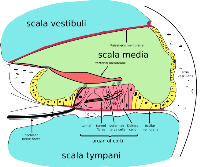
Cochlear Cross-Section
Stapes movement turns fluid pressure into basilar membrane movement, which bends hair cells.
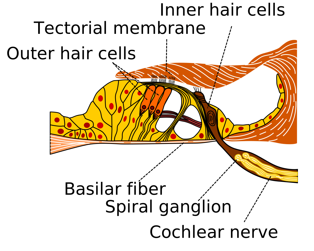
Organ of Corti
Hair-cell damage is sensorineural loss. Noise and aging injure this detector, especially high-frequency regions.
Solve it like a bedside localization problem.
Right-click or long-press to cross out. Double-click or double-tap to mark. Single-click to answer.
Three rules that save the question.
If these stay in memory, the table stops being a table.
1. Bad conductive ear gets louder Weber.
A blocked ear hears its own skull vibration better because outside sound is reduced. Conductive loss traps internal vibration.
2. Bad sensorineural ear loses Weber.
If the cochlea or CN VIII cannot detect well, vibration is weaker in that ear. Weber goes to the better ear.
3. Rinne AC > BC does not mean normal.
Sensorineural loss keeps AC > BC because air conduction still beats bone conduction. Both are just reduced.