Six cells build the whole thing. The board question is always which one, where it lives, and the single disease that gives it away.
Start here. A 29-year-old woman is evaluated for two weeks of blurred vision in one eye and an odd electric shock down her spine when she bends her neck. MRI shows several bright oval lesions in the brain and spinal cord white matter. The cell being destroyed is the one that wraps insulation around central nervous system axons, and one of these cells wraps many axons at once. Which cell is under attack?
Schwann cell
Oligodendrocyte
Astrocyte
Good instinct picking Schwann. Both Schwann cells and oligodendrocytes make myelin, so they feel interchangeable. Think of it like two electricians who both wrap wires: one only works inside the house (central nervous system), the other only outside on the street (peripheral nerves). This patient is losing insulation in the brain and cord, which is inside the house. That is the oligodendrocyte, and it is the cell that lets one worker insulate about thirty wires at once. The Schwann cell works the street and covers only one wire each, and it is the one that fails in Guillain-Barre, not this. Central white matter demyelination, many axons per cell, means oligodendrocyte every time.
The Cell Lineup
Open each card for the mechanism and the one disease that gives the cell away. Location and job show on the surface.
CNS & PNS · the worker
Neuron
The cell that actually thinks
Conducts the action potential and releases neurotransmitter. Everything else on this page exists to support it.
Nissl bodies (rough ER) mark the cell body
see the mechanism →
Why it matters
How it works
Sodium rushing in depolarizes the membrane. At the terminal, calcium entry triggers neurotransmitter release. Potassium leaving resets it.
Board clue
Nissl substance (rough ER) sits in the cell body and dendrites, never in the axon. Central chromatolysis after injury = Nissl breaks up.
CNS · the caretaker
Astrocyte
The most common CNS glia
Supports neurons, helps build the blood-brain barrier, mops up leftover neurotransmitter, and forms the scar after injury.
GFAP positive. This is the one they test.
see the mechanism →
Why it matters
How it works
After brain injury it swells and multiplies, called reactive gliosis, and lays down the glial scar (the brain version of a fibrous scar).
Board clue
GFAP positive. Its tumor is the astrocytoma. When a stain lights up around a brain lesion wall, think astrocyte.
CNS · the immune cell
Microglia
The brain's own macrophage
The resident scavenger. After injury or infection it turns on and eats debris. It comes from a different origin than the other glia.
Mesoderm origin, not neural tube
see the mechanism →
Why it matters
How it works
It is the immune arm of the brain. In advanced HIV, infected microglia fuse into multinucleated giant cells and form microglial nodules.
Board clue
Multinucleated giant cells in the brain plus dementia in an HIV patient = microglia. It is the brain's HIV reservoir.
CNS · white matter
Oligodendrocyte
CNS myelin, one cell wraps many
Makes the myelin insulation in the central nervous system. A single oligodendrocyte reaches out and wraps about thirty different axons.
Destroyed in multiple sclerosis
see the mechanism →
Why it matters
How it works
One cell, many axons. Lose it and central signals slow or stop. That is multiple sclerosis. The JC virus also infects it, causing PML.
Board clue
Fried-egg look on histology, predominant glia of white matter. CNS demyelination points here.
PNS · peripheral nerves
Schwann cell
PNS myelin, one cell wraps one
Makes the myelin in peripheral nerves. One Schwann cell insulates exactly one segment of one axon, and it helps peripheral nerves regrow.
Attacked in Guillain-Barre
see the mechanism →
Why it matters
How it works
One cell, one axon. It also guides peripheral axons back after injury, which is why nerves in the arm can recover but the cord cannot.
Board clue
S100 positive. Its tumor is the schwannoma; on CN VIII it is the acoustic neuroma. Ascending weakness after infection = Guillain-Barre.
CNS · the lining
Ependymal cell
Lines the fluid spaces
Ciliated cells that line the ventricles and the central canal of the cord. The cilia keep cerebrospinal fluid moving.
Its tumor: ependymoma
see the mechanism →
Why it matters
How it works
Its beating cilia circulate CSF through the ventricles. Specialized ependymal cells form the choroid plexus that makes CSF.
Board clue
A tumor inside the fourth ventricle in a child, causing hydrocephalus, is the ependymoma.
Neuron cell body. The dark clumps are Nissl bodies (rough ER), found in the soma and dendrites, never the axon. tap to enlarge
Board trap
GFAP versus the CNS macrophage. Astrocyte is GFAP positive. The stain that lights up the scar wall around a brain lesion is GFAP, so it is astrocyte, not microglia. Microglia is the phagocyte and the HIV reservoir. Two different cells, two different questions.
Action Potential Theater
Meet the axon of a single neuron. At each moment, predict which channel is doing the work before the membrane answers.
ONE NEURON · ONE FIRING
Resting membrane sits at -70 mV.
Phase 1: the membrane starts to climb toward zero. What opens?
Board trap
The blocker question. Local anesthetics and tetrodotoxin both block the voltage-gated sodium channel. No sodium in means no depolarization, so the nerve cannot fire and cannot signal pain. Pufferfish poisoning is the classic tetrodotoxin stem.
One Cook, Many Pizzas
The single highest-yield distinction on this page. Compare the two myelin makers, then race a signal down a bare axon against a myelinated one.
Oligodendrocyte (CNS)
Schwann (PNS)
Oligodendrocyte
WhereCentral nervous system
Axons per cellMany (about 30)
Classic diseaseMultiple sclerosis
Also hit byJC virus (PML)
RegenerationPoor. Astrocyte scar blocks it
Schwann cell
WherePeripheral nerves
Axons per cellOne (a single segment)
Classic diseaseGuillain-Barre
AlsoSchwannoma / acoustic neuroma
RegenerationBetter. Schwann guides regrowth
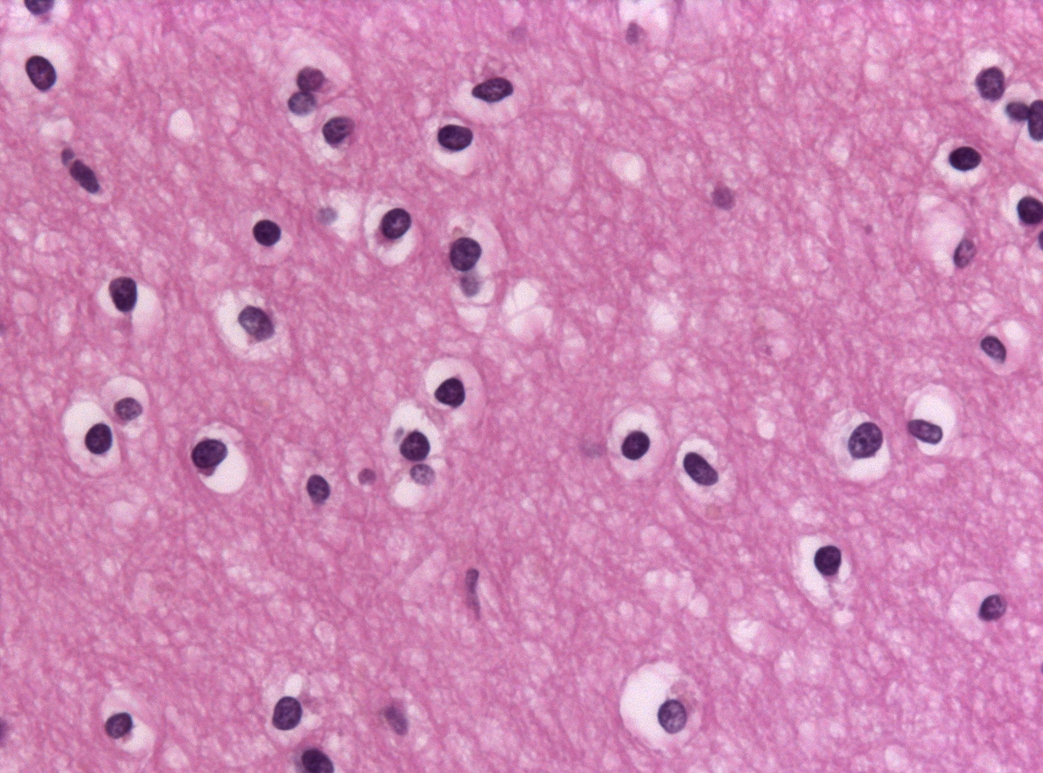
Oligodendrocytes on H and E stain. These are the CNS myelin makers; one cell wraps many axons at once. tap to enlarge
CHAPTER I · THE CONDUCTION LAB
MYELIN, PHD
RACE MASTER, NODE 12
Two axons, same neuron, same distance. One is bare. One is wrapped by oligodendrocyte myelin with gaps called Nodes of Ranvier. Predict who reaches the finish first, then watch the signal run.
Which axon wins the race?
Pick one, then the signal runs.
PATTERN LOCKED
SALTATORY CONDUCTION
Myelin insulates the axon so current cannot leak. The signal regenerates only at the Nodes of Ranvier and skips the wrapped stretches in between.
RouteNode to node, hopping the myelin
Why fastSodium channels cluster at the nodes; it regenerates only there
PearlStrip the myelin (MS) and the hop breaks. Signals slow or die.
Which cell, which disease?
Commit to each branch before the answer shows.
1
A patient is losing myelin. First question: is the damage inside the brain and spinal cord, or out in the limb nerves?
Central: brain and spinal cord white matter
Peripheral: nerves in the arms and legs
2
Central it is. The myelin maker there wraps many axons at once. Which cell, and which classic disease destroys it?
Oligodendrocyte, hit in multiple sclerosis
Schwann cell, hit in Guillain-Barre
2
Peripheral it is. Ascending weakness a few weeks after a gut or respiratory infection. Which cell is the target?
Schwann cell, the PNS myelin maker
Oligodendrocyte, the CNS myelin maker
Sort the Sensors
Drag each receptor to what it senses. On a phone, tap a receptor then tap its home. Then watch the fibers race by speed.
Light touch, superficial
Rapidly adapting, hairless skin
Vibration, deep pressure
Rapidly adapting, deep skin and joints
Sustained pressure, shape
Slowly adapting, fingertips
Pain and temperature
Bare endings, no capsule
4 receptors to place.
The nerve fiber speed lane
Bigger and more myelin means faster. Press go and watch them finish in order.
Small, lightly myelinated. Cold and sharp, fast, first pain.
C
Small, unmyelinated. Warmth and dull, slow, second pain. Slowest.
Pacinian corpuscle. The onion-ring layers are the giveaway. It sits deep and senses vibration. tap to enlarge
Board trap
First pain versus second pain. A-delta carries the fast, sharp, well-localized first pain because it is myelinated. C fibers carry the slow, dull, aching second pain because they are unmyelinated. Same pinprick, two waves of pain arriving at different speeds.
Lock the receptor distinctions
🧠
Meissner vs Pacinian
Meissner is shallow and light (think a feather on a fingertip). Pacinian is deep with onion rings and feels vibration. Shallow-soft vs deep-buzz.
tap to reveal
⏱
Merkel = the slow one
Merkel discs adapt slowly, so they hold sustained pressure and read shape and edges. Meissner and Pacinian both adapt fast and fade quickly.
tap to reveal
✈
A-Delta plane is fast
A-Delta plane is fast (myelinated, sharp first pain). A taxi-C is slow (unmyelinated, dull second pain). The plane beats the taxi.
tap to reveal
🧵
GFAP vs macrophage
Astrocyte glows GFAP and builds the scar. Microglia is the brain macrophage and HIV giant cells. Scar stain = astrocyte, eater = microglia.
tap to reveal
Board Walkthrough
Twenty-five board-style clinical vignettes, one at a time, shuffled and never repeating. Right-click or long-press to cross out a choice. Double-click to highlight.
Myelin stain of white matter. Pale zones are lost myelin, the histologic signature of a demyelinating lesion. tap to enlarge