Cerebellar Lesions
The little brain is not a strength machine. It is a timing, tone, balance, and correction machine. If the limb overshoots, think lateral cerebellar hemisphere. If the trunk cannot stay upright, think vermis. If the route is input in or output out, the peduncles tell you where the signal is traveling.
The cerebellum edits movement while it is happening.
The board move is localization, not memorizing every fiber name.
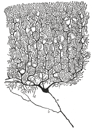
Cerebellar cortex compares intended motion with incoming sensory feedback. Purkinje cells then put an inhibitory GABA brake on deep nuclei. The deep nuclei send the corrected motor plan back toward motor systems.
That gives you the exam split: lateral hemisphere lesions make an ipsilateral limb overshoot, shake on approach, and miss finger-to-nose. Vermis lesions make the midline body fail, so gait gets wide and the patient cannot sit or stand without falling.

Input
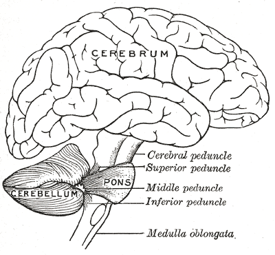
Inferior peduncle brings spinal, vestibular, and olivary input. Middle peduncle brings pontine cortical copies.
Processing
Mossy fibers are most inputs. Climbing fibers come from the inferior olive. Purkinje cells convert cortex math into GABA brake.
Output
Deep nuclei leave mainly through the superior peduncle toward red nucleus and thalamus, then motor cortex.
Inferior and middle bring the story in. Superior sends the verdict out.
Nia is routing a movement signal.
Loading route...
Inferior and middle are the inbound doors. Superior is the major outbound door.
Three doors, one clean mental image.
Inferior peduncle: spinal cord proprioception, vestibular input, and inferior olive signals enter here.
Middle peduncle: pontine input enters here. The pons is carrying the cortex copy into the cerebellum.
Superior peduncle: deep nuclei output leaves here to red nucleus and thalamus.

Click the region and predict the body failure.
Pick a region.
The cerebellum tells you what failed: limb targeting, trunk balance, or vestibular eye-body stability.

Purkinje cells brake. Deep nuclei speak.
The cell logic is simple.
Mossy fibers carry most incoming information. Climbing fibers come from the inferior olive and give a powerful teaching signal. Purkinje cells integrate the cortical math and inhibit the deep nuclei with GABA.
Then the deep nuclei decide the output: dentate for motor planning through thalamus, interposed nuclei for red nucleus limb correction, fastigial for vestibular and reticular balance systems.
Dentate
Lateral planning, distal limb coordination, thalamus route.
Interposed
Globose plus emboliform, limb correction toward red nucleus.
Fastigial
Midline balance through vestibular and reticular systems.


Romberg, trunk, limb: three questions localize most stems.
Eyes closed makes it much worse?
If cerebellar, what fails hardest?
Vertigo and nystagmus dominate?
Commit before reveal.
Use the button stack like a patient encounter. Eyes closed worse points to sensory ataxia. Eyes open and closed both bad points cerebellar. Then decide trunk versus limb.
Compare the traps.
Four traps to remove before the exam can hurt you.
The one-sentence lock.
A lateral cerebellar hemisphere lesion makes the ipsilateral limb inaccurate; a vermis lesion makes the trunk unreliable; a dorsal column lesion waits until the eyes close to punish balance.