Every blood cell you have, the bacteria killer, the antibody factory, the red cell carrying oxygen, traces back to one ancestor in the marrow. Learn the family tree once and the markers, the leukemias, and the lineage traps fall into place.
Medically reviewed by Fatima Ali, DO & Kaitlyn Cocuzzo, MD
A medical student is shown a simplified blood cell family tree and asked one question. Red blood cells and platelets keep oxygen moving and plug leaks, so they feel like a separate non-immune crew. But every blood cell shares one origin. Which progenitor gives rise to both the red blood cells and the platelets?
One cell in the marrow can become any blood cell. It does two things: copies itself and commits to a path. Watch it decide.

The hematopoietic stem cellHSC. The master blood stem cell. It self-renews (makes more of itself) and differentiates (commits down a path). It carries the marker CD34, which is how transplant teams find and collect it. (HSC) is the founder. It is multipotent: it can build every blood and immune cell. Its badge is CD34, the marker collected for a stem cell transplant.
When the HSC commits, it picks one of two roads. That single fork explains most of the lineage map: common myeloid progenitor (the big, broad crew) or common lymphoid progenitor (the small, exclusive crew). Tap a road below and watch the path draw.
The full map, top to bottom. Tap a node to see its one job, its key markers, and the board trap that hides in it.
The split is the whole game. Left road (myeloid) is crowded: it builds the granulocytes, the monocyte, the mast cell, the platelet maker, and the red cell. Right road (lymphoid) is tiny: B, T, NK, and a dendritic subset. Dendritic cells come from BOTH roads, which is why they get their own trap later.
Three granulocytes look alike under the scope until you learn the giveaway. Tap each tab and read the picture.
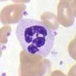
The most abundant white blood cell (40 to 70 percent). First responder to bacteria. Nucleus has 2 to 5 connected lobes, so it is also called a segmented cell or a poly.
Giveaway: many lobes, pale pink granules, and it dominates the smear. Acute bacterial infection drives the count up.

Two lobes (a bilobed nucleus) and bright red-orange granules. Rises with parasites and allergy. Driven by IL-5.
Giveaway: the red granules look like a bag of cherries. Think worms, wheezing, and weird drug reactions.
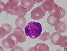
The rarest white blood cell (under 1 percent). Carries histamine and heparin in dark blue-black granules. Binds IgE and joins allergic reactions.
Giveaway: granules so dark you can barely see the nucleus. Basophilia is a clue for chronic myeloid leukemia.
These three feel like outsiders, but they are pure myeloid. This is where the points hide.

Largest white cell, CD14 positive, horseshoe nucleus. In tissue it becomes a macrophage and can become a dendritic cell.
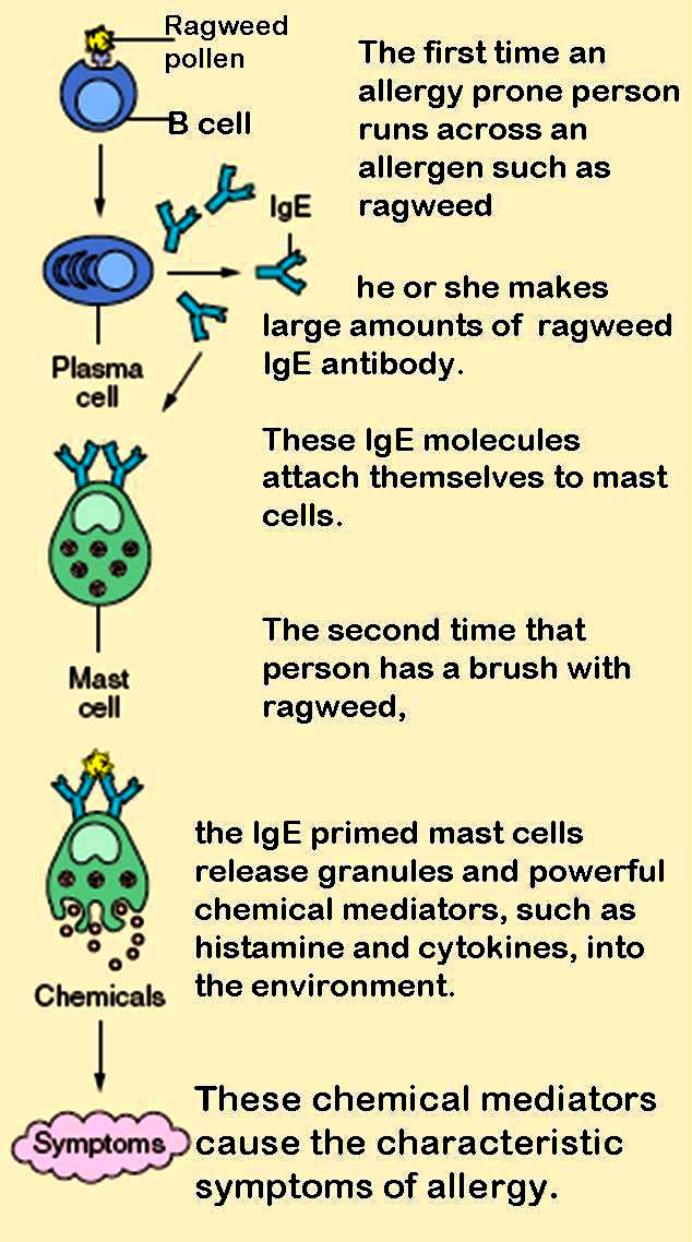
Lives resident in tissue (skin, gut, airway). Loaded with histamine, binds IgE. Myeloid, and NOT a basophil that moved into tissue.

The megakaryocyte sheds platelets; the red blood cell carries oxygen. Both come from the myeloid road.
Three core cells, two famous traps. Then prove you have it with a challenge and an elimination round.

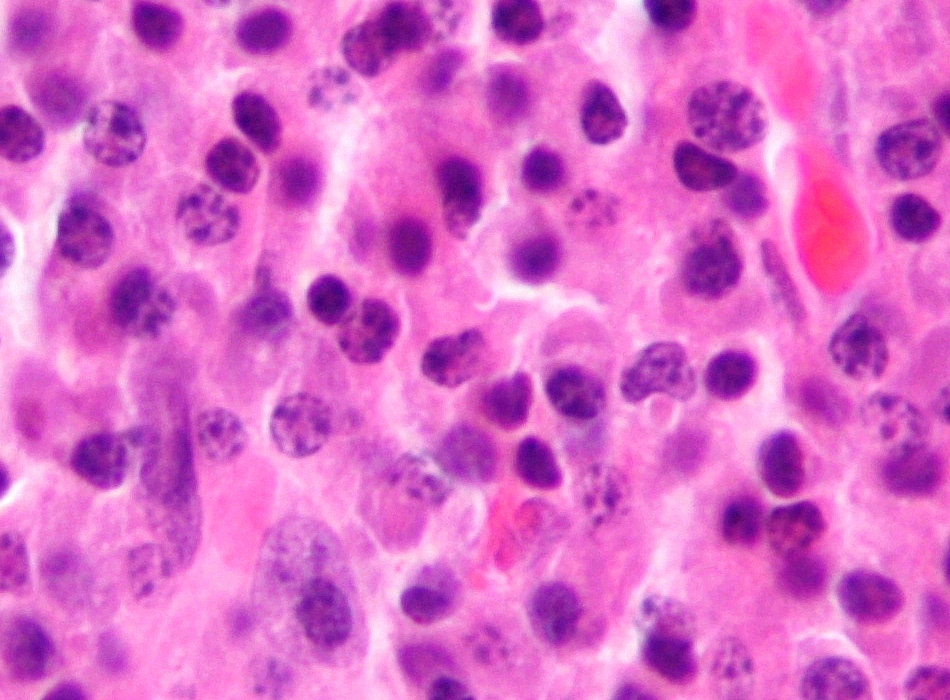
Matures in bone marrow. Markers CD19, CD20. When activated it becomes a plasma cell, the antibody factory.
Matures in the thymus. Marker CD3. Splits into CD4 helper (the coordinator) and CD8 cytotoxic (the killer).
Marker CD16, CD56. Lymphoid by birth, but it kills on sight with no antigen needed. It reads missing selfHealthy cells display MHC class I. Virus-infected and tumor cells often drop it. The NK cell kills any cell that fails to show its MHC class I, so it is on patrol for missing-self.: a cell that drops its MHC class I gets killed.
Elimination round. Clues drop one at a time. Knock out the cell that does not fit until one is left standing.
Five questions pulled fresh each visit, answer order shuffled. Low pressure. Just checking it stuck.
Real board-style cases, one at a time. Double tap to highlight a clue, long press to cross out an option. Tap Remix to reshuffle the deck.
References: standard histology, hematology, and immunology board texts. Images: Wikimedia Commons (see each lightbox for attribution).
Medically reviewed by Fatima Ali, DO and Kaitlyn Cocuzzo, MD. Vignettes are original clinical teaching cases; demographics, values, and answer order are written for practice. Confirm management against current references at the point of care.
