Immunology · Lymphatic System

Organs, Histology & Drainage

The lymphatic system is half plumbing, half army base. It returns leaked fluid to the blood AND builds the cells that fight infection. The board tests both. Let's map it.

Medically reviewed by Fatima Ali, DO & Kaitlyn Cocuzzo, MD

A 61-year-old man is brought to the office by his wife because of 3 months of vague upper abdominal fullness and a 15 lb weight loss. On examination, a firm, fixed, nontender lymph node is palpable just above the left clavicle. Abdominal exam reveals mild epigastric tenderness. Which structure most directly explains why an abdominal cancer surfaced as a node in his left neck?

Right lymphatic duct
Thoracic duct
Cisterna chyli only
Azygos vein
That left supraclavicular node is the Virchow node, and a palpable one is the Troisier sign. Here's the plumbing: the thoracic duct drains the entire abdomen and pelvis, then empties into the left venous angle right under that node. So a gastric or pancreatic tumor can float its cells up the duct and beach them in the left neck. The right lymphatic duct only drains the right upper quarter, so it can't carry belly cancer. Left neck node + weight loss = look in the belly.
Begin
The Big Picture

Two Jobs in One System

The lymphatic system belongs to the circulatory system AND the adaptive immune system. Every board fact below traces back to one of its two jobs.
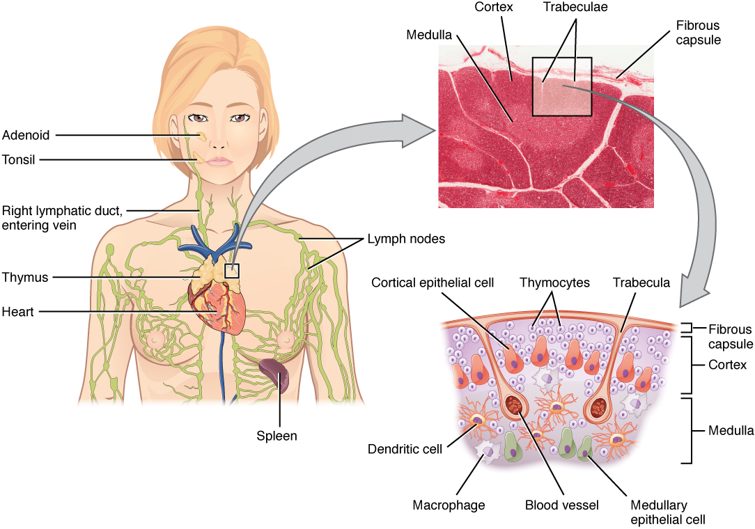
Thymus location and histology with the cortex and medulla labeled
Thymus · dark cortex, light medulla
Hassall corpuscle in thymic medulla
Hassall Corpuscle · thymic medulla
Lymph node follicle with germinal center
Lymph Node Follicle · germinal center
Reactive follicular hyperplasia of a lymph node
Reactive Hyperplasia · enlarged node
Splenic white pulp around a central arteriole
Splenic White Pulp · PALS + follicle
Peyer patch in ileal mucosa
Peyer Patch · ileal MALT
Cervical lymphadenopathy of the right neck
Cervical Lymphadenopathy
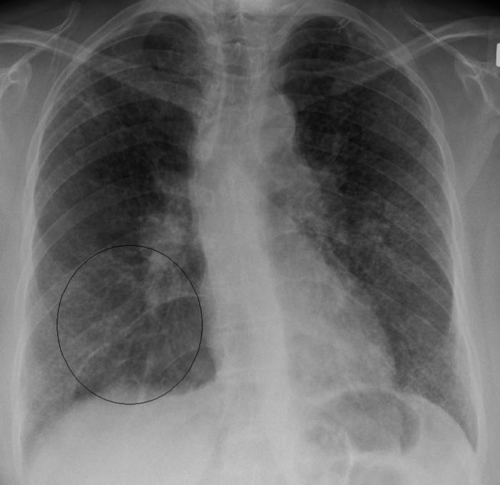
Chest x-ray showing bilateral hilar lymphadenopathy in sarcoidosis
Bilateral Hilar LAD · sarcoidosis
Overview of the human lymphatic system: vessels, nodes, thymus, spleen, tonsils
The whole system: capillaries and vessels (the plumbing) plus organs (the army base) draining back to the great veins at the base of the neck.

Job 1 (plumbing). Your capillaries leak fluid into tissues all day. Lymphatic capillaries soak that interstitial fluidThe fluid sitting between cells. Blood capillaries push it out faster than they reabsorb it, so something has to collect the leftovers, or you swell. back up (plus stray proteins and cell debris) and return it to the bloodstream. Miss this and the tissue swells, which is edema.

Job 2 (army base). The same vessels ferry white cells, mostly lymphocytesB cells and T cells, the cells of the adaptive immune system. They are stored in nodes and shuttled between marrow and nodes through lymphatic vessels., and carry antigen-presenting cellsDendritic cells and macrophages that pick up a piece of a pathogen and carry it to a node to show the lymphocytes, kicking off the immune response. into nodes to start the immune response. The node is where the antigen meets the lymphocyte.

The third job: fat transport Lymph from the gut also carries dietary fat as chyleLymph plus absorbed triglycerides, picked up by lacteals in the small intestine during digestion. It is fatty, hence milky.. Fresh interstitial lymph is clear, but as it picks up proteins, fats, debris, and leukocytes it turns milky. That is why a torn thoracic duct leaks a milky fluid into the chest, which is a chylothorax.

Follow One Cell From Birth

B cells and T cells both start in bone marrow. The split: B stays home, T ships out to the thymus. Pick a cell and watch it grow up.

BONE MARROW B + T are born here THYMUS T cells mature STAY IN MARROW B cells mature here SECONDARY ORGAN node / spleen / MALT
Pick a cell. A T cell ships to the thymus to grow up; a B cell stays in the marrow. Both end up working in a secondary organ.

Putting it together: antigen presentation drives B-cell proliferation and T-cell activation; the now-mature lymphocytes become effector cells out in the secondary organs (nodes, spleen, MALT). Marrow and thymus build the soldiers; the secondary organs are where the fighting happens. 🧠Bone marrow = B cells. Thymus = T cells. The letter is the location.

Eponym swaps the board loves Same structures, fancy old names. The cisterna chyli is the cistern of Pecquet. The thoracic duct is Van Hoorne's duct (a.k.a. the left lymphatic or alimentary duct). A palpable Virchow node is the Troisier sign. The board renames them to see if you still recognize the structure.

Trunks feed the ducts. Regional lymph collects into named lymphatic trunksThe large collecting channels that empty into the two terminal ducts: jugular, subclavian, and bronchomediastinal (paired), plus lumbar and intestinal (the intestinal trunk is unpaired and carries chyle). first: jugular, subclavian, bronchomediastinal, lumbar, and the unpaired intestinal trunk. Those trunks then drain into the thoracic duct or the right lymphatic duct. We map the whole route in subpage 4.

Where Lymphocytes Are Made

Primary Organs: Marrow & Thymus

Primary lymphoid organs are the factories. Bone marrow makes everything; the thymus is the finishing school for T cells.

Bone Marrow

The factory floor

B and T lymphocytes both arise from pluripotent hematopoietic stem cellsThe master blood stem cell. It can become any blood cell, including both lymphocyte lines. Lives in red marrow. in the marrow.

  • B cells finish maturing right here, then migrate out to secondary organs to form follicles and proliferate.
  • T cells are born here but leave immature, traveling to the thymus to mature.

Bone marrow = B-cell maturation.

Thymus

The T-cell finishing school

Function: maturation and differentiation of T lymphocytes.

Location: superior anterior mediastinumHigh in the chest, behind the sternum (retrosternal / precordial), in front of the great vessels. This is why thymic masses show up as an anterior mediastinal mass on chest x-ray., retrosternal. Bilobar, wrapped in a fibrous capsule.

Thymus = T-cell maturation.
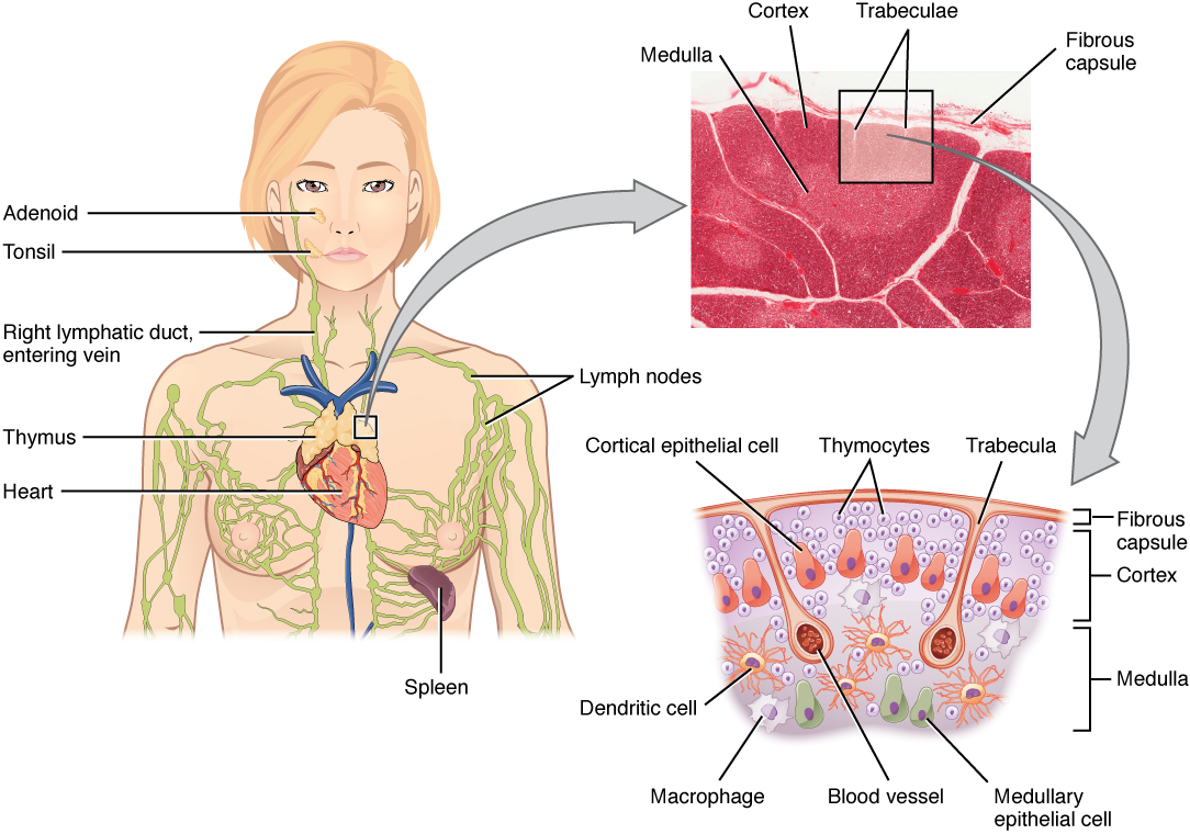
Location and histology of the thymus in the anterior mediastinum with cortex and medulla
The thymus sits behind the sternum, in front of the heart and great vessels. A mass here is an anterior mediastinal mass.
Big in babies, gone in grown-ups The thymus is largest and most active in neonates and infants, casting a thymic shadowA normal soft-tissue density in the upper chest of a baby. Do not mistake it for a mass. on chest x-ray, the thymic sail sign. InvolutionAge-related shrinkage. The thymus is steadily replaced by fat starting around age 1. begins around 1 year of age. In adults it is atrophic and largely replaced by fat. So a prominent thymus in a newborn is normal; a thymic mass in a 50-year-old is not.

Thymus Histology: Cortex vs Medulla

One question shows up over and over: which region has Hassall corpuscles? Tap each tab and lock it in.

Cortex (outer)
Medulla (inner)

Cortex · dark, dense, immature

Peripheral region. Packed with densely arranged immature T lymphocytes called thymocytesDeveloping T cells. So many nuclei crammed together that the region stains dark on H&E.. The cell density is why it looks dark.

This is where T cells are tested and most of them die (selection). Survivors move inward.

Medulla · light, sparse, mature

Central region. Fewer cells, so it stains light. Contains mature T lymphocytes ready to leave.

Hallmark: Hassall corpuscles, concentric onion-like whorls of eosinophilic epithelial reticular cells. If you see Hassall corpuscles, you are in the medulla. 🧠Hassall = Has all the mature T cells. Onion-ring whorls in the medulla.

Who else lives there: the thymic stroma is built from thymic epithelial cellsThey form the scaffold and educate developing T cells about self vs non-self., dendritic cells, and macrophages.

Embryology: the 3rd pouch The thymic epithelium comes from the 3rd pharyngeal pouch (endoderm). The thymic lymphocytes come from mesoderm. The 3rd pouch detail is the setup for the big clinical association below.

When the Thymus Goes Wrong

DiGeorge syndrome

Failure of the 3rd AND 4th pharyngeal pouches to develop. Result: no thymus + no parathyroids.

  • No parathyroids → hypocalcemia (tetany, seizures).
  • No thymus → no mature T cells → viral and fungal infection susceptibility.

3rd + 4th pouch = thymus + parathyroid gone. 🧠CATCH-22: Cardiac, Abnormal facies, Thymic aplasia, Cleft palate, Hypocalcemia, chromosome 22.

Hypoplasia / aplasia

Too little or no thymic tissue. Drives DiGeorge and SCIDSevere Combined Immunodeficiency. T-cell (and often B-cell) function is absent, so the child gets every kind of infection early in life.. Without a thymus, T cells never mature.

Thymic hyperplasia (two flavors)

True hyperplasia: rebound enlargement, classically after steroids or chemotherapy.

Lymphoid hyperplasia: autoimmune. Seen with myasthenia gravis, SLE, and rheumatoid arthritis.

Thymoma: the anterior mediastinal mass A thymoma is a tumor of thymic epithelial cells. Associations the board wants: myasthenia gravis, pure red cell aplasia, and immunodeficiency (Good syndrome). Often asymptomatic, found as an incidental anterior mediastinal mass on chest x-ray. When symptomatic, it is usually a 40 to 60-year-old with chest pain, cough, dyspnea, SVC syndromeSuperior vena cava compression: facial swelling, distended neck veins, headache. A big anterior mediastinal mass can squeeze the SVC., or a paraneoplastic syndrome. Treatment: surgery, with chemo and radiation as needed.

Worse cousin: thymic carcinomaA frankly malignant thymic tumor, distinct from the more indolent thymoma. is more aggressive, invades the mediastinum, and metastasizes outside the chest. Thymoma is usually contained; thymic carcinoma breaks out.

Where the Fighting Happens

Secondary Organs: Spleen, Node, MALT

Secondary lymphoid organs are where antigen meets lymphocyte. Each has a zone for B cells and a zone for T cells. Learn the zones and the histology answers fall out.

The Spleen

Function: filters old and misshapen RBCs and plateletsThe red pulp is a blood filter. Old or abnormal red cells get culled here by macrophages., and mounts the humoral immune response using B and T cells. Location: under the left diaphragm, anterolateral to the left kidney, tucked behind ribs 9 to 11.

White pulp (immune)
Red pulp (blood filter)

White pulp · antigen filtration & presentation

The immune zone, organized around arterioles:

  • Lymphoid follicles = the B-cell area.
  • PALSPeriarteriolar lymphatic sheath: the cuff of T cells wrapped directly around the central arteriole. (periarteriolar lymphatic sheath) = the T-cell area, wrapped around the central arteriole.

PALS = T cells. Follicles = B cells. 🧠PALS are your T-cell buddies hugging the arteriole. Follicle = B.

Red pulp · blood filtration

The plumbing zone. This is where blood is filtered and old or damaged red cells and platelets are removed by macrophages in the cords and sinusoids.

Red pulp = mechanical blood filter. White pulp = immune surveillance. Two different jobs in one organ.

Splenic white pulp: lymphoid follicle and PALS around a central arteriole
Splenic white pulp: the central arteriole wears a T-cell sleeve (PALS) with a B-cell follicle budding off the side.

The Lymph Node, Zone by Zone

Function: nonspecific filtration by macrophages, storage and circulation of B and T cells, and immune activation (antigen presentation → B proliferation + T activation). Structure: bean-shaped, fibrous capsule with trabeculae. Lymph enters through many afferent vessels at the cortex, percolates from the subcapsular sinus through the trabecular sinus to the medullary sinus, and exits at the hilumThe notch where blood vessels and the few efferent lymphatics enter and leave. Many afferents in, few efferents out. via few efferent vessels. Blood vessels branch into high endothelial venulesHEVs: specialized post-capillary venules with plump endothelium that let B and T cells hop between blood and node. (HEV).

Lymph node follicle with a pale germinal center surrounded by a dark mantle
A secondary follicle: a pale germinal center (active B cells) ringed by a dark mantle of small naive B cells.
Cortex
Paracortex
Medulla

Cortex · the B-cell zone

Outermost layer. Holds the lymphoid follicles:

  • Secondary follicle (active): a dark mantle zoneA rim of small, dark, naive B cells pushed to the edge. around a pale germinal center where B cells proliferate and mature.
  • Primary follicle (inactive): a dense ball of naive B cells, no germinal center yet.

A pale germinal center means the node is busy fighting something.

Paracortex · the T-cell zone

Sits between cortex and medulla. Contains T cells and the HEVs that let lymphocytes enter and leave the bloodstream. This is the site of T-cell activation.

  • Enlarges in cell-mediated (viral) responses. A florid paracortical hyperplasia is classic in EBV mononucleosis.
  • Depleted / underdeveloped in DiGeorge (no T cells to fill it).

Medulla · cords + sinuses

Innermost region:

  • Medullary cords: plasma cells and lymphocytes.
  • Medullary sinus: macrophages and reticular cells; drains into the efferent lymphatics.

Plasma cells finish here and dump antibody into the exiting lymph.

The zone map in one line Follicle (cortex) = B. Paracortex = T. Medulla = plasma cells + macrophages. Mono swells the paracortex (T zone); DiGeorge empties it. The spleen uses the same logic: follicle = B, PALS = T.

MALT: Mucosa-Associated Lymphoid Tissue

MALT (tonsils, Peyer patches, solitary mucosal follicles) guards the wet surfaces. It looks like other secondary organs but adds a specialized follicle-associated epitheliumThe epithelial layer over a mucosal follicle. In the gut (GALT) it contains M cells.. In the gut (GALT), that epithelium contains M cells, which transcytose antigen from the lumen into the lamina propria.

MALT lymphoid nodule in a mucosal wall
A MALT nodule sitting right under a mucosal surface, sampling whatever passes through the lumen.

Peyer patches · the IgA factory

Location: the ileum (lamina propria + submucosa). Histology: aggregated lymphoid follicles with dendritic cells under a follicle-associated epithelium containing M cells.

The pathway the board tests:

  • M cell grabs luminal antigen → hands it to a macrophage (APC).
  • APC stimulates germinal-center B cells → plasma cells.
  • Plasma cells secrete IgA, which picks up a secretory componentA protein added as IgA crosses the epithelium. It protects IgA from digestion and lets it work on the luminal surface..
  • Result: secretory IgA released onto the gut surface.

🧠M cell = the Mail slot. It posts antigen inward so B cells can make secretory IgA.

iBALT · the lung's on-demand tissue

Inducible bronchus-associated lymphoid tissueLymphoid tissue that forms in the airway walls only after inflammation or infection, not present at baseline. forms in the walls of the upper and lower respiratory tract (perivascular) after inflammation or infection.

Central large B-cell follicles with adjacent T-cell areas, follicular dendritic cells, specialized stroma, lymphatics, and HEVs. Lacks the M-cell dome epithelium that Peyer patches have.

Tonsils · Waldeyer's Ring (first defense for inhaled/ingested bugs)

Four tonsil groups form a ring at the back of the throat. Tap each header cell to reveal its details and self-test.

Tonsil Location Epithelium Crypts
Pharyngeal (adenoids) Roof / posterior nasopharynx Respiratory (ciliated) Small folds, no crypts; fibrous capsule
Palatine Tonsillar fossa of oropharynx Non-keratinized stratified squamous 10 to 20 deep branched crypts
Lingual Base of tongue (near skeletal muscle) Non-keratinized stratified squamous Shallow crypts
Tubal Lateral nasopharynx wall (near eustachian tube) Respiratory (ciliated) None described

🧠Palatine = the one with deep branched crypts, and the one usually pulled in a tonsillectomy. The palatine tonsil is the classic "tonsil" you see when someone says "ah," and the one with the deepest crypts.

The Two Exit Pipes

Lymphatic Drainage & the Ducts

All lymph ends up back in the blood through just two terminal ducts. Knowing which duct drains which territory is the single highest-yield map on this page.

Lymph is collected by capillaries, runs through vessels and secondary organs, and re-enters the blood via the thoracic duct and the right lymphatic duct. During digestion, gut lymph carrying triglycerides (chyle) joins the thoracic duct and reaches the venous system.

The 75/25 Split

The thoracic duct drains the cyan area: everything except the right arm, right thorax, and right head/neck. The right lymphatic duct drains only that small orange right-upper-quarter. Both empty into their own venous angle at the base of the neck.
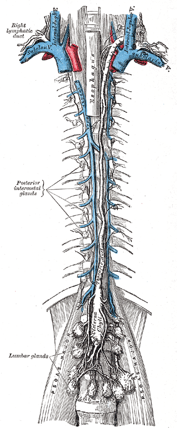
Thoracic duct ascending the posterior mediastinum to the left venous angle
The thoracic duct rises from the cisterna chyli up the posterior mediastinum and arches into the left venous angle.
Thoracic Duct

The big pipe

  • Drains: everything except the right arm, right thorax, and right head/neck (so the entire left side + everything below the diaphragm).
  • Origin: the cisterna chyliA dilated sac near the left crus of the diaphragm where the lumbar and intestinal trunks meet. The cistern of Pecquet., a dilated sac near the left crus of the diaphragm.
  • Course: ascends the posterior mediastinum (thoracic aorta on its left).
  • Ends: the left venous angle (left subclavian + left internal jugular junction).
  • Volume: up to about 4 L/day.
  • Injury: chylothorax (milky pleural fluid).
Right Lymphatic Duct

The little pipe

  • Drains: right arm, right thorax, right head/neck.
  • Formed by: the junction of the right subclavian, right bronchomediastinal, and right jugular trunks.
  • Ends: the right venous angle (right subclavian + right internal jugular).

🧠Right duct = Right arm + Right chest + Right head. Everything else is thoracic duct.

The classic duct trap A stem describes a left-sided supraclavicular problem or an abdominal cancer and lists "right lymphatic duct" as a tempting option. It can't be: the right duct never touches the abdomen or the left neck. Belly and left side travel the thoracic duct. Only the right upper quarter uses the right duct.
When drainage fails: lymphedema If the plumbing backs up, protein-rich fluid stays in the tissue and you get lymphedemaSwelling from impaired lymph drainage. Unlike venous edema, it is protein-rich and classically nonpitting once chronic.. Primary: Milroy disease (a VEGFR3 mutation, congenital). Secondary (more common): the worldwide leader is lymphatic filariasis (Wuchereria bancrofti); in the developed world it is post-mastectomy axillary node dissection, radiation, or tumor. The anatomy on this page is what gets obstructed. For how to physically move the lymph (pumps, thoracic inlet, contraindications), see the OMT treatment companion.

Test the Rule

Commit before you peek. These two challenges drill the duct split the way the board asks it.

A penetrating chest injury on the left side leaks a steady stream of milky fluid into the pleural space. Which duct was torn?
Right lymphatic duct
Thoracic duct
Azygos vein
A node enlarges just above the right clavicle in a patient with a right lung tumor. Which terminal duct drains that territory?
Thoracic duct
Right lymphatic duct
Cisterna chyli
The Signature Interactive

Node Clusters & Their Differentials

Tap a glowing node cluster on the body. Each one tells you where it sits, what region it drains, and what an enlarged one means. This is how the board hides a diagnosis inside an anatomy stem.

Cervical nodes Left supraclavicular (Virchow) Right supraclavicular Axillary nodes Epitrochlear node Hilar / mediastinal nodes Periumbilical (Sister Mary Joseph) Pre-aortic: celiac / SMA / IMA Para-aortic / lumbar Iliac nodes (pelvic) Inguinal nodes Popliteal node tap any glowing node
Pick a node cluster

Each cluster is color-coded. Tap one and this panel fills with its location, the body region it drains, and the differential a swollen one suggests.

Detective: Which Node Is It?

Four node groups are on the table. Each clue eliminates one. Tap the node the clue rules out. Last one standing is the answer.

A patient has an isolated enlarged node and you must place the primary. Read each clue, then tap the node it eliminates.

Right supraclavicular
right chest, lung, esophagus
Left supraclavicular
Virchow node
Axillary
breast, arm
Superficial inguinal
lower limb, perineum
Loading clue...

Full Regional Reference

Everything the node map covers, written out by region. The drainage area is the "what feeds this node," and the differential is "what a swollen one means."

Head & Neck
  • Parotid: ear, cheek, forehead skin → infections.
  • Preauricular: parotid, nose, eyelids → conjunctivitis, herpes zoster.
  • Retroauricular (postauricular): back of head, outer ear → rubella.
  • Submandibular: tongue, gum, cheek, lips → oral tumors, tonsillitis.
  • Submental: floor of mouth, tongue tip, lower lip.
  • Occipital: scalp → lice, fungal, rubella, measles.
  • Deep cervical: along the internal jugular in the carotid triangle; drains all superficial head/neck → URI, mono, Kawasaki, head/neck malignancy. Drains to both the right lymphatic and thoracic ducts.
  • Posterior triangle: bordered by SCM, trapezius, clavicle.
  • Formal staging uses cervical levels I to VIIA surgical numbering of neck nodes from submental (I) down to superior mediastinal (VII). The Virchow node is level IV..
Supraclavicular (the money nodes)
  • Right supraclavicular: drains via the right lymphatic duct → neck, right thorax, right upper limb, right mediastinum, lungs, esophagus.
  • Left supraclavicular = Virchow node: drains via the thoracic duct → left thorax, lungs, left upper limb, and the entire abdomen and pelvis.
  • A palpable left supraclavicular node is the Troisier sign → abdominopelvic malignancy, classically metastatic gastric or pancreatic carcinoma. Also lung carcinoma and sarcoidosis.

🧠Virchow = on the V, think the belly. Left supraclavicular, gastric until proven otherwise.

Upper Extremity
  • Axillary, central group: receives anterior, posterior, and lateral groups → drains to apical. Mastitis, breast cancer.
  • Anterior (pectoral): mammary/pectoral region, skin above the umbilicus.
  • Posterior (subscapular): upper back, posterior neck.
  • Lateral (brachial): most of the upper limb.
  • Apical (subclavicular / infraclavicular): from the central group and upper-outer breast quadrant → right lymphatic + thoracic ducts.
  • Epitrochlear (supratrochlear): ~3 cm above the medial epicondyle; ulnar forearm/hand → axillary. Think secondary syphilis, tularemia, melanoma, lymphoproliferative disease, hand/forearm infection.
  • Breast extras: subareolar Sappey plexusThe lymphatic plexus under the areola where breast lymph collects before heading to the axilla.; interpectoral Rotter nodesNodes between pectoralis major and minor (axillary level II), a metastatic route for breast cancer..
Abdomen & Lower Extremity
  • Periumbilical: umbilical abdomen/pelvis → axillary, inguinal, para-aortic. The Sister Mary Joseph nodule = a periumbilical metastasis from abdominopelvic malignancy (gastric, pancreatic, ovarian).
  • Superficial inguinal (T-shape in the femoral triangle):
    • Horizontal group: lower abdominal wall below the umbilicus, gluteal skin, scrotum/vulva, distal vagina/penis (not the glans), and the distal anal canal below the pectinate line.
    • Vertical group: the lower limb (except gluteal, popliteal, and lateral leg).
    → drains to deep inguinal. Differential: STDs (LGV, herpes, chancroid, syphilis), anal/vulvar cancer, lower-limb infection, melanoma.
  • Deep inguinal: medial femoral triangle; lower limb, glans penis/clitoris, from superficial inguinal + popliteal → external iliac. The most superior is the node of CloquetThe highest deep inguinal node, sitting in the femoral canal. Drains the glans/clitoris..
  • Popliteal: popliteal fossa; dorsolateral foot + posterior calf → deep inguinal.

High-yield: the testes, epididymis, and seminal ducts drain to the para-aortic (lumbar) nodes, NOT inguinal. They follow their embryologic descent. 🧠Gonads stay with their embryo origin: testes/ovaries → para-aortic L1-L2. Scrotal SKIN goes inguinal.

Thoracic (nonpalpable)
  • Mediastinal / paratracheal: mediastinal structures → thoracic duct. Lung/esophageal carcinoma, Hodgkin lymphoma, metastases, granulomatous disease.
  • Hilar: the main-stem bronchus and lungs. Unilateral hilar nodes → TB or lung carcinoma. Bilateral hilar nodes → sarcoidosis (also histoplasmosis).

🧠Bilateral hilar LAD in a young patient = sarcoidosis until proven otherwise.

Abdominal (pre-aortic, by gut region)
  • Celiac (foregut): stomach, upper duodenum, pancreas, spleen, liver/biliary.
  • SMA (midgut): lower duodenum through the splenic flexure.
  • IMA (hindgut): splenic flexure through the upper rectum.
  • All three drain to the cisterna chyli via the intestinal trunk.
  • Para-aortic / lumbar: kidneys, adrenals, ovaries/uterus/tubes, testes; from the common iliac → cisterna chyli via the lumbar trunk. Endometrial, ovarian, and testicular cancer; metastases.
Pelvic (iliac chain)
  • Internal iliac: lower rectum + anal canal above the pectinate line, bladder (except fundus), cervix/lower uterus/proximal vagina, prostate, corpora cavernosa → common iliac. Bladder/cervical/prostate cancer, STIs.
  • External iliac: uterine body, bladder fundus, cervix, upper vagina, from the deep inguinal → common iliac. STIs, metastases.
  • Common iliac: from internal + external iliac → para-aortic.
The pectinate line split (memorize this line) Above the pectinate line drains to the internal iliac (visceral). Below the pectinate line drains to the superficial inguinal (somatic). Same rule sets up vagina (upper 2/3 vs lower 1/3) and the anal canal. This is why an anal cancer below the line can show up as a groin node. 🧠Above the dent = internal iliac (and portal venous). Below the dent = inguinal (and systemic).
Two more named metastasis signs Sister Mary Joseph nodule = periumbilical met (gastric, pancreatic, ovarian). Krukenberg tumor = bilateral ovarian metastasis from a GI primary (classically gastric signet-ring cell). Both, like Virchow node, mean a hidden abdominal malignancy has spread.
Prove It

clinical Walkthrough

Original full clinical vignettes, one at a time. Shuffled, never-repeat. Answer first, then every choice gets explained and the reasoning chain unlocks. Eight patients walked in. Don't miss the node.

Keep Going

Treatment companion
Lymphatic OMT & Pumps
Now that you know the anatomy, see how to MOVE the lymph: thoracic inlet, Miller and pedal pumps, Galbreath, contraindications.
Up one level
Immunology
T cells, B cells, complement, hypersensitivities, and the immunodeficiencies in clinical practice.
Related
Immunodeficiencies
DiGeorge, SCID, and the rest. The thymus failures from this page, in full.
Home
All Topics
Pick another deep dive.

References: Gray's Anatomy; standard histology and immunology board texts. Images: Wikimedia Commons (see each lightbox for attribution) and Bone Wizardry clinical archive.

Medically reviewed by Fatima Ali, DO and Kaitlyn Cocuzzo, MD. Vignettes are original clinical teaching cases; demographics, values, and answer order are written for practice. Confirm management against current references at the point of care.

Bone Wizardry is an independent educational resource for visual learning in the medical sciences. It is not affiliated with, endorsed by, or sponsored by any licensing or examination board, contains no real or recalled examination questions, and does not guarantee any educational or examination outcome.