Heme deep dive
Bleeding Disorder Decoder
A bleeding patient is a panel waiting to be read. Set platelets, PT, PTT, fibrinogen, D-dimer, and the smear, and the diagnosis falls out. The next move is to read the surface and the consumption trio before you panic about the name.
Toggle the labs. Watch the diagnosis resolve.
This is the move the board rewards: stop guessing the disease and start reading the panel. Set each value, and the decoder names the disorder and tells you which clue clinched it. Try the sepsis preset first.
Two doors: platelet plug or fibrin clot.
Every bleeding disorder enters through one of two doors. Primary hemostasis is the platelet plug at the mucosa and skin. Secondary hemostasis is the fibrin clot that holds in deep tissue. The bleeding surface tells you which door before any lab returns.
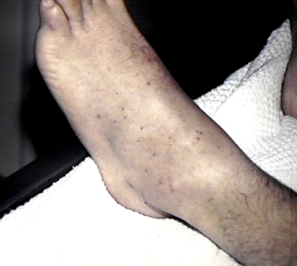
Primary hemostasis: the platelet plug
Platelets and von Willebrand factor seal small surface breaks fast. When that fails, bleeding is mucocutaneous and immediate: petechiae, purpura, gum bleeding, epistaxis, and heavy menses. Split it into quantity and quality. Quantity problems are ITP, TTP, and DIC. Quality problems are von Willebrand disease, Bernard-Soulier, and Glanzmann.
Secondary hemostasis: the fibrin clot
Clotting factors weave fibrin so the plug holds under pressure. When a factor is missing, the first plug forms then fails, so bleeding is deep and delayed: hemarthrosis, muscle hematomas, and rebleeding hours after a procedure. Hemophilia A is factor VIII, hemophilia B is factor IX, and vitamin K deficiency starves factors II, VII, IX, and X.
Which hemostasis is broken?
For each finding, call the door before you reveal the reason. Mucocutaneous and immediate is primary. Deep and delayed is secondary.
DIC eats the clotting system alive.
This is the case that scares students into the wrong answer. A septic patient bleeds and clots at the same time. Watch the consumption happen: a trigger fires tissue factor everywhere, microthrombi consume platelets and fibrinogen, red cells shear into schistocytes, and the leftover system has nothing left to stop bleeding while fibrinolysis floods the blood with D-dimer.
Trigger: sepsis, trauma, obstetric disaster, or cancer activates tissue factor across the whole vasculature.
The same shear shows on the smear
Red cells forced through the fibrin mesh get sliced into fragments called schistocytes. That is microangiopathic hemolysis. Both DIC and TTP produce it, so the smear alone does not separate them. The coags do.