Different arteries water different brain real estate. Kill the artery, kill the function of the land it feeds. Know the map, read the deficits.
Start with one clue.
A patient has sudden aphasia, right lower-face droop, and right arm weakness worse than right leg weakness. Which territory should you map first?
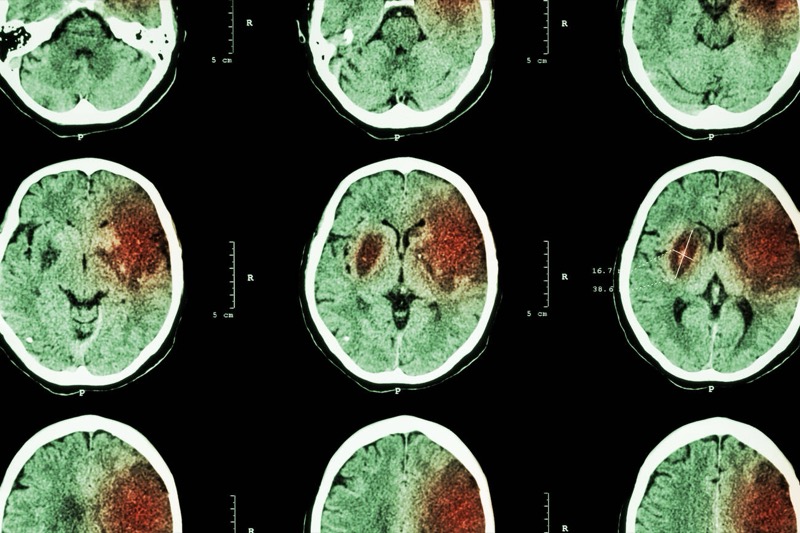
Clinical Images
📷 CT: hypodense area of ischemic infarction · tap to expand
Vessel Territory Map
Select a territory to see deficits
The Vascular Atlas
CHAPTER I · THE BRAIN'S WATERSHED
The Vascular Atlas
Six arteries, six syndromes. Each territory bleeds out a signature: face-arm, leg, vision, swallow, deafness, locked-in. The map IS the diagnosis.
Inferior view · Circle of Willis · Frontal lobes up
Tap any territory
ARTERY
Syndrome name
Tagline.
Route
Pattern
Pearl
Board Pearl
Crossed deficits = brainstem. Ipsilateral cranial nerve finding plus contralateral body finding can only happen in the brainstem, where cranial nerve nuclei live on the same side as the lesion but the body tracts have already crossed. In the cortex, everything is contralateral.
Global Hypoxic-Ischemic Encephalopathy
This one is not a vessel problem. It is a neuron problem.
The Clue
Every syndrome on the last page came from ONE artery going out and taking a matching piece of cortex with it. This one is different. The whole brain loses flow at once, so the map does not decide who dies. The neurons themselves decide, because some neurons simply cannot survive being cut off, and others can hang on a little longer.
CHAPTER II · THE FRAGILE FEW
Global Hypoxic-Ischemic Encephalopathy
Cut off ALL blood flow to the brain (shock, severe anemia, a crashing glucose) and consciousness is gone in under 10 seconds. Neurons carry no glycogen of their own, so the clock starts immediately. But the damage does not land evenly. It picks favorites.
Rank the fragility, most vulnerable first
Tap the four structures below in the order they die, from FIRST casualty of global ischemia to the one that can hang on the longest. No wrong-guessing penalty, just commit to an order.
The real order
Notice where watershed landed. It rides along in the same board mnemonic, but for a completely different reason. Hippocampus, neocortex, and Purkinje cells die because those specific NEURON POPULATIONS are intrinsically fragile: high metabolic demand, sparse collateral blood supply, receptors that flood with calcium the instant oxygen drops. Watershed territory dies because of GEOGRAPHY: it sits at the far edge of two arterial supply zones, so when perfusion PRESSURE drops (not total flow arrest, just a weak pump), it is simply the last place blood reaches. Same list, two different mechanisms, one board trap.
Memory hooks
🐠
Vulnerable hippos need pure water
The classic board mnemonic for order of injury in global ischemia: hippocampus, neocortex, Purkinje cells, watershed. Say it out loud once and the ranking interactive above stops being a guess.
tap to reveal
⏲
No glycogen, no grace period
Neurons cannot store their own glucose. Muscle and liver bank glycogen for a rainy day; neurons run hand to mouth on whatever the blood delivers right now. Cut the delivery and the clock is already running before the patient hits the floor.
tap to reveal
⏰
10 seconds to lights out, 4 minutes to permanent
Total loss of cerebral perfusion drops consciousness in under 10 seconds (think: cardiac arrest syncope). Permanent neuronal injury starts once that ischemia passes about 4 minutes. Between those two numbers is the entire resuscitation window.
tap to reveal
💩
Not just cardiac arrest
Boards love non-arrest causes of global hypoxic injury: severe shock of any flavor, severe anemia (blood present but not enough oxygen-carrying capacity), and repeated or severe hypoglycemia (fuel present but not the fuel neurons can burn). Any of the three can produce the same hippocampus-first injury pattern.
tap to reveal
🛑
Even survival is not free
Outcomes span a spectrum: full recovery is rare once injury is established, most land in coma, and prolonged global injury can progress to a persistent vegetative state where brainstem reflexes survive but the cortex does not.
tap to reveal
Board Pearl
Global versus focal is the first fork in the road. One artery down with a matching neurologic deficit is a stroke syndrome: read the map. Sudden collapse with NO focal deficit, a story of shock, severe anemia, or a hypoglycemic episode, and a pattern of amnesia plus ataxia once the patient wakes up: that is global hypoxic-ischemic injury hitting the hippocampus and cerebellum. Do not go hunting for a single occluded vessel when the whole brain lost supply at once.
Challenge before reveal
A 58-year-old man is resuscitated after 6 minutes of pulseless cardiac arrest. He regains a pulse and is extubated two days later. He is awake, follows commands, and moves all four limbs normally, but he cannot form new memories and cannot walk a straight line without swaying. His neurologic exam shows no focal weakness, no facial droop, no sensory level. What structures explain this exact combination, and why did the cortex controlling his limb strength come through relatively unscathed?
Hippocampal (CA1) and cerebellar (Purkinje cell) injury from global hypoxic-ischemic encephalopathy.
Step 1: No focal weakness, no facial droop, no sensory level rules out a single-artery stroke syndrome.
Step 2: A cardiac arrest with 6 minutes down time means the ENTIRE brain lost perfusion together, past the roughly 4-minute threshold for permanent injury.
Step 3: New memory loss with intact language and motor strength localizes to the hippocampus, the most vulnerable structure in the brain to global ischemia.
Step 4: Gait ataxia without weakness localizes to the cerebellum, specifically the Purkinje cells, the second structure down the fragility list.
Step 5: Motor cortex neurons are comparatively more ischemia-tolerant than hippocampal CA1 and Purkinje cells, so a short global insult can spare strength while still wiping out memory and coordination. Selective vulnerability, not diffuse damage, is the tested concept.
Middle Cerebral Artery
The most common stroke. Face and arm, not leg.
The Clue
Face droops, arm is weak, speech is gone. That is MCA territory. The MCA feeds the lateral cortex: motor and sensory strip for face and arm, Broca's area, Wernicke's area, and the internal capsule.
Superior Division
Inferior Division
Complete MCA
Broca's Aphasia + Right Hemiplegia
Superior division. Frontal motor strip plus Broca's area.
Speech
Non-fluent aphasia. Short broken phrases. Patient understands you and is frustrated.
Motor
Contralateral face and arm weakness. Leg largely spared.
Sensation
Contralateral face and arm numbness.
Side
Left hemisphere (Broca's is dominant hemisphere in right-handers).
High yield: They cannot get words out but understand everything. Trapped inside a broken speaker.
Wernicke's Aphasia
Inferior division. Posterior superior temporal gyrus.
Speech
Fluent aphasia. Word salad. Comprehension impaired.
Motor
Often minimal weakness. Lesion is posterior to motor strip.
Insight
Patient does NOT realize they sound wrong.
Vision
May have right superior quadrantanopia (Meyer's loop).
Discriminator: Broca = angry, can't talk, understands. Wernicke = chatty, makes no sense, no clue.
Total MCA Syndrome
Complete MCA occlusion. The cortical apocalypse.
Gaze
Eyes deviate TOWARD the lesion (away from the weak side).
Motor
Contralateral face, arm, and leg weakness (all three).
Sensory
Contralateral hemianesthesia.
Vision
Contralateral homonymous hemianopia. NO macular sparing.
Cortical gaze rule: eyes look TOWARD lesion in cortex; AWAY from lesion in pons. Cortex looks at its own hand. Pons is wired backwards.
Tap to reveal each finding
Face droop pattern
Contralateral lower face only. Forehead spared because upper face has bilateral cortical input.
tap to reveal
Arm vs leg
Arm and face hit hard. Leg largely spared. Leg lives on the medial surface (ACA territory).
tap to reveal
Right hemisphere variant
Non-dominant MCA: hemineglect, anosognosia. Patient ignores left side. May deny weakness.
tap to reveal
Internal capsule branch
Lenticulostriate occlusion: pure motor lacunar stroke. Equal face, arm, leg weakness, no cortical signs.
tap to reveal
Memory hooks
🔊
MCA = the loud one
Most common stroke you will see. Face droops, arm dies, words go. If a board question gives sudden aphasia with arm weakness, write MCA before you finish reading.
tap to reveal
😎
Broca = Broken speaker
B for Broken: can't say it, understands it. Frontal lobe = front of mouth = motor production. Patient is angry, frustrated, points at things.
tap to reveal
💬
Wernicke = Word salad
W for Word salad: talks fluently, total nonsense, no insight. Posterior temporal lobe = back of brain = comprehension. Patient happy, confused.
tap to reveal
👁️
Eyes look at the lesion
Cortex: eyes deviate TOWARD the dead hemisphere (away from the weak side). The healthy frontal eye field wins the tug of war.
tap to reveal
Challenge before reveal
A 65-year-old woman with atrial fibrillation suddenly cannot speak in full sentences. Her husband says she understands him, but she can only force out single words and looks furious. Her right face droops. Her right arm is weak. Her right leg is full strength. Which artery? Walk through the chain.
Left MCA superior division.
Step 1: Aphasia plus contralateral motor deficit = cortical, dominant hemisphere (left).
Step 2: Non-fluent + intact comprehension + frustration = Broca's aphasia.
Step 3: Face and arm weak, leg spared = lateral cortex, not medial. MCA territory, not ACA.
Step 4: Broca's area is fed by the SUPERIOR division of the MCA.
Atrial fibrillation explains the embolic source. This is a classic cardioembolic Broca aphasia.
Anterior Cerebral Artery
The leg stroke. Face and arm are fine.
The Clue
Leg is weak. Arm and face are normal. That is ACA. The ACA feeds the medial surface of the hemisphere, which handles the leg region of the motor homunculus. The leg literally hangs over the midline where the ACA lives.
Classic ACA
ACA vs MCA
Contralateral Leg Weakness
Medial frontal cortex. The leg stroke.
Motor
Contralateral leg weakness. Arm and face largely spared.
Sensory
Contralateral leg numbness.
Behavior
Abulia: no initiative, flat affect (medial frontal lobe).
clinical medicine buzzword: contralateral leg weakness with arm and face spared, plus urinary incontinence.
ACA vs MCA Discriminator
Two cortical arteries. Opposite distributions.
ACA hits
Leg dominant. Medial cortex. Behavioral changes. Incontinence.
MCA hits
Face and arm dominant. Lateral cortex. Aphasia or neglect.
Same
Both contralateral. Both cortical. Both can have UMN facial pattern.
Tell
Where does the weakness pile up? Leg = ACA. Arm/face = MCA.
One sentence: leg dominant = ACA; face and arm dominant = MCA. They live on opposite surfaces of the hemisphere.
Tap to reveal each finding
Why the leg?
The motor homunculus drapes over the brain. The leg region drops down the medial surface, which is exactly where the ACA runs.
tap to reveal
Abulia explained
Medial prefrontal cortex drives initiative. Damage = patient stops starting things. Awake but does nothing.
tap to reveal
Bladder mechanism
Medial frontal cortex inhibits the pontine micturition center. Lose the inhibitor = urge incontinence.
tap to reveal
Bilateral ACA pearl
Both ACAs from one anterior communicating artery aneurysm rupture: paraplegia plus profound abulia. Looks like a spinal cord lesion.
tap to reveal
Memory hooks
🦵
ACA = A leg, no Arm
A for Anterior, A for the part not affected: the Arm. Hits the leg, spares the arm. Opposite of MCA. Single mnemonic, both arteries.
tap to reveal
🚽
Wet pants = ACA
Urinary incontinence in a stroke patient = think medial frontal cortex = ACA. The bladder control sits right in ACA territory.
tap to reveal
😶
Abulia = won't start anything
Frontal flat affect, no spontaneous speech, no spontaneous movement. They will answer if you push. They will not initiate.
tap to reveal
Challenge before reveal
A 70-year-old man develops sudden left leg weakness. His left arm is full strength. His face moves symmetrically. His wife says he has been quiet and disinterested all morning, and his pants are wet. No aphasia, no visual field cut. Which artery? Walk through it.
Right ACA.
Step 1: Isolated leg weakness with spared arm and face = medial cortex distribution.
Step 2: No cortical aphasia, no neglect, no hemianopia rules out a big MCA stroke.
Step 3: Abulia (quiet, disinterested) + urinary incontinence = medial frontal lobe. ACA territory.
Step 4: Left-sided weakness = right hemisphere lesion = right ACA.
Posterior Cerebral Artery
The vision stroke. Hemianopia with macular sparing.
The Clue
Half the visual field is gone in both eyes, but central vision is still intact. That is PCA. The PCA feeds the occipital cortex. The macula gets dual supply from both MCA and PCA, so it survives even when the PCA is occluded.
Classic PCA
Deep Branch (Thalamus)
Top of Basilar
Homonymous Hemianopia with Macular Sparing
Cortical PCA. Occipital cortex.
Vision
Contralateral homonymous hemianopia. Same half missing in both eyes.
Macula
SPARED. They can read. The peripheral half is gone.
Motor
No weakness. Motor cortex is MCA territory.
Side
Dominant PCA: alexia without agraphia (splenium plus left occipital lesion).
One-shot rule: if the stem says "macular sparing," stop reading and write PCA.
Thalamic Pain Syndrome (Dejerine-Roussy)
Deep PCA branch to thalamic VPL.
Acute
Contralateral hemisensory loss across all modalities.
Weeks later
Burning, lancinating central pain on the same numb side.
Bilateral PCA territory infarct from basilar tip embolus.
Vision
Cortical blindness. Both occipital lobes gone. Macular sparing may preserve a tiny central window.
Anton
Anton syndrome: cortical blindness with denial of blindness. Patient confabulates what they "see."
Memory
Bilateral hippocampi infarcted = severe anterograde amnesia.
Pupils
Pupils still react. The afferent and efferent pupillary loops are subcortical.
The trap: Anton syndrome = patient bumps into walls but insists they see fine. Cortical blindness with preserved pupillary reflex.
Tap to reveal each finding
Why macular sparing?
The occipital pole (macula tip) is dual-supplied by PCA + a watershed MCA branch. PCA dies, MCA backup keeps the macula alive.
tap to reveal
Alexia without agraphia
Left PCA hits left occipital + splenium of corpus callosum. Right occipital still sees, but visual info cannot reach left language cortex. Reads nothing, writes fine.
tap to reveal
No weakness clue
PCA is posterior. Motor cortex is anterior (MCA territory). A pure visual field deficit with full strength is the PCA signature.
tap to reveal
MCA hemianopia trap
MCA strokes can cause hemianopia from optic radiation involvement, but NEVER with macular sparing. MCA has no occipital pole backup.
tap to reveal
Memory hooks
👁️
PCA = Posterior = Pictures
Posterior cortex = visual cortex. PCA = picture artery. If the question is about vision, PCA is your first guess every time.
tap to reveal
📖
Macular sparing = MCA backup
The occipital tip has dual blood supply. PCA dies but MCA keeps the macula alive. Only PCA strokes have macular sparing.
tap to reveal
🕵️
Anton = blind in denial
Bilateral PCA from basilar tip embolus = cortical blindness, but the patient swears they see. They confabulate. Walking into walls but talking like nothing is wrong.
tap to reveal
Challenge before reveal
A 72-year-old woman with hypertension reports she "cannot see to her right" in either eye. Reading is fine. She bumps into things on her right when walking. No weakness, no aphasia, normal speech. Which artery, and why is the reading preserved? Walk through it.
Left PCA.
Step 1: Same half (right) missing in both eyes = right homonymous hemianopia. Defect is post-chiasmal, contralateral to the lesion (left).
Step 2: No motor weakness, no aphasia = not MCA cortex.
Step 3: She can still read = macular sparing. Central vision is preserved because the occipital tip has dual MCA + PCA supply.
Step 4: Macular sparing locks in PCA. MCA hemianopia does not spare the macula.
PICA: Wallenberg Syndrome
Lateral medulla. Crossed everything. No limb weakness.
The Clue
One side of the face is numb. The other side of the body is numb. Patient cannot swallow. One eyelid droops with a tiny pupil. They are dizzy and falling toward one side. No arm or leg weakness. That is Wallenberg. The dead giveaway is the CROSSED sensory pattern.
Full Wallenberg
The Cross
PICA vs AICA
Wallenberg: The Six-Pack
Lateral medulla. Six findings. Zero limb weakness.
Face sensation
IPSILATERAL face numb (CN V nucleus is in the lateral medulla).
Body sensation
CONTRALATERAL body numb (spinothalamic crossed in cord).
Horner's
IPSILATERAL: ptosis + tiny pupil + no sweating (descending sympathetics).
CN IX, X
IPSILATERAL dysphagia and hoarseness.
Ataxia
IPSILATERAL: inferior cerebellar peduncle. Falls toward the lesion.
Limbs
NORMAL strength. Corticospinal tract is medial, not lateral.
clinical medicine buzzword: crossed sensory loss + Horner + dysphagia, no limb weakness.
Why the Cross Happens
Two pain pathways. Two crossing points. One lesion.
Face pain
Synapses in the lateral medulla SAME SIDE as the stroke. Numb on the lesion side.
Body pain
Spinothalamic already crossed BELOW in the cord. Numb on the OPPOSITE side.
Result
One lateral medullary lesion hits face on its side, body on the other side.
Why important
Crossed pattern is anatomically only possible in the brainstem. Cortex is all-contralateral.
Same crossed picture. Two extra cranial nerves separate them.
PICA = medulla
No facial droop. No deafness. CN V, IX, X only.
AICA = pons
All PICA findings PLUS CN VII (facial droop) and CN VIII (deafness, vertigo).
CN VII clue
Ipsilateral facial weakness includes the forehead (LMN pattern). LMN = pons or below.
CN VIII clue
Sudden deafness with brainstem signs = AICA, not PICA.
Killer line: if you see facial droop or deafness with brainstem signs, you have left the medulla. The artery is AICA.
Tap to reveal each finding
Horner mechanism
Descending sympathetic fibers from hypothalamus pass through the lateral medulla. Cut the fibers, lose sympathetic tone on that side: ptosis, miosis, anhidrosis.
tap to reveal
Falls toward the lesion
Inferior cerebellar peduncle damaged on one side = ipsilateral cerebellar dysfunction. Cerebellum coordinates the SAME side, so they list toward the dead side.
tap to reveal
Why no limb weakness
The corticospinal tract is in the MEDIAL medulla (ASA territory). PICA feeds the LATERAL medulla. Different highway, different artery.
tap to reveal
Dysphagia mechanism
CN IX (glossopharyngeal) and CN X (vagus) nuclei live in the lateral medulla. Damage = soft palate weakness, hoarseness, swallowing failure on the lesion side.
tap to reveal
Vertical etiology
Most common cause = vertebral artery dissection or atherosclerosis. PICA is a branch of the vertebral artery.
tap to reveal
Hiccups + Wallenberg
Intractable hiccups can accompany Wallenberg. The respiratory pattern generator sits in the lateral medulla.
tap to reveal
Memory hooks
❌
Cross your X = brainstem
Ipsilateral face + contralateral body sensory loss makes an X. That X only happens in the brainstem. Cortex is one-sided.
tap to reveal
🐶
PICA = AICA's little cousin
Same crossed picture, one floor down (medulla). PICA does not get face droop or deafness because CN VII and CN VIII live upstairs in the pons.
tap to reveal
😀
No face droop, no deafness
PICA spares CN VII and CN VIII. If the patient has facial droop or hearing loss with brainstem signs, you are in the pons (AICA), not the medulla (PICA).
tap to reveal
🍸
Drunk and crooked
Patient walks like they had five drinks (cerebellar ataxia), one eye half closed with a tiny pupil (Horner), and cannot swallow (CN IX, X). Picture stays. Wallenberg.
tap to reveal
Challenge before reveal
A 58-year-old man on warfarin develops sudden vertigo, hoarseness, and trouble swallowing. Exam: right facial pinprick is decreased. Left arm and leg pinprick is decreased. Right ptosis with a 2 mm right pupil. He veers right when walking. Strength is 5/5 everywhere. No facial droop, hearing intact. Which artery? Walk the chain.
Right PICA. Wallenberg syndrome.
Step 1: Crossed sensory loss (right face + left body) = brainstem, not cortex.
Step 2: Right Horner (ptosis, miosis) + dysphagia + ipsilateral ataxia = lateral medulla anatomy.
Step 3: Strength preserved = corticospinal spared = lateral, not medial, medulla.
Step 4: No facial droop and no deafness = still in medulla, not pons. PICA, not AICA.
Step 5: Side = right (face pain on right, ipsilateral findings on right). Right PICA.
Brainstem: AICA, Weber, Basilar, ASA
Same crossed logic, different levels of the brainstem.
The Map
Brainstem strokes follow one rule: cranial nerves stay on the side of the lesion, body tracts already crossed. So you get an ipsilateral cranial nerve plus a contralateral body deficit. The level of the lesion tells you which cranial nerve.
The four levels at a glance
Lateral pons (AICA)
Wallenberg findings PLUS CN VII (face droop) and CN VIII (deafness, vertigo). Pons floor adds two cranial nerves.
tap to reveal
Ventromedial midbrain (Weber)
Ipsilateral CN III (down-and-out, blown pupil) + contralateral hemiplegia. Pure motor plus oculomotor.
tap to reveal
Ventral pons (Basilar trunk)
Locked-in syndrome. Quadriplegia, mute, conscious. Vertical gaze and blink only.
tap to reveal
Medial medulla (ASA)
Contralateral limb weakness + ipsilateral tongue deviation + contralateral vibration loss. Sparing pain and temp (those are lateral, PICA territory).
tap to reveal
Memory hooks for brainstem strokes
🔑
Crossed = brainstem
Ipsilateral cranial nerve + contralateral body deficit can ONLY happen in the brainstem. CN nuclei sit on the lesion side; body tracts already crossed.
tap to reveal
🏋️
Climb the brainstem, add the nerves
Medulla = CN IX, X, XII. Pons = adds CN V, VI, VII, VIII. Midbrain = adds CN III, IV. Going up the brainstem means more cranial nerves on the menu.
tap to reveal
👁️
Pupil tells the level
Big fixed pupil = CN III = midbrain (Weber). Small constricted pupil = Horner = lateral medulla or pons (PICA, AICA). Pupils preserved with quadriplegia = ventral pons (Basilar).
tap to reveal
🔁
Eyes look AWAY from lesion in pons
Pontine PPRF damage: eyes deviate AWAY from the lesion (toward the weak side). Cortex is opposite (eyes toward the lesion). Don't flip them.
tap to reveal
Decision tree: localize the artery
A patient with sudden focal neurologic deficit. Walk through the questions. Each answer narrows the territory.
1
First question: are the deficits crossed (cranial nerve on one side, body on the other) or all on the same side?
All same side: cortical pattern
Crossed: cranial nerve here, body there
Pure motor or pure sensory, no cortical signs at all
2
Cortical. Where does the weakness pile up?
Face and arm dominant, plus aphasia or neglect
Leg dominant, plus abulia or incontinence
No motor deficit. Visual field cut with macular sparing
3
Brainstem. Which cranial nerves are involved?
CN V (face pain), IX, X (dysphagia) + Horner. NO facial droop, NO deafness
All PICA findings PLUS CN VII (face droop) and CN VIII (deafness)
CN III only (down-and-out eye, blown pupil) + contralateral hemiplegia
Quadriplegia + preserved consciousness + vertical gaze only
CN XII (tongue deviates) + contralateral limb weakness + contralateral vibration loss
4
No cortical signs, isolated motor or sensory. Which pattern?
Equal face, arm, leg weakness. No sensory, no aphasia
All sensory modalities lost on one side. No motor
Hemiparesis + hemisensory loss together, no cortical signs
AICA: Lateral Pontine Syndrome
Wallenberg + face droop + deafness. One floor up.
Full Picture
vs PICA
AICA: Everything PICA Has, Plus Two
Lateral pons. Adds CN VII and CN VIII to Wallenberg.
All PICA findings
Crossed sensory loss, Horner's, ataxia, dysphagia all still present.
CN VII added
IPSILATERAL facial droop (CN VII nucleus is in the pons). LMN pattern, includes forehead.
CN VIII added
IPSILATERAL deafness and vertigo. The labyrinthine artery branches off AICA.
Gaze palsy
IPSILATERAL horizontal gaze palsy (PPRF and CN VI in the pons).
Same crossed logic. Two extra cranial nerves separate them.
PICA
Lateral medulla. CN V, IX, X. No facial droop. No deafness.
AICA
Lateral pons. Adds CN VII and CN VIII.
Single discriminator
Facial droop OR sudden deafness with brainstem signs = AICA.
Trace It
CN VII nucleus and the labyrinthine artery (CN VIII supply) live in the pons, one level above the medulla.
clinical medicine trap: if a Wallenberg-like stem adds "ipsilateral hearing loss," switch your answer from PICA to AICA. Don't miss it.
Challenge before reveal
A 64-year-old man presents with sudden vertigo and hoarseness. Exam: left face droops including the forehead. Hearing is gone in the left ear. Right arm and leg pinprick decreased. Left ptosis, miosis. Veers left walking. Strength 5/5. Which artery? Walk the chain.
Left AICA.
Step 1: Crossed sensory (right body, left ipsilateral findings) = brainstem.
Step 2: Horner + ipsilateral ataxia + hoarseness = lateral medullary findings.
Step 3: BUT add LMN facial droop (CN VII) and ipsilateral hearing loss (CN VIII): you are in the pons.
Step 4: Lateral pons = AICA. PICA does not include CN VII or VIII. Side: left.
Weber Syndrome: Midbrain
Down-and-out eye + opposite hemiplegia. Two structures, one lesion.
Full Picture
Pupil Discriminator
Weber: CN III + Contralateral Hemiplegia
Ventromedial midbrain. CN III root + corticospinal tract.
CN III
IPSILATERAL CN III palsy: eye down and out, dilated fixed pupil, ptosis.
Motor
CONTRALATERAL hemiplegia (corticospinal tract crosses below the lesion).
Face motor
CONTRALATERAL lower face weakness (UMN pattern, forehead spared).
Sensory
Intact. Pure motor + CN III palsy only.
Anatomy lock: CN III nerve root and the corticospinal tract run side by side through the ventral midbrain. One lesion takes both.
Weber CN III vs Horner: The Pupil Tells
Both have ptosis. Pupils point opposite directions.
Weber (CN III)
LARGE fixed pupil. Parasympathetic gone. Eye down and out.
Horner (PICA/AICA)
SMALL constricted pupil. Sympathetic gone. Eye position normal.
Lid level
Both droop. CN III ptosis is more severe (upper lid muscle is direct CN III).
A 68-year-old woman with hypertension wakes with a droopy right eyelid. Her right eye is deviated down and out, the right pupil is dilated and fixed, and she has 2/5 strength in the left arm and leg. No sensory deficit. Where is the lesion, and which artery branch?
Right ventromedial midbrain. Paramedian branch of the right PCA. Weber syndrome.
Step 1: Ipsilateral CN III palsy (down-and-out, blown pupil) + contralateral hemiplegia. Pure motor, no sensory.
Step 2: That combination can only happen where CN III root and the corticospinal tract run together: ventral midbrain.
Step 3: Blood supply = paramedian (perforator) branches of the PCA. Side = right (CN III on right, hemiplegia on left).
Step 4: Differential: blown pupil rules out Horner (small pupil); preserved sensation rules out a lateral brainstem syndrome.
Basilar Artery: Locked-In Syndrome
Awake. Aware. Quadriplegic. Blink and vertical gaze only.
Locked-In
Top of Basilar
vs Coma
Locked-In: Ventral Pontine Catastrophe
Bilateral ventral pons infarct. Both corticospinal and corticobulbar tracts gone.
PRESERVED. CN III lives in the midbrain above the lesion.
Communication
Blink and vertical eye movement only. That is the entire motor output.
The visceral detail: they are fully awake. They hear, think, feel, understand. They just cannot move or speak. Locked-in is not a coma; it is the opposite of a coma.
Top-of-the-Basilar: Bilateral PCA
Embolus to the basilar tip. Hits both PCAs at once.
Vision
Cortical blindness. Both occipital lobes infarcted.
Anton
Patient denies blindness. Confabulates what they "see."
Memory
Bilateral hippocampal infarcts = severe anterograde amnesia.
Pupils
Asymmetric or fixed; midbrain pretectum involvement.
Different lesion, same artery family: top of basilar = bilateral PCA territory. Locked-in = ventral pons. Both come from the basilar system.
Locked-In vs Coma vs Persistent Vegetative State
All three look immobile. All three are different.
Locked-in
Conscious + aware. Vertical gaze and blink only. Ventral pons.
Coma
Unconscious. No purposeful movement. Reticular activating system OR bihemispheric injury.
PVS
Eyes open, sleep-wake cycles, NO awareness. Cortical death with brainstem alive.
Test
Ask for vertical gaze or blink to command. Locked-in patients DO IT. Coma and PVS do not.
Bedside maneuver: "Look up if you understand me." If they look up, they are awake and trapped. That is locked-in until proven otherwise.
Challenge before reveal
A 52-year-old man is brought in unresponsive after collapsing. On exam he cannot move any limb and cannot speak. He cannot move his eyes side to side. When you ask him to look up, his eyes track upward and he blinks once for yes. Brainstem reflexes (pupils, corneal) are present. Which vessel? Walk the chain.
Step 1: Quadriplegia + mute = bilateral corticospinal AND corticobulbar tract failure. That requires a midline lesion.
Step 2: He is conscious (tracks vertical, blinks to command). Reticular formation (dorsal pons) is intact.
Step 3: He cannot move horizontally but CAN move vertically. PPRF/CN VI in the pons are out; CN III in the midbrain is intact.
Step 4: Bilateral ventral pons infarct = basilar trunk occlusion. Time-critical: thrombectomy window.
ASA: Medial Medullary Syndrome
Tongue deviates toward the lesion. Limbs weak on the other side.
Full Picture
ASA vs PICA
Medial Medullary Syndrome
Anterior spinal artery. Medial medulla.
Motor
CONTRALATERAL limb weakness (corticospinal tract is medial in the medulla).
CN XII
IPSILATERAL tongue deviation (hypoglossal nucleus is medial). Tongue points TOWARD the lesion.
Vibration / proprioception
CONTRALATERAL loss (medial lemniscus is medial).
Pain / temp
INTACT. Spinothalamic tract is lateral (PICA territory, not here).
Pain/temp loss = lateral (PICA). Vibration/proprioception loss = medial (ASA).
The medulla is split in half: medial = motor + DCML + CN XII; lateral = pain/temp + autonomics + cerebellum + CN V/IX/X. One artery for each half.
Challenge before reveal
A 71-year-old woman with diabetes presents with sudden right arm and leg weakness. Pain and temperature sensation are intact. Vibration sense is decreased on the right arm and leg. When she sticks her tongue out, it deviates to the left. Which artery? Walk the chain.
Left ASA (anterior spinal artery), medial medulla.
Step 1: Right hemiparesis + right vibration loss = contralateral, so the lesion is on the LEFT side of the medulla.
Step 2: Pain/temp intact = lateral spinothalamic tract is fine. Lesion is medial, not lateral. PICA ruled out.
Step 3: Tongue deviates LEFT = ipsilateral CN XII palsy = hypoglossal nucleus on the LEFT.
Step 4: Medial medulla = ASA territory. Side: left.
Lacunar Strokes
Small vessel disease. Pure syndromes. No cortical signs.
The Clue
Pure motor OR pure sensory deficit with no aphasia, no neglect, no hemianopia. That is lacunar. Tiny perforating arteries deep in the brain get clogged by lipohyalinosis: years of hypertension slowly destroying their walls until they seal shut.
Pure Motor
Pure Sensory
Sensorimotor
Ataxic Hemi / Dysarthria
Pure Motor: Internal Capsule Lacune
Posterior limb of internal capsule (lenticulostriate arteries).
Location
Posterior limb of internal capsule.
Motor
Contralateral face, arm, leg weakness. EQUAL distribution.
Sensory
INTACT. Zero sensory deficit.
Cortical signs
None. No aphasia, no neglect, no hemianopia. Just weakness.
Why equal: the internal capsule packs face, arm, and leg motor fibers into a tiny corridor. One small lacune knocks them all out together.
Pure Sensory: Thalamic VPL Lacune
Thalamogeniculate arteries from PCA.
Location
VPL nucleus of thalamus.
Sensory
Contralateral hemisensory loss. ALL modalities: pain, temp, vibration, proprioception.
Motor
INTACT. Zero motor deficit.
Late finding
Dejerine-Roussy: weeks later, central neuropathic pain on the numb side.
Thalamus = sensory relay: all sensory modalities funnel through VPL. One small lacune wipes them all out at once.
Sensorimotor: Thalamocapsular Lacune
Lacune at the junction of thalamus and posterior internal capsule.
Motor
Contralateral hemiplegia (equal face, arm, leg).
Sensory
Contralateral hemisensory loss (all modalities).
Cortical signs
None. No aphasia, no neglect, no hemianopia.
Why both
The lacune sits between motor (capsule) and sensory (thalamus) tracks.
Looks like an MCA stroke, but the absence of cortical signs gives it away. Cortical strokes bring aphasia, neglect, or hemianopia. Lacunar does not.
Ataxic Hemiparesis & Dysarthria-Clumsy Hand
Two more classic lacunar syndromes (Fisher).
Ataxic hemi
Contralateral leg weakness + ipsilateral cerebellar-type ataxia. Posterior limb internal capsule or basis pontis.
Dysarthria-clumsy hand
Slurred speech + clumsy contralateral hand. Genu of internal capsule or basis pontis.
Common theme
Small vessel + HTN. No cortical signs.
Imaging
MRI shows tiny (3-15 mm) deep lesions. CT often misses early.
Five classic lacunar syndromes (Fisher): pure motor, pure sensory, sensorimotor, ataxic hemiparesis, dysarthria-clumsy hand.
Tap to reveal each finding
Lipohyalinosis
Chronic HTN damages tiny perforating arteries. Walls thicken, lumen seals. The deep tissue downstream infarcts in a tiny pocket = a lacune.
tap to reveal
No cortical signs
Lacunes are deep. They never reach cortex. So aphasia, neglect, or hemianopia immediately rule out lacunar.
tap to reveal
Pure = the magic word
Pure motor or pure sensory with no other findings = lacunar until proven otherwise. Mixed pictures with cortical signs are not lacunar.
tap to reveal
Risk factors
Hypertension, diabetes, smoking, age. Cardioembolic source is rare for lacunes (those favor cortical strokes).
tap to reveal
Memory hooks
🔍
Pure means lacunar
Pure motor, pure sensory, ataxic hemiparesis, dysarthria-clumsy hand. Five Fisher syndromes. All deep, all small, all from chronic HTN.
tap to reveal
🔩
No cortex, no cortical signs
Lacunes never reach the surface. So no aphasia, no neglect, no hemianopia. Any of those signs = not lacunar, look elsewhere.
tap to reveal
🌊
Equal face, arm, leg = capsule
Cortex distributes motor regions across a wide strip. Internal capsule packs them all together. Equal weakness of all three points to capsule (lacunar), not cortex (MCA).
tap to reveal
Challenge before reveal
A 78-year-old man with 30 years of HTN develops sudden left face, arm, and leg weakness, all roughly equal. Sensation is normal. Speech is normal. No visual field cut. CT shows no hemorrhage. MRI later shows a 4 mm deep lesion. Which artery and where? Walk the chain.
Right lenticulostriate (MCA branch). Posterior limb of right internal capsule. Pure motor lacunar infarct.
Step 1: Equal face, arm, leg weakness on one side = motor fibers packed together. Cortex spaces them out; capsule packs them.
Step 2: No cortical signs (no aphasia, no neglect, no hemianopia) rules out cortical MCA.
Step 3: Sensory entirely intact rules out thalamic and sensorimotor lacunes.
Step 4: Chronic HTN + tiny deep MRI lesion fits lipohyalinosis. Pure motor lacune is classic.
Original clinical vignettes. Shuffled, never-repeat, full explanations for every choice.
Medically reviewed by Kaitlyn Cocuzzo, MD and Fatima Ali, DO · Last updated July 7, 2026 at 7:42 PM ET
Bone Wizardry is an independent educational resource for visual learning in the medical sciences. It is not affiliated with, endorsed by, or sponsored by any licensing or examination board, contains no real or recalled examination questions, and does not guarantee any educational or examination outcome.