The most common malignant brain tumor in kids. It hides in the back of the head, drops cells down the spine, and shows up on the slide as little blue flowers. Learn the four clues that win it at 3am.
Brain tumors overview · Pilocytic astrocytoma · Drop metastases
Medulloblastoma comes with the same four fingerprints every time. Spot any one of them in a vignette and you should be reaching for this diagnosis. Tap through them.
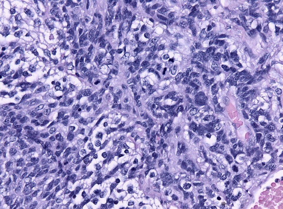
Small blue round cells holding hands around a pink center
Under the microscope, medulloblastoma is a small blue round cell tumor: tightly packed cells with dark nuclei and barely any cytoplasm. The signature move is a rosette: a ring of these cells arranged around a fuzzy pink core of cell processes (neurofibrillary stuff, not a blood vessel).
The rosette tells you this is a primitive neuroectodermal tumor (PNET). Same family as neuroblastoma in the adrenal. Different location, same flower.
Midline, infratentorial, fourth-ventricle squeezer
Medulloblastoma plants itself in the cerebellar vermis: the midline strip of cerebellum that runs trunk balance. This puts it directly on top of the fourth ventricle.
Compare to pilocytic astrocytoma, which prefers a cerebellar hemisphere off to one side and causes appendicular ataxia (one arm misses). Midline = medulloblastoma. Off to the side = pilocytic.
Brain tumor + colon polyps in the same patient
If the vignette throws in a family history of colon polyposis or a teenager with both a brain tumor and a colonoscopy full of polyps, think Turcot syndrome. Brain CA + colon CA in one body.
The other flavor of Turcot involves mismatch repair mutations (Lynch variant) and pairs polyps with glioblastoma instead. The discriminator: if it is a medulloblastoma in a kid, think APC. See the Turcot section below.
Tumor cells fall down the spinal canal like sediment
Medulloblastoma sits right next to the fourth ventricle, so cells shed straight into the CSF and ride the current down. They settle on the spinal cord and cauda equina. The name for this: drop metastases.
This is why staging requires a full spine MRI plus CSF cytology, not just the brain MRI. And why treatment is craniospinal radiation: you have to hit the whole CSF column, not just the cerebellum.
Pediatric brain tumors split sharply by the tentorium. Tap a tumor below to highlight where it lives. Anything below the tentorium is posterior fossa, which is medulloblastoma country.
Left: real H&E micrograph of a small-blue-round-cell tumor with Homer Wright rosettes. Right: a drawn schematic so you know exactly what to look for. Tap the photo to enlarge.
When the vignette pairs a brain tumor with colon polyposis, the answer is Turcot. There are two flavors. Same name, different gene, different brain tumor.
Three rosettes show up in clinical practice. They look similar through a microscope and they are easy to mix up at midnight. The trick: what is in the center.
| Rosette | What is in the center | Cells arrange around | Tumor | Memory hook |
|---|---|---|---|---|
| Homer Wright PNET | Eosinophilic fibrillary tangle (cell processes). No vessel. | A pink swirl of neurites | Medulloblastoma, neuroblastoma, retinoblastoma (less common) | Cells thinking: processes in the middle, neurons trying to remember themselves |
| Perivascular pseudorosette EPEN | A blood vessel. Cell processes radiate toward the vessel wall. | A capillary in the dead center | Ependymoma (any age, often fourth ventricle in kids) | Cells drinking: everyone wants a sip from the central vessel |
| Flexner-Wintersteiner RETINO | An empty lumen (a tiny central hole, no vessel, no fibrils) | A clear central space, like a primitive photoreceptor | Retinoblastoma | Cells looking: arranged around an empty hole like a tiny eye |
3am rule: The center wins. Fibrils = Homer Wright. Vessel = perivascular pseudorosette. Empty hole = Flexner-Wintersteiner. If you can identify what is in the middle, you have the diagnosis.
Five clinical stems. Pick, get the explanation, learn the discriminator. No timer.
