An adult walks in with a single liver mass on imaging. No cirrhosis. No hepatitis. What is the most common malignant tumor found in the liver?
Each site has a reason. Tap a card to flip it and see the mechanism.
Given a primary cancer, pick where it most likely spreads. Guess before the reveal drops.
4 clinical scenarios. Tap your answer before you see the explanation. Some have a specific twist. Yes, prostate is in here.
Prostate Cancer
A 68-year-old man with known prostate cancer has back pain and an elevated PSA. His spine X-ray shows dense, ivory-white vertebrae. What type of bone lesion is this?
Osteolytic lesions
Osteoblastic lesions
Mixed lytic-blastic
Exactly right. Prostate is the one cancer that loves to MAKE bone. Ivory vertebrae on X-ray = osteoblastic = prostate cancer. The board will try to confuse you with the pain, but the dense white bone is the give-away.
Lytic lesions eat bone: breast, kidney, lung, thyroid. Prostate does the opposite. Dense white bone on X-ray = osteoblastic. The cancer is stimulating osteoblasts to overproduce bone.
Mixed is technically possible for some cancers, but for prostate the clinical medicine want osteoblastic. Dense white vertebrae with an elevated PSA = prostate = blastic. That is the pairing to lock in.
Prostate = osteoblastic only. Elevated PSA + dense bone = prostate until proven otherwise. Every other common cancer does lytic.
Colon Cancer
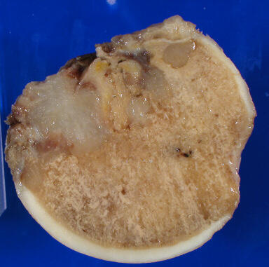
A 56-year-old woman undergoes staging CT after her colon cancer diagnosis. Imaging shows multiple nodular lesions in one specific organ. Which organ is the most classic first-stop met site for colon cancer?
The portal vein. Colon drains into portal circulation, which flows straight to the liver. The liver is the first capillary bed colon cancer hits. That is why hepatic mets from colorectal cancer are so common.
Lung is a common met site overall, but colon cancer hits the liver first. The portal vein takes venous drainage from the GI tract directly to the liver before any other site. Liver then lung is the classic colon cancer pattern.
Brain mets happen with colon cancer, but late. The first-stop met via portal drainage is the liver. If clinical medicine say colon cancer + one new lesion on staging, they want liver.
Bone mets occur but are not the classic first-stop for colon cancer. Portal drainage means liver gets first dibs. Bone involvement is typically later or part of widespread disease.
Colon cancer met = liver first, portal vein explains why. This is the most tested colon cancer met in clinical practice.
Renal Cell Carcinoma
A 44-year-old man presents with hematuria and flank pain. CT shows a renal mass. Chest X-ray reveals bilateral rounded pulmonary nodules. What is the classic term for this CXR pattern?
Cannonball mets
Miliary pattern
Bat-wing infiltrates
Kerley B lines
Cannonball mets. Large, round, bilateral pulmonary nodules that look like someone fired artillery through both lungs. Renal cell carcinoma is the board classic for this pattern, along with choriocarcinoma.
Good instinct but miliary is tiny, uniform, millet-seed-sized nodules: think TB, histoplasmosis, or miliary TB. Renal cell cannonball mets are large, round, obvious. Miliary anything = infectious, not renal cancer.
Bat-wing infiltrates are bilateral perihilar haze: pulmonary edema, not mets. Bat-wing pattern means fluid overload or CHF, not cancer. Renal cancer makes round solid nodules, not fluffy infiltrates.
Kerley B lines are small horizontal lines at the lung periphery from lymphatic engorgement: pulmonary congestion, not mets. Kerley B = fluid backing up. Renal mets = solid cannonball nodules.
Renal cell + bilateral round nodules = cannonball mets. Large, well-circumscribed, bilateral. If you see this combo, say cannonball and say renal cell.
Lung Cancer
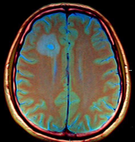
A 62-year-old woman with lung cancer develops progressive confusion, severe headache, and new focal neurologic deficits. MRI shows multiple ring-enhancing lesions at the grey-white junction. Why specifically at that location?
Blood vessels narrow there, trapping tumor cells
Grey matter has thinner BBB
Rich lymphatic drainage there
Random distribution, coincidence
The grey-white junction is where vessels abruptly change caliber. Larger cortical vessels transition to smaller white-matter vessels. Circulating tumor cells hit this bottleneck, slow down, and get stuck. Like a piece of debris hitting a drain screen.
The blood-brain barrier is actually present throughout the brain. The vascular caliber change at the junction is the real answer. The BBB is not thinner there; the vessels are just narrower, which creates a physical trap for tumor emboli.
The brain has essentially no lymphatic drainage in the traditional sense (it uses cerebrospinal fluid and glymphatic pathways). Lymphatic drainage does not explain the grey-white junction preference. Vessel caliber change does.
Not random. The grey-white junction preference is reproducible and mechanistically explained by vascular anatomy. Recognizing this pattern is exactly the board pearl they test. It is never random with mets.
Grey-white junction = vessel caliber abruptly narrows = tumor emboli get stuck. Ring-enhancing lesions at that location in a cancer patient = met. Done.
Tap each hook to reveal the sticky version. One tap = the mnemonic drops.
🔑 Which cancer does osteoBLASTic?
Prostate BLASTs bone. Pro-BLAST-ate. One cancer does blastic. That one is prostate. Everything else eats bone (lytic).
🔑 Most common brain tumor overall?
Mets, not primary. Think of the brain as a guest house for other organs' problems. It rents out rooms to cancer from everywhere else. Glioblastoma is the most common PRIMARY, but most brain tumors total are mets.
🔑 Colon cancer goes to liver first. Why?
Portal vein is the highway. Colon drains into the portal vein, which goes to the liver before anywhere else. The liver is at the end of the colon's exit ramp. Tumor cells riding that highway make their first stop there.
🔑 Bilateral adrenal mets = what crisis?
Adrenal insufficiency. Bilateral destruction = no cortisol. Hypotension, hyponatremia, hyperkalemia. Cancer patient crashing with those electrolytes: check the adrenals.
🔑 Pericardial met complication?
Tamponade. Beck's Triad. Cancer seeds the pericardium, fluid builds up, heart gets squeezed. Muffled sounds + hypotension + elevated JVD in a cancer patient = malignant tamponade. Drain it.
🔑 Most common cancer IN the liver?
Metastatic disease, not HCC. HCC = most common PRIMARY liver cancer. But if you count all tumors found in liver tissue, metastatic disease wins by a landslide. clinical medicine love this distinction.
5 patients. Each one is testing a different met concept. Let's see which ones stick.
Bone Wizardry is an independent educational resource for visual learning in the medical sciences. It is not affiliated with, endorsed by, or sponsored by any licensing or examination board, contains no real or recalled examination questions, and does not guarantee any educational or examination outcome.