Your neutrophils manufacture literal household bleach to kill bacteria. When the factory breaks (CGD), catalase-positive bugs walk free.
Board question: A 4-year-old boy is brought in for his third serious infection this year. He had a liver abscess from Staphylococcus aureus at age 2, then a lung mass from Aspergillus last spring. His labs show normal lymphocyte counts, normal complement, and normal immunoglobulins. A nitroblue tetrazolium test comes back negative. What is the defect?
Good instinct if you went for Bruton's or DiGeorge. Those are the two immunodeficiencies everyone learns first, so they feel right. But think about it: his immunoglobulins are normal (not Bruton's), his lymphocytes are normal (not DiGeorge), and his complement is normal. The negative NBT test is the key: normal neutrophils turn the dye blue by producing reactive oxygen species. His neutrophils can't. The organism pattern seals it: Staph and Aspergillus are both catalase-positive, the exact organisms that exploit CGD. Break it down: negative NBT + catalase-positive organisms = CGD every time.
Section 1
The Bleach Circuit
Tap a step or hit Play to watch the factory light up. Each node is a molecule. Each arrow is an enzyme.
Plain English
A free radical is a molecule missing one electron from its outer shell. That makes it chemically desperate. It rips electrons out of whatever it touches: cell membranes, proteins, DNA. Your neutrophils weaponize that desperation. They build free radicals on purpose and aim them at bacteria.
Tap any node above. Walk the path: oxygen becomes a radical, the radical becomes peroxide, peroxide becomes literal household bleach inside the phagosome.
What a Free Radical Does to a Cell
Same chemistry, two outcomes: weapon (when aimed at a bug) or weapon-of-mass-destruction (when aimed at you).
🎯
1 · Pierces cell membrane
Steals electrons from membrane lipids. Lipid bilayer fragments.
↓
🧐
2 · Pierces nuclear membrane
Same trick on the inner envelope. Nucleus is now exposed.
↓
🧬
3 · Damages DNA
Oxidizes bases, breaks strands, scrambles the code.
↓
⚖️
4 · Apoptosis OR mutation
Clean exit (cell dies) or dirty survival (becomes cancer). Sources: infections (viral #1 overall), sulfa drugsSulfa drugs are the most common drug cause of free radical formation. They generate oxidative stress, which is why G6PD-deficient patients hemolyze on them., ionizing radiation, normal aerobic respiration.
Normal Neutrophil
CGD Neutrophil
Normal Neutrophil
NADPH oxidaseFunctional
Respiratory burstActive
H2O2 productionYes
HOCl (bleach) productionYes
NBT test resultPositive (turns blue)
Kills catalase-positive bugsYes
CGD Neutrophil
NADPH oxidaseAbsent / defective
Respiratory burstAbsent
H2O2 productionNone
HOCl (bleach) productionNone
NBT test resultNegative (stays yellow)
Kills catalase-positive bugsNo
The Failure, In One Picture
A CGD neutrophil meets Staph aureus. Watch the chain collapse.
🧹
Neutrophil · broken
Phagocytoses the bug just fine. But NADPH oxidase is missing, so no superoxide, no H2O2, no bleach. The phagosome is a holding cell, not a death chamber.
🦧
Staph aureus · surviving
Catalase-positive. Makes its own H2O2 as a byproduct, but instantly destroys it. Even if a tiny bit of peroxide leaks in from elsewhere, Staph wipes it. Bug stays alive inside a neutrophil that can't fight back.
CauseNADPH oxidase absent (X-linked, gp91phox most common)
↓ thenNo superoxide → no H2O2 → no HOCl
↓ thenCatalase-positive bugs destroy any leftover H2O2
↓ thenPhagocytosed bacteria survive inside the neutrophil
EffectRecurrent abscesses + granulomas. Frustrated macrophages wall off bugs they can't kill.
⚠️
Board Trap: G6PD vs CGD
G6PD deficiency also involves NADPH, but the mechanism is different and the target is different. G6PD is needed to regenerate NADPH in red blood cells (via the pentose phosphate pathway). Without NADPH, glutathione can't neutralize free radicals in RBCs, and sulfa drugsSulfa drugs are the most common drug cause of free radical formation. They generate oxidative stress that RBCs can't handle without enough glutathione. trigger hemolytic anemia. G6PD affects RBCs. CGD affects neutrophil killing. Same molecule (NADPH), completely different problem.
Section 2
The Lab Bench
Two visuals every CGD board question hinges on.
Nitroblue Tetrazolium (NBT) Test
A yellow dye that turns blue only if neutrophils make free radicals. Tap each tube to flip.
Normal patient
Blue
Positive test
Burst is working
tap to flip
Why it turns blue
Neutrophil makes superoxide. Superoxide is a free radical, hungry for electrons. It rips electrons off the yellow NBT dye, reducing it to blue formazan crystals you can see under the microscope. Blue = burst.
CGD patient
Yellow
Negative test
No burst, no color
tap to flip
Why it stays yellow
No NADPH oxidase → no superoxide → no electron thief → no reduction. The dye sits there unchanged. Yellow = broken. Modern equivalent: dihydrorhodamine (DHR) flow cytometry, same idea, more sensitive.
Catalase Sort Game. Drop each organism into the right bin. Catalase-positive bugs are the ones that hurt CGD patients (they destroy any H2O2 the broken neutrophil scrapes together). Tap an organism, then tap a bin.
Tap an organism, then the bin where it belongs.
Section 3
Diagnose the Defect
Read the clue. Tap the diagnosis that DOESN'T fit. Last one standing is the answer.
The scenario: A child with recurrent serious infections. Four possible diagnoses are on the board. Clues will appear one at a time. Eliminate the ones that don't fit. You want to tap the card that the new clue rules out.
Bruton's
X-linked, absent B cells
DiGeorge
Absent T cells, thymus
CGD
NADPH oxidase defect
C3 Deficiency
No opsonization
Loading first clue...
Section 4
The Catalase-Positive Lineup
These organisms survive in a CGD patient because they destroy the only thing a broken neutrophil had left. Tap to flip.
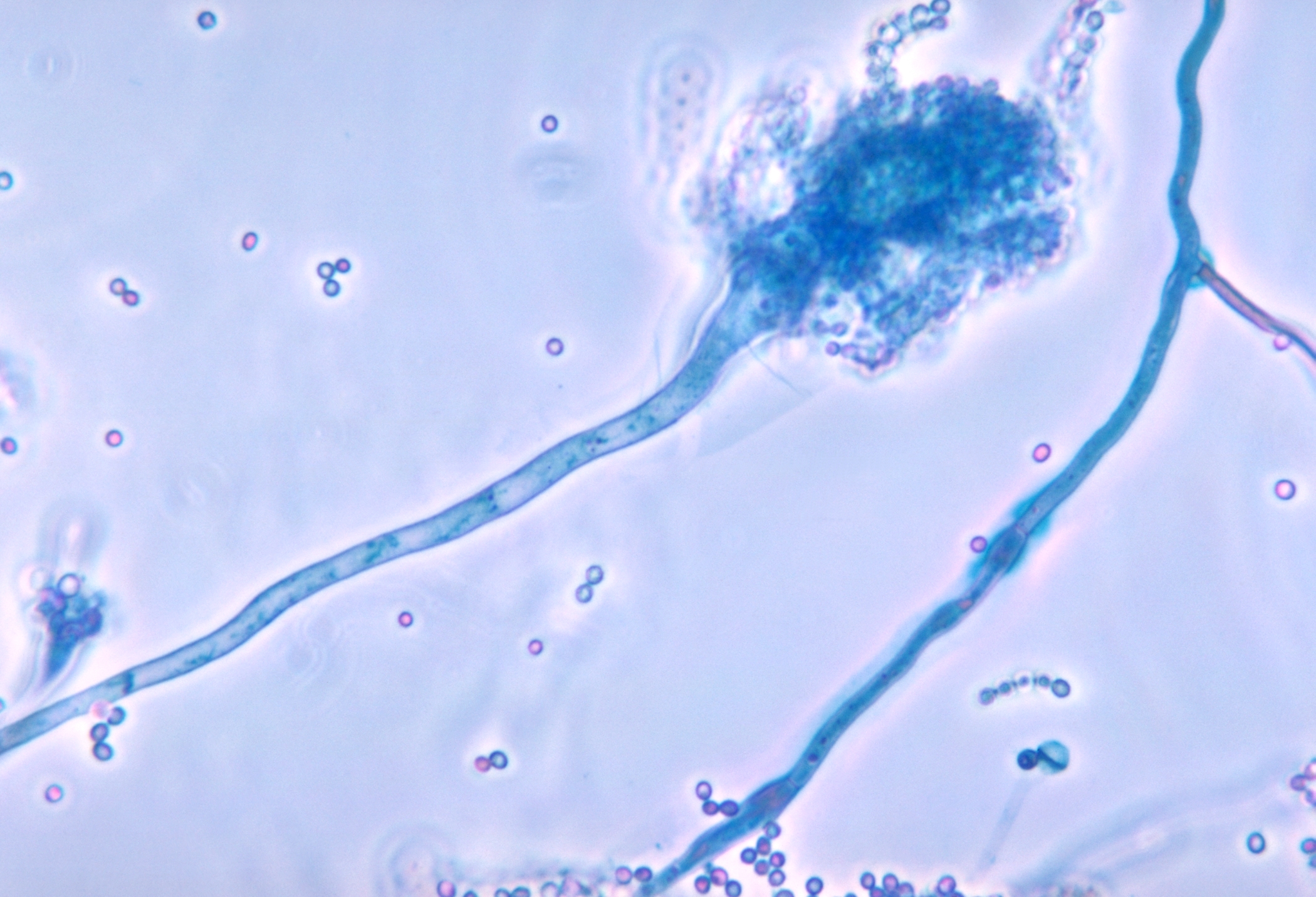
Aspergillus fumigatus conidiophore under microscopy, the mold whose septate hyphae and 45-degree branching CGD neutrophils cannot clear. Tap to expand.
💥
Staph aureus
Gram-positive coccus in clusters
🔥 #1 cause of CGD infections overall. Skin, liver abscesses.
tap to flip →
Why Catalase Matters
The trick
Normal bacteria produce their own H2O2 as a waste product. In a normal neutrophil, that H2O2 gets hijacked and turned into bleach. Staph aureus makes catalase, which destroys H2O2 before the neutrophil can use it. In a CGD patient who already can't make H2O2, Staph thrives unchecked.
Board clue
Liver abscess or lymphadenitis in a boy under 5 = think CGD. Staph aureus lymphadenitis in a toddler is a classic CGD opener on boards.
🍴
Aspergillus
Septate mold with 45-degree branching
🔥 Pulmonary mass or cavitary lesion in a CGD kid. Lethal if missed.
tap to flip →
Why Catalase Matters
The trick
Aspergillus is heavily catalase-positive. It produces massive quantities of catalase, wiping out any H2O2 the neutrophil tries to accumulate. In immunocompetent people, the respiratory burst clears Aspergillus spores easily. In CGD, the spores germinate into invasive hyphae with nothing to stop them.
Board clue
Aspergillus in a young child with no other immunocompromise clue = CGD until proven otherwise. In adults, think steroids or leukemia first.
💫
Serratia marcescens
Gram-negative rod, pink/red pigment
🔥 Classic CGD organism. Often mentioned alongside Staph in board questions.
tap to flip →
Why Catalase Matters
The trick
Serratia is another robust catalase producer. You might see it as a CGD clue in a board question without the Staph setup. It causes pneumonia, urinary tract infections, and septicemia in CGD patients.
The others
Also memorize: Nocardia (branching gram-positive rod, pulmonary) and Pseudomonas aeruginosa (green pus, gram-negative rod). All catalase-positive. Any of these in a young male child = CGD on your differential.
Pulmonary aspergilloma in a gross lung specimen, the lung mass that emerges when Aspergillus spores germinate inside a CGD neutrophil that cannot kill them. Tap to expand.
Section 5
Memory Hooks
Tap to unblur. Test yourself before looking.
🔨
CGD: Why catalase-positive?
Normal bacteria donate their own H2O2 to the neutrophil's killing machine. A catalase-positive bug destroys that gift before it can be used. In CGD, there's no backup plan. The neutrophil already can't make H2O2, and the bug destroys what little it gets from elsewhere. Empty factory, hostile supplier.
tap to reveal
🧪
NBT Test: What's normal?
Normal neutrophils turn nitroblue tetrazolium (NBT) from yellow to blue by generating superoxide. No respiratory burst = no blue = negative test. CGD neutrophils stay yellow. Remember: Blue = working burst. Yellow = broken.
tap to reveal
👨💋
CGD Inheritance
X-linked recessive. NADPH oxidase lives on the X chromosome. Boys get it, girls are carriers. The classic board patient is a young boy with recurrent Staph and Aspergillus infections. When you see a girl with CGD, it's the rarer autosomal recessive form.
tap to reveal
💊
G6PD vs CGD: Same molecule, different problem
NADPH appears in two separate high-yield board scenarios. In CGD: neutrophils can't make NADPH oxidase, so no superoxide, so no bleach, so catalase-positive bugs survive. In G6PD deficiency: RBCs can't regenerate NADPH, so glutathione runs out, so sulfa drugs trigger hemolysis. CGD = neutrophils. G6PD = red cells. Don't cross them.
tap to reveal
🔌
Free Radicals: The Pierce-and-Destroy Arc
Free radicals pierce the cell membrane, then the nuclear membrane, then damage DNA. Two outcomes: apoptosis (clean death) or mutation (which can become cancer). Most common source in the body: infections. Most common drug source: sulfa drugs. When you see an ionizing radiation question, remember the damage is the same mechanism.
tap to reveal
Section 7
Prove It
Five questions, drawn at random each load. Explanations after every answer.