Hematology · Oncology

Multiple Myeloma & MGUS

Plasma cell dyscrasia from benign paraprotein to end-organ disaster

Opening Challenge

A 68-year-old man presents with back pain for 3 months, fatigue, and new confusion. Labs: Ca 12.4, Creatinine 2.8, Hgb 9.2, total protein elevated with M-spike on SPEP. X-ray shows multiple lytic "punched-out" lesions in the skull and vertebrae. Bone marrow biopsy shows 35% plasma cells.

A) MGUS (Monoclonal Gammopathy of Undetermined Significance)
B) Multiple Myeloma
C) Smoldering Myeloma
D) Waldenstrom Macroglobulinemia
Multiple Myeloma. This patient has all four CRAB criteria: HyperCalcemia (Ca 12.4), Renal failure (Cr 2.8), Anemia (Hgb 9.2), and Bone lesions (lytic punched-out lesions). Plus plasma cells >10% in marrow (35% here). The M-spike on SPEP = monoclonal protein (usually IgG or IgA). Bence-Jones protein (free light chains) would appear on UPEP.

Why not the others? MGUS has NO CRAB and <10% plasma cells. Smoldering has >10% plasma cells or high M-protein but NO CRAB by definition. Waldenstrom has IgM paraprotein and lymphoplasmacytic lymphoma pattern, not lytic bone lesions.
01 · Diagnostic Framework

CRAB Criteria

End-organ damage from malignant plasma cells. One letter at a time.

C
HyperCalcemia · Ca >11 mg/dL
Mechanism: Myeloma cells secrete osteoclast-activating factors (RANKL, IL-6, MIP-1alpha) that drive osteoclast activity without matching osteoblast response. Bone resorption releases calcium into blood. PTH is low (PTH-independent hypercalcemia).

Symptoms: Stones, bones, groans, psychic moans (nephrolithiasis, bone pain, constipation/nausea, confusion/lethargy).

Treatment: IV fluids (saline diuresis) + bisphosphonates (inhibit osteoclasts) + corticosteroids.
PTH-independent RANKL driven Ca >11 IV hydration
R
Renal Failure · Cr >2 mg/dL
Primary mechanism: Bence-Jones protein (free light chains from malignant plasma cells) are directly nephrotoxic. They precipitate in distal tubules and collecting ducts, forming tubular casts that obstruct and cause inflammation. This is called "myeloma kidney" or cast nephropathy.

Secondary: Hypercalcemia causes prerenal azotemia and direct tubular damage. Hyperuricemia from cell turnover can cause urate nephropathy. AL amyloid deposits in glomeruli.

Warning: Avoid IV contrast (iodinated contrast agents are acutely nephrotoxic in the setting of cast nephropathy and proteinuria).
Bence-Jones nephrotoxic Cast nephropathy Avoid IV contrast AL amyloid
A
Anemia · Hgb <10 g/dL
Mechanism: Malignant plasma cells crowd out normal hematopoietic precursors in the bone marrow (myelophthisis). Cytokines (IL-6, TNF-alpha) also suppress erythropoiesis directly.

Type: Normocytic, normochromic. Not iron deficiency, not megaloblastic.

Peripheral smear: Rouleaux formation (RBCs stack like coins because elevated M-protein increases serum viscosity and reduces RBC surface charge). ESR is markedly elevated for the same reason.

Treatment: Erythropoiesis-stimulating agents (EPO) + treat underlying myeloma.
Normocytic normochromic Rouleaux formation ESR elevated Myelophthisis
B
Bone Disease · Lytic Lesions
Mechanism: Same osteoclast activation (RANKL, no osteoblast counterpart) creates purely lytic lesions. No new bone is laid down. Classic locations: skull (pepper-pot skull), vertebrae (compression fractures, back pain), ribs, pelvis.

Board differentiator: Myeloma = purely LYTIC. Prostate cancer mets = BLASTIC. Breast cancer mets = mixed.

Imaging: DEXA scan is NOT useful (lytic lesions don't show as low density on DEXA). Use whole-body low-dose CT or PET/CT instead.

Treatment: Bisphosphonates (zoledronic acid, pamidronate) throughout treatment course.
Punched-out lytic Vertebral fractures Pepper-pot skull Bisphosphonates No DEXA
Myeloma vs. metastatic cancer bone lesions: Myeloma bone disease = purely LYTIC. Prostate cancer mets = BLASTIC (bone forming, dense on X-ray). Breast cancer mets = mixed lytic and blastic. This distinction is a frequent board topic.
Malignant plasma cells RANKL / IL-6 / MIP-1a Osteoclast activation Bone resorption Ca release + lytic lesions
02 · The Continuum

Spectrum: MGUS to Myeloma

Same biology, different threshold. Diagnosis lives in the numbers and organ damage.

Condition Plasma Cells M-Protein End-Organ Damage Progression Risk
MGUS <10% <3 g/dL, no Bence-Jones None ~1%/year to myeloma
Smoldering Myeloma 10-60% ≥3 g/dL None (by definition) ~10%/year (initial years)
Multiple Myeloma >10% plus CRAB Any Yes (CRAB criteria) Active disease
Plasma Cell Leukemia >20% in blood Variable Severe, multi-organ Very poor prognosis
Benign end MGUS in Detail
Monoclonal Gammopathy of Undetermined Significance. The most common plasma cell dyscrasia. Affects ~3% of adults over 50, ~5% over 70. Incidental finding on SPEP obtained for other reasons.

Diagnostic criteria (all three required):
  • M-protein <3 g/dL on SPEP
  • Plasma cells <10% on bone marrow biopsy
  • No CRAB end-organ damage

Management: No treatment. Annual monitoring with SPEP, CBC, creatinine, and calcium. Educate patient about symptoms of progression (bone pain, anemia symptoms, confusion). Risk of progression ~1%/year to multiple myeloma, Waldenstrom, or AL amyloidosis.
Workup Diagnostic Workup for Suspected Myeloma
SPEP (serum protein electrophoresis): identifies M-spike. Usual order IgG > IgA > IgM by frequency. IgD and IgE rare.

UPEP (urine protein electrophoresis): detects Bence-Jones protein (free kappa or lambda light chains in urine). Negative UPEP does not exclude myeloma.

Serum free light chain ratio (kappa/lambda): abnormal ratio indicates clonal light chain production.

Bone marrow biopsy: quantifies plasma cell percentage. Needed to distinguish MGUS vs. smoldering vs. active myeloma.

Skeletal imaging: whole-body low-dose CT or PET/CT preferred over plain skeletal survey. DEXA is NOT useful.

Prognostic labs: LDH (elevated = aggressive disease), beta-2 microglobulin (elevated = advanced disease, ISS stage), albumin (inverse prognostic marker).

FISH cytogenetics on marrow: del(17p), t(4;14), t(14;16) = high-risk disease. t(11;14) is common but not high-risk.
Treatment Treatment Principles
Novel agents era: Standard induction is VRd: Bortezomib + Lenalidomide + Dexamethasone.

Key drug mechanisms:
  • Bortezomib: proteasome inhibitor. Plasma cells depend on protein turnover (produce massive amounts of immunoglobulin); blocking the proteasome triggers ER stress and apoptosis.
  • Lenalidomide: immunomodulatory drug (IMiD), thalidomide analogue. Activates T cells and NK cells, promotes myeloma cell apoptosis via cereblon pathway.
  • Daratumumab: anti-CD38 monoclonal antibody. Plasma cells overexpress CD38. Used in frontline and relapsed settings.

Transplant-eligible patients: Induction with VRd, then autologous stem cell transplant (ASCT), then lenalidomide maintenance indefinitely.

Bone disease management throughout: Zoledronic acid (IV bisphosphonate) reduces skeletal-related events for ALL myeloma patients regardless of visible lesions.
The key rule: MGUS and smoldering myeloma are defined by the ABSENCE of CRAB. The moment any CRAB criterion appears, the disease becomes active myeloma requiring treatment. Plasma cell percentage alone doesn't flip the diagnosis without end-organ damage.
03 · Beyond CRAB

Complications Drill

Three classic complications that extend beyond the four CRAB letters.

Free light chains AL Amyloidosis
Mechanism: Free light chains (usually lambda) misfold and polymerize into amyloid fibrils that deposit in organ extracellular spaces. Unlike cast nephropathy (tubular obstruction), amyloid infiltrates organ architecture diffusely.

Organs affected and their clues:
  • Kidney: nephrotic syndrome (massive proteinuria, edema). Most common organ.
  • Heart: restrictive cardiomyopathy. Thickened walls on echo but LOW voltage on EKG (the amyloid does not conduct electricity well). Granular sparkling appearance on echo.
  • Liver: hepatomegaly (enlarged but non-tender).
  • Tongue: macroglossia (classic exam finding).
  • Peripheral nerves: polyneuropathy (length-dependent sensorimotor).
  • Soft tissue: "shoulder pad sign" (amyloid deposits around shoulders causing fullness).

Diagnosis: Congo red stain shows apple-green birefringence under polarized light on any affected tissue biopsy. Fat pad aspiration is a less invasive first step.

Treatment: Treat the underlying plasma cell dyscrasia to eliminate the source of light chains.
High protein Hyperviscosity Syndrome
Classic association: IgM paraprotein in Waldenstrom macroglobulinemia (IgM is a pentamer, very large molecule). Also occurs with very high IgA or IgG levels in myeloma.

Mechanism: Excess monoclonal protein dramatically increases serum viscosity. Blood sludges in small vessels.

Symptoms:
  • Eyes: blurred vision, dilated tortuous retinal veins with "sausage-link" appearance on fundoscopy
  • Neuro: headache, mental status changes, stroke-like symptoms
  • Bleeding: mucosal bleeding (proteins coat platelets, impairing function)

Emergency treatment: Plasmapheresis (removes the excess protein from blood immediately). This is a time-sensitive intervention. Treat the underlying dyscrasia after stabilization.
Immunity Infections in Myeloma
Mechanism: Multiple myeloma causes humoral immunodeficiency. Malignant plasma cells produce only one useless monoclonal protein; normal B cells and plasma cells are suppressed. Functional antibodies (IgG, IgA against pathogens) are depleted despite high total protein.

Most common infections:
  • Encapsulated bacteria: Streptococcus pneumoniae and Haemophilus influenzae. These require opsonizing antibodies for clearance, which are absent.
  • Herpes zoster reactivation (especially with bortezomib and lenalidomide, which further suppress cellular immunity)

Prophylaxis:
  • Acyclovir (or valacyclovir): zoster prophylaxis for all patients on bortezomib or lenalidomide
  • IVIG: consider for patients with recurrent severe bacterial infections and documented hypogammaglobulinemia
  • Pneumococcal and flu vaccines before starting immunosuppressive therapy
High-yield board trap: AL amyloid heart involvement = thickened walls on ECHO but LOW voltage on EKG. This combination (thick walls + low EKG voltage) is pathognomonic. Do not confuse with LVH (which gives HIGH voltage). Congo red + apple-green birefringence = amyloid anywhere in the body.
04 · Differential Diagnosis

Elimination Game

Four diagnoses. Two clues. One survives.

A 55-year-old woman has an incidental finding on routine bloodwork: total protein 8.9 g/dL. SPEP shows a small M-spike of 1.8 g/dL. UPEP is negative. Bone marrow biopsy shows 5% plasma cells. She has no bone pain, calcium is normal, creatinine is normal, and hemoglobin is 13.2 g/dL. She feels entirely well.
Multiple Myeloma
Smoldering Myeloma
MGUS
AL Amyloidosis
Clue 1: No CRAB criteria present. Calcium normal, creatinine normal, hemoglobin 13.2 (not anemic), no bone lesions on imaging. By definition, active multiple myeloma requires at least one CRAB criterion. This patient has none. Multiple Myeloma is eliminated.
Clue 2: Plasma cells are only 5% (less than 10%) AND M-protein is only 1.8 g/dL (less than 3 g/dL). Smoldering myeloma requires plasma cells 10-60% OR M-protein ≥3 g/dL. Neither threshold is met. There is also no organ involvement suggesting amyloid (no nephrotic syndrome, no thickened heart walls, no macroglossia). Smoldering Myeloma and AL Amyloidosis are eliminated.
MGUS Plasma cells <10%, M-protein <3 g/dL, no end-organ damage. Annual monitoring only. No treatment needed.
05 · Retrieval Practice

Quiz

Four board-style questions. Original vignettes. Pick your answer before reading the explanation.

Question 1 of 4

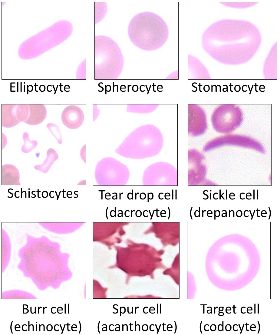
A 72-year-old woman with recently diagnosed multiple myeloma has a peripheral blood smear reviewed. The red blood cells appear stacked in linear arrangements, resembling coins in a stack.

What protein abnormality in her serum is directly responsible for this morphologic finding?
AElevated ferritin due to anemia of chronic disease
BElevated monoclonal immunoglobulin reducing the negative charge between red blood cells
CLow albumin causing decreased colloid oncotic pressure
DElevated fibrinogen from hepatic acute-phase response
Tempting to pick fibrinogen since fibrinogen is the classic cause of rouleaux in inflammatory states. But myeloma has a specific protein causing this, not a general inflammatory response. Think of RBCs as magnets that normally repel each other because they have the same charge. M-protein is a chemical neutralizer that strips the negative charge off the surface, letting the magnets suddenly flip and stack face-to-face. Albumin works in the opposite direction (stabilizes charge repulsion). Fibrinogen is the inflammatory rouleaux cause, M-protein is the myeloma-specific cause. Correct: B.

Rouleaux formation is caused by elevated monoclonal immunoglobulin (M-protein) in the serum. Normally, red blood cells repel each other because of their negative surface charge. High concentrations of immunoglobulins and other proteins reduce the zeta potential (surface charge repulsion), allowing RBCs to stack into rouleaux. This is also why the ESR is dramatically elevated in myeloma. Albumin would have the opposite effect on ESR. Fibrinogen can contribute in other inflammatory states but the question is pointing to the M-protein as the specific cause in myeloma.

Break it down: M-protein reduces zeta potential (surface charge) on RBCs, allowing rouleaux stacking; ESR dramatically elevated in myeloma because M-protein promotes the same stacking that slows RBC sedimentation rate; albumin has the opposite effect on ESR; fibrinogen = inflammatory rouleaux, M-protein = myeloma-specific rouleaux.
Question 2 of 4

A 66-year-old man with multiple myeloma and a creatinine of 3.1 mg/dL is scheduled for CT imaging of the chest, abdomen, and pelvis for staging purposes.

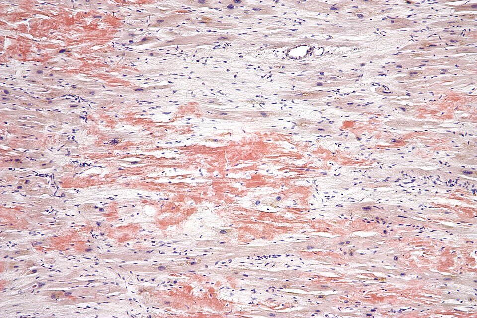
Why should iodinated IV contrast be avoided in this patient?
AContrast agents chelate calcium and worsen myeloma-related hypercalcemia
BContrast acts as a precipitant in tubules already obstructed by Bence-Jones protein casts, accelerating renal failure
CContrast interferes with SPEP interpretation and obscures the M-spike
DContrast activates RANKL and accelerates osteoclast-mediated bone destruction
Tempting to use contrast CT since imaging the spine for cord compression seems urgent enough to accept the risk. But in myeloma kidney, the tubules are not working at baseline and the contrast interaction is not just the usual risk. Think of the tubules as partially clogged pipes: Bence-Jones protein casts are already blocking them. Iodinated contrast interacts with the Tamm-Horsfall protein inside the clogged pipes, precipitating more solid material on top of the existing blockage. The pipes go from partially clogged to acutely obstructed. Correct: B.

In myeloma kidney (cast nephropathy), tubules are already partially obstructed by Bence-Jones protein (free light chain) casts. Iodinated contrast agents interact with the Tamm-Horsfall protein and precipitated light chains in the tubular lumen, forming additional obstructing casts. This can precipitate acute tubular necrosis on top of existing cast nephropathy. The combination is acutely nephrotoxic beyond the baseline contrast-induced nephropathy risk. This is a well-recognized contraindication. If cross-sectional imaging is needed, use non-contrast CT (which is actually preferred for lytic bone lesions anyway) or MRI.

Break it down: myeloma kidney = Bence-Jones protein (free light chain) casts in tubules; iodinated contrast interacts with Tamm-Horsfall protein to form additional casts; leads to acute tubular necrosis on top of cast nephropathy; use non-contrast CT or MRI instead; iodinated contrast is absolutely contraindicated in cast nephropathy.
Question 3 of 4

A 61-year-old man with known AL amyloidosis develops progressive exertional dyspnea and lower extremity edema. Echocardiogram shows thickened left ventricular walls with a "granular sparkling" appearance and impaired relaxation. His ejection fraction is preserved at 55%.

What finding on his 12-lead EKG would be characteristic despite the thickened ventricle walls?
ATall R waves in lateral leads consistent with left ventricular hypertrophy
BDelta waves and short PR interval from accessory pathway conduction
CLow voltage complexes throughout all leads despite thickened walls
DRight bundle branch block from amyloid infiltration of the right bundle
Tempting to pick tall R waves (LVH pattern) since the walls are thick on echo. Thick walls normally mean more muscle mass which normally means more electrical amplitude. Cardiac amyloidosis is the exception that paradoxically goes in the opposite direction. Think of amyloid fibrils as an electrical insulator woven into the wall of the heart: the myocytes generate their normal signal, but the amyloid blanket absorbs it before the signal reaches the EKG leads. The wall is thick, but the signal is faint. That discordance is the fingerprint. Correct: C.

This is the pathognomonic combination: thick walls on echo + LOW voltage on EKG. Amyloid fibrils infiltrate the myocardium and do not conduct electricity effectively (they are non-conducting protein deposits). The electrical signal generated by myocytes is absorbed and dampened by the amyloid before reaching the surface EKG leads. This is the exact opposite of true left ventricular hypertrophy (which gives high voltage). The discordance between wall thickness on echo and EKG voltage is the key diagnostic clue for cardiac amyloidosis. Tall R waves (choice A) would suggest LVH, not amyloid. Delta waves (choice B) are for Wolff-Parkinson-White.

Break it down: cardiac amyloidosis = thick walls on echo PLUS low voltage on EKG (paradoxical discordance); amyloid = electrical insulator that absorbs the signal; true LVH = thick walls plus HIGH voltage; low voltage plus thick walls = amyloid until proven otherwise.
Question 4 of 4

A 58-year-old woman is found to have an M-spike on SPEP obtained during workup for fatigue. SPEP shows M-protein of 2.1 g/dL. Bone marrow biopsy shows 8% plasma cells. She has normal calcium, creatinine, hemoglobin, and no bone lesions on imaging. Her only complaint is mild fatigue, which workup attributes to iron deficiency rather than marrow infiltration.

Which single criterion most definitively distinguishes her diagnosis from multiple myeloma?
AThe IgG subtype of her M-protein rather than IgA
BThe absence of Bence-Jones protein on UPEP
CThe absence of end-organ damage (no CRAB criteria)
DPlasma cell percentage below 10%
Tempting to diagnose active myeloma based on plasma cell percentage since that is the biopsy finding and it seems high enough. But the biopsy number alone is not enough without CRAB. Think of CRAB criteria as the bridge between "abnormal test" and "disease requiring treatment": the plasma cells are the production factory and the CRAB findings are evidence that the factory is actively damaging the building. No CRAB damage = the factory is running but the building is still standing. Treat MGUS with observation, not chemotherapy. Correct: C.

The single most important distinction between MGUS and active myeloma is the presence or absence of CRAB end-organ damage. This patient has none (calcium normal, creatinine normal, hemoglobin normal by marrow criteria, no bone lesions). The CRAB criteria define when a plasma cell dyscrasia becomes clinically significant and requires treatment.

Why not D? Plasma cell percentage alone is important, but it's the combination with CRAB that defines active myeloma. A patient with 15% plasma cells but no CRAB is smoldering myeloma, not active myeloma. A patient with even 11% plasma cells plus any CRAB criterion meets the diagnosis of active myeloma. CRAB is the single definitional dividing line between "watch" and "treat."

Break it down: CRAB = hyperCalcemia, Renal failure, Anemia, Bone lesions; CRAB present = active myeloma, treat; CRAB absent = MGUS or smoldering, observe; plasma cell percentage alone does not define active myeloma without end-organ damage.
0/4
quiz complete
Board-Style Walkthrough

Board-Style Walkthrough

Original board-style vignettes. Shuffled, never-repeat, full explanations for every choice.