An alpha cell tumor that eats you alive: diabetes, dermatitis, DVT, depression, and a rash so specific that it IS the diagnosis.
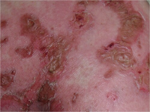
A 58-year-old man presents with new-onset diabetes, a 15-lb weight loss over 3 months, chronic diarrhea, and a painful, itchy rash on his groin and legs that has areas of blistering with central crusting. The rash appears to migrate, with older lesions healing centrally while new vesicles form at the edges.
What is the most likely diagnosis?
📷 NECROLYTIC MIGRATORY ERYTHEMA: painful, itchy vesicles with central crusting · tap to expand
★If you see necrolytic migratory erythema (NME) on a board exam, the answer is ALWAYS glucagonoma. This rash is pathognomonic. No other condition produces it.
What is NME?
Necrolytic migratory erythema is the signature rash of glucagonoma. It is painful, itchy, and it migrates. Here is what makes it unique:
Starts as erythematous patches that develop vesicles (small blisters)
The center of each lesion crusts and heals while the edges keep spreading
Favors the groin, perineum, buttocks, and lower extremities
Caused by amino acid deficiency: glucagon breaks down proteins so aggressively that the skin literally cannot maintain itself
THE SYNDROME
The 4 D's + Weight Loss + Diarrhea
Glucagonoma is an alpha cell tumor, usually in the tail of the pancreas. It secretes excess glucagon, and that excess eats you alive.
Understanding what glucagon normally does makes the tumor syndrome obvious:
Source: Alpha cells of the pancreatic islets (periphery of the islet)
Main stimulus: Hypoglycemia and stress (glucagon is the "starvation hormone")
Primary target:LiverThe liver is glucagon's main target. Glucagon activates hepatic gluconeogenesis (making new glucose from amino acids) and glycogenolysis (breaking down glycogen stores). It also promotes lipolysis in adipose tissue.
Opponent: Insulin (beta cells, opposite effects on blood sugar)
💡The weight loss in glucagonoma is NOT from poor appetite alone. Glucagon forces the body to break down proteins and fats for gluconeogenesis. The body is literally consuming its own muscle and fat to make glucose it does not need.
Cell to Hormone
Match each islet cell type to its product. Tap the correct answer.
Alpha cells produce:
Glucagon's second messenger is:
THE ASSOCIATION
MEN1 · Diagnosis · Rarity
One of the rarest tumors you will ever see on a board exam. But when the stem gives you the clues, you will not miss it.
MEN1 Association
Glucagonoma can occur in MEN1Multiple Endocrine Neoplasia type 1 is an autosomal dominant syndrome caused by mutation of the menin gene on chromosome 11. It causes tumors in the pituitary, parathyroid, and pancreas. Remember: "3 P's." (Multiple Endocrine Neoplasia type 1), the syndrome of:
Pituitary tumors (prolactinoma most common)
Parathyroid hyperplasia (most common manifestation overall)
Pancreatic islet cell tumors
⚠️
Board Trap: Most Common Pancreatic Tumor in MEN1
The most common pancreatic tumor in MEN1 is gastrinoma (causes Zollinger-Ellison syndrome), NOT glucagonoma. Glucagonoma CAN appear in MEN1, but gastrinoma is far more frequent. If the question asks "most common pancreatic tumor in MEN1," the answer is gastrinoma.
Imaging: CT or MRI showing a pancreatic mass, usually in the tail
Biopsy: Confirms alpha cell origin with immunohistochemistry
Rarity: Approximately 1 in 20 million. Extremely rare, but extremely testable because the presentation is so distinctive
Treatment
Surgical resection is the definitive treatment (distal pancreatectomy for tail lesions)
Octreotide (somatostatin analog) controls symptoms by suppressing glucagon secretion
Address nutritional deficiency: amino acid and zinc supplementation to help the skin heal
Anticoagulation for DVT risk
📷 OCTREOTIDE: somatostatin analog that suppresses glucagon secretion · tap to expand
💡Glucagonoma is one of the few tumors where the skin finding alone is diagnostic. NME on the exam = glucagonoma, period. You do not need to wait for labs or imaging to answer the question.
CLINICAL REASONING
Walk the Glucagonoma Case
Each tap reveals the next step. Trace the clinical logic before you look at the answer.
The Diagnostic Chase
A 55-year-old man is referred to your clinic. His dermatologist treated him for "eczema" for 8 months with no improvement. He now has new-onset diabetes and has lost 18 lbs.
The rash is painful, blistering, and appears to migrate: old lesions crust centrally while new vesicles form at the edges. It started in the groin. What is the most likely identity of this rash?
Yes. The pattern is textbook NME: vesicles at the advancing edge, central crusting, groin-to-legs migration. No eczema migrates like this. Pemphigus makes flaccid blisters but does not migrate with central clearing. This is NME until proven otherwise.
Atopic eczema does not produce the migratory wave pattern. NME has a biological driver: excess glucagon consuming amino acids. The skin cannot rebuild fast enough, so each lesion crusts and the deficiency front moves outward. Eczema stays put.
Pemphigus attacks the desmoglein cement between skin cells, causing flaccid blisters that rupture on pressure. It does not migrate in a wave pattern. NME migrates because amino acid depletion moves through different skin territories as depletion deepens. Different mechanism, different shape.
NME is now on your differential. This patient also has new-onset diabetes and 18 lbs of weight loss. What single laboratory test confirms the diagnosis?
Exactly. Serum glucagon above 500 pg/mL (often above 1000) confirms glucagonoma. HbA1c tells you how long the diabetes has been running but not WHY. CT finds the mass but a high glucagon level proves the etiology. The lab comes first, imaging follows.
HbA1c confirms sustained hyperglycemia but cannot distinguish glucagonoma from type 2 diabetes, steroid-induced diabetes, or any other cause. You already know the sugar is up. The question is: why? Only the glucagon level answers that.
CT will find the pancreatic tail mass, but it cannot tell you if the mass is actually secreting glucagon versus being a non-functional NET. Lab confirmation of glucagon elevation establishes the hormonal diagnosis. Lab first, then image to locate the tumor.
Glucagon returns at 1,100 pg/mL (normal less than 200). CT shows a 6 cm pancreatic tail mass with two liver metastases. Which treatment comes FIRST, before considering surgery?
Right. Octreotide binds somatostatin receptors (SSTR2) on the tumor cells, suppressing glucagon output. This stabilizes the NME, improves glucose control, and reduces catabolic wasting before the patient undergoes the stress of surgery. Nutritional repletion (amino acids, zinc) runs in parallel. Debulking surgery follows once the patient is optimized.
Streptozocin-based chemotherapy is reserved for metastatic disease that does not respond to somatostatin analogs and cannot be debulked. Starting with chemotherapy before symptom control would expose a severely catabolic, malnourished patient to cytotoxic agents without first reducing the hormonal chaos.
Insulin counters hyperglycemia but does nothing about the amino acid catabolism driving NME and wasting. The problem is too much glucagon, not too little insulin. Suppressing glucagon at its source (octreotide) is superior to trying to counterbalance it downstream.
MEMORY
Memory Hooks
Tap to unblur. Try to recall before you look.
🩹
The 4 D's of Glucagonoma
Dermatitis (NME) + Diabetes + DVT + Depression. Plus weight loss and diarrhea. Every D is directly caused by excess glucagon. The rash is pathognomonic: NME = glucagonoma, period.
tap to reveal
🔑
Why the Rash Migrates
Glucagon forces the liver to convert amino acids into glucose nonstop. The skin, which turns over every 28 days, runs out of amino acid building blocks. The depletion front moves, so the rash moves. Old lesions crust as the deficiency advances elsewhere.
tap to reveal
💊
Octreotide: The Brake
Somatostatin = STOP. Octreotide mimics somatostatin at SSTR2 receptors on the tumor. Less glucagon out of the tumor = less amino acid destruction = less NME + less diabetes. Surgery follows, but octreotide stabilizes first.
Most common functioning NET in MEN1: gastrinoma (causes Zollinger-Ellison syndrome). Glucagonoma CAN appear but is rare. Non-functioning NETs are most common overall. Know the ranking: gastrinoma > insulinoma > glucagonoma in MEN1.
tap to reveal
📍
Why the Pancreatic Tail?
Alpha cells concentrate in the body and tail of the pancreas. Glucagonoma follows its origin cells: 70 to 80% in the body/tail. Gastrinoma goes to the head and duodenum (the gastrinoma triangle). Location follows cell distribution.
tap to reveal
BOARD CHALLENGE
Board-Style Walkthrough
Full 3rd-order vignettes with progressive teaching chains. Shuffled every session. Never repeat until all are done.