The three-hormone axis that governs every bone, kidney, and gut calcium decision
Opening Challenge
A 55-year-old woman presents with a history of kidney stones, fatigue, constipation, and depression. Labs: Ca 11.4 mg/dL (high), PTH 145 pg/mL (high), phosphorus 2.1 mg/dL (low), alkaline phosphatase elevated.
A) Primary hyperparathyroidism
B) Humoral hypercalcemia of malignancy
C) Vitamin D toxicity
D) Sarcoidosis
Primary hyperparathyroidism. The defining clue: high calcium AND high PTH together. In every other cause of hypercalcemia, PTH is suppressed low because the body is trying to turn it off. Only primary hyperPTH keeps PTH elevated when calcium is already high, because the gland itself is the problem (adenoma 80%). PTH raises calcium AND lowers phosphorus. The classic presentation: "stones, bones, groans, and psychic moans" -- kidney stones, subperiosteal bone resorption, abdominal pain/constipation, psychiatric symptoms (fatigue, depression, confusion).
01 · The Framework
The Calcium Thermostat
Tap each state to trace the feedback loop from sensor to response. Three players. One thermostat.
Calcium Thermostat · Interactive Feedback Loop
Select a calcium state above to trace the feedback loop.
Trigger
Low Ca²⁺
CaSR on parathyroid chief cells detects the drop
→
Parathyroid
PTH Released
Bone + kidney + activates Vitamin D
→
Kidney
1,25-(OH)₂D Made
1-alpha-hydroxylase converts 25-OH-D to active calcitriol
→
Net Effect
Ca UP
Bone resorption + kidney Ca retention + gut absorption
Source: Chief cells of the parathyroid glands (4 glands embedded in thyroid capsule).
Released when: Serum Ca²⁺ falls. Also released when Mg²⁺ is low (but severe hypoMg paradoxically blocks PTH -- more on that in Section 3).
Bone: Activates osteoclasts indirectly (via osteoblast signaling) -- Ca and phosphorus released from bone matrix.
Kidney: (1) DCT -- promotes Ca reabsorption. (2) Proximal tubule -- blocks phosphate reabsorption (phosphaturia). (3) Activates 1-alpha-hydroxylase -- converts 25-OH-VitD to active 1,25-(OH)₂D.
Gut: Indirect only, via the VitD it activates.
Net result: Calcium UP, Phosphorus DOWN.
Ca UPPhos DOWNVitD ActivationKidney Ca retentionOsteoclast activation
CaSR (Calcium-Sensing Receptor): When Ca²⁺ rises, CaSR on parathyroid cells fires and shuts off PTH secretion. This is the negative feedback loop. In familial hypocalciuric hypercalcemia (FHH), a CaSR mutation makes the sensor less sensitive, so PTH stays elevated even when Ca is high -- this mimics primary hyperPTH on labs.
Low Ca²⁺→PTH secreted→Bone resorption + Kidney Ca retained + Phos excreted→Ca normalizes→CaSR detects high Ca→PTH shuts off
Skin + Liver + Kidney · Three-Step Activation
Vitamin D
Step 1 -- Skin: UV light converts 7-dehydrocholesterol to cholecalciferol (D3). Ergocalciferol (D2) comes from dietary plants.
Step 2 -- Liver: 25-hydroxylase converts D3 to 25-OH-VitD (calcidiol). This is the storage form. This is what you measure clinically to check VitD status -- NOT the active form.
Step 3 -- Kidney: 1-alpha-hydroxylase converts 25-OH-VitD to 1,25-(OH)₂-VitD (calcitriol). This is the active form. PTH stimulates this step. So PTH controls how much active VitD gets made.
Actions of calcitriol: Intestine (Ca AND phosphorus absorption UP), Bone (Ca release at high levels), Kidney (Ca and Phos reabsorption).
Deficiency consequences: Kids = rickets (soft bones, bowing of legs, rachitic rosary at costochondral junctions, craniotabes). Adults = osteomalacia (bone pain, proximal muscle weakness, pseudofractures on X-ray).
25-OH-VitD: check this clinically1,25-(OH)₂D: active formPTH controls kidney stepGut Ca + Phos absorptionDeficiency: rickets / osteomalacia
Board trap: You measure 25-OH-VitD to screen for deficiency (it's the storage pool). The active form (1,25-OH₂D) has a short half-life and is tightly regulated -- measuring it doesn't tell you if someone is VitD deficient. In sarcoidosis, 1,25-OH₂D is elevated (not 25-OH-D) because macrophages are making active VitD directly.
Source: Parafollicular C cells of the thyroid gland (NOT the parathyroid glands).
Released when: Serum Ca²⁺ is high. Opposes PTH. Lowers calcium by inhibiting osteoclasts and promoting renal Ca excretion.
Clinical relevance #1 -- Tumor marker: Calcitonin is the tumor marker for medullary thyroid carcinoma (MTC). MTC arises from C cells. Elevated calcitonin = MTC until proven otherwise. MTC is associated with MEN 2A (MTC + pheochromocytoma + primary hyperPTH) and MEN 2B (MTC + pheo + mucosal neuromas + marfanoid body).
Clinical relevance #2 -- Hypercalcemia treatment: Calcitonin (intranasal or SC) is used for acute hypercalcemia. Rapid onset (hours), but tachyphylaxis develops within 48 hours. Used as a bridge until bisphosphonates (zoledronic acid) kick in (takes 2-4 days).
Clinical relevance #3 -- Osteoporosis: Intranasal salmon calcitonin for postmenopausal osteoporosis (less used now that bisphosphonates dominate).
Ca DOWNMTC tumor markerMEN 2A / MEN 2BTachyphylaxis within 48hBridge to bisphosphonates
MTC and MEN 2: Any patient with medullary thyroid carcinoma requires genetic testing for RET proto-oncogene mutation. MEN 2A = MTC + pheo + primary hyperPTH. MEN 2B = MTC + pheo + mucosal neuromas + marfanoid habitus (NO primary hyperPTH in 2B). Calcitonin and CEA are both tumor markers for MTC.
02 · Too Much Calcium
Hypercalcemia Causes
High Ca is common in clinical practice. The PTH level splits the diagnosis in two.
First move every time: Check PTH. If PTH is HIGH (or even inappropriately normal) with high Ca, think primary hyperPTH or FHH. If PTH is LOW (suppressed), think everything else.
Parathyroid Adenoma
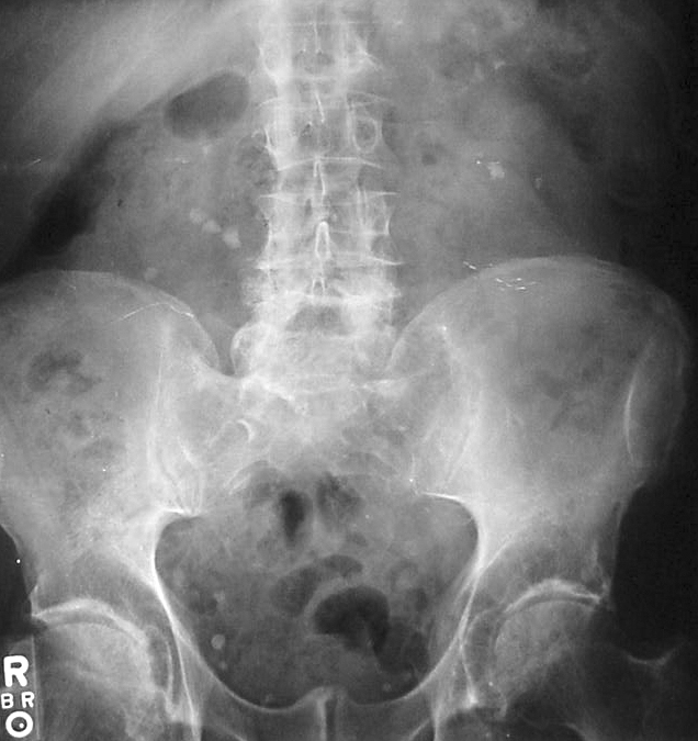
Calcium Oxalate Stone
Nephrolithiasis X-ray
Mnemonic
CHIMPANZEES
C -- Cancer (malignancy, HHM)
H -- Hyperparathyroidism (primary)
I -- Iatrogenic / milk-alkali syndrome (CaCO₃ excess)
M -- Multiple myeloma
P -- Paget's disease of bone
A -- Addison's disease
N -- Neoplasm (same as Cancer, but emphasizes PTHrP tumors)
Z -- Zollinger-Ellison (rare association)
E -- Excess Vitamin D (supplementation, granulomas)
E -- Excess Vitamin A
S -- Sarcoidosis / granulomatous disease, thiazide diuretics
Primary HyperPTH vs. Humoral Hypercalcemia of Malignancy (HHM)
Feature
Primary HyperPTH
HHM (Malignancy)
PTH
HIGH (the gland is broken)
LOW (suppressed by high Ca)
PTHrP
Normal
HIGH (the tumor is secreting it)
Phosphorus
Low (PTH promotes phosphaturia)
Low (PTHrP acts like PTH)
Cl/Phos ratio
> 33 (classic)
< 33
Cancer type
None
Squamous cell (lung/head-neck), RCC, breast
Trace It
Parathyroid adenoma (80%), hyperplasia (15%)
PTHrP mimics PTH at the PTH receptor
Urine Ca
HIGH (24h urine Ca elevated)
HIGH
FHH trap: Familial hypocalciuric hypercalcemia (FHH) looks exactly like primary hyperPTH on basic labs: high Ca, high/normal PTH. The differentiator is the 24-hour urine calcium -- FHH has LOW urine Ca (the kidney holds onto calcium because of the CaSR mutation). Primary hyperPTH has HIGH urine Ca. This is why the 24-hour urine calcium is done before any parathyroidectomy.
Granulomatous Disease · PTH-Independent Mechanism
Sarcoidosis Hypercalcemia
Mechanism: Activated macrophages inside granulomas express 1-alpha-hydroxylase. They make 1,25-(OH)₂-VitD unregulated -- independent of PTH control. Result: excessive VitD action in the gut (Ca absorption up) and bone.
Pattern: High Ca, low PTH (suppressed), high 1,25-OH₂D (NOT 25-OH-D), elevated ACE level.
Treatment: Glucocorticoids. Steroids block the macrophage 1-alpha-hydroxylase activity. This is why sarcoidosis hypercalcemia responds to steroids but NOT to the treatments used for primary hyperPTH. Also treat with hydration and avoid sunlight/VitD supplementation.
Macrophage 1-alpha-hydroxylasePTH suppressedACE level highSteroids work
Multiple Myeloma · Osteoclast Activation
Malignancy-Associated Hypercalcemia
Two mechanisms:
1. HHM (Humoral): PTHrP secreted by tumor acts like PTH systemically. Squamous cell lung, RCC, breast. PTH is suppressed.
2. Local osteolysis: Tumor in bone directly activates osteoclasts via cytokines (IL-1, IL-6, TNF). Multiple myeloma uses this mechanism (osteoclast activating factors from plasma cells).
Treatment of acute hypercalcemia: IV saline (volume expansion + calciuresis) + bisphosphonates (zoledronic acid -- takes 2-4 days, but durable) + calcitonin (rapid onset, bridge, tachyphylaxis) + furosemide (loop diuretic promotes calciuresis, only after volume-repleted).
PTH suppressedPTHrP elevated (HHM)IV saline firstBisphosphonates definitiveCalcitonin: bridge only
03 · Too Little PTH
Hypoparathyroidism
Low PTH, low calcium, high phosphorus. Three scenarios, one pattern.
Trousseau Sign · carpal spasm
Calcium Stone
Parathyroid Tissue
Hypoparathyroidism pattern: PTH down, Ca down, Phos up. The reverse of what PTH normally does. All three point the same direction -- once you know that PTH raises Ca and lowers Phos, the low-PTH state is just the mirror image.
Primary Hypoparathyroidism
PTH glands are absent or destroyed -- PTH = LOW
►
Causes: Post-thyroidectomy (#1 cause -- surgical removal or damage to parathyroids). Autoimmune hypoparathyroidism. DiGeorge syndrome (22q11.2 deletion: absent parathyroids + absent thymus = no PTH + no T cells). Hemochromatosis. Wilson's disease (copper infiltration).
Labs: Low Ca, High Phos, Low PTH.
Symptoms of hypocalcemia:
- Chvostek sign: tap the facial nerve anterior to the ear -- facial twitch (tetany of facial muscles)
- Trousseau sign: inflate BP cuff above systolic for 3 min -- carpal spasm (most specific sign)
- Tetany and perioral numbness (paresthesias around mouth)
- Seizures (hypocalcemia lowers seizure threshold)
- Prolonged QT interval on ECG (can cause torsades de pointes, fatal arrhythmia)
Treatment: Oral calcium carbonate or calcium citrate + calcitriol (active VitD, because PTH is absent so the kidney cannot make calcitriol on its own).
Post-thyroid surgery #1DiGeorge 22q11.2Chvostek + TrousseauLong QT -- torsades riskTreat: Ca + calcitriol
Pseudohypoparathyroidism (PHP)
PTH is HIGH but organs don't respond -- receptor defect
►
Key distinction: Same lab pattern as primary hypoparathyroidism (low Ca, high Phos) BUT PTH is ELEVATED. The body is making PTH correctly -- the target organs just cannot respond because of a defective G-protein (Gs-alpha subunit mutation). So the parathyroids keep screaming louder, but the kidney and bone are deaf.
Type 1a (Albright hereditary osteodystrophy -- AHO): Gs-alpha mutation. Phenotype: short stature, shortened 4th and 5th metacarpals (ring and pinky fingers look shorter), round face, intellectual disability, obesity. This phenotype is AHO. If AHO is present AND labs show hypocalcemia/hyperphosphatemia with high PTH = PHP Type 1a.
Pseudo-pseudohypoparathyroidism: AHO phenotype but NORMAL calcium levels (the Gs-alpha mutation is on the non-imprinted allele so there is no PTH resistance). Confusing name, normal labs.
PHP Type 1b: Normal AHO phenotype, but PTH resistance in kidney only (renal-specific methylation defect).
Treatment: Same as primary hypoparathyroidism -- calcium + calcitriol.
PTH HIGH (organs deaf)Same Ca/Phos as hypoParathShort 4th/5th metacarpalsGs-alpha mutationAHO phenotype
Magnesium and PTH
HypoMg blocks PTH at two levels -- check Mg before anything else
►
Two-level block: Magnesium is required for PTH secretion (Mg-dependent exocytosis from chief cells) AND for PTH receptor signaling at the G-protein level. Severe hypomagnesemia shuts down both steps -- no PTH is released, and even if some is present, target organs cannot respond.
Result: Functional hypoparathyroidism with hypocalcemia that does NOT respond to calcium or VitD supplementation.
Clinical context: Most common in alcoholics (poor nutrition, GI losses). Also in patients on proton pump inhibitors long-term, cisplatin, amphotericin, prolonged diarrhea.
Board rule: Refractory hypocalcemia that doesn't correct with calcium and calcitriol supplementation = check and replace magnesium first. You cannot correct hypocalcemia if the magnesium is depleted. Mg must come first.
Treatment: IV or oral magnesium repletion. Ca and VitD will then work once Mg is restored.
Refractory hypocalcemiaAlcoholics #1Check Mg firstPPIs, cisplatin, ampho-BFix Mg, then Ca corrects
QT prolongation = emergency: Hypocalcemia prolongs the QT interval and can cause torsades de pointes. Any patient with symptomatic hypocalcemia (seizures, tetany, prolonged QT on ECG) needs IV calcium gluconate immediately, not oral. Oral calcium is for chronic maintenance.
04 · Diagnostic Reasoning
PTH Decision Tree
Given Ca and PTH levels, derive the diagnosis one branch at a time.
The two-move approach: Step 1: Is Ca high or low? Step 2: Is PTH appropriately responding or going the wrong direction? The lab pattern determines the diagnosis before any imaging.
Step 1: The patient has serum calcium of 11.4 mg/dL (elevated). What is the PTH?
PTH is 142 pg/mL (elevated)
PTH is 8 pg/mL (suppressed)
High Ca + High PTH: Parathyroid is not responding to feedback. The gland keeps secreting despite high Ca. This is either primary hyperPTH (autonomous adenoma) or FHH (CaSR mutation). Next discriminator: 24-hour urine calcium.
High Ca + Low PTH: Parathyroid correctly senses high Ca and shuts off. The problem is coming from somewhere else: malignancy (PTHrP), VitD toxicity, granulomatous disease, immobilization.
Step 2A: 24-hour urine calcium result?
24h urine Ca: 520 mg/day (elevated)
24h urine Ca: 45 mg/day (low); Ca/Cr ratio less than 0.01
Primary Hyperparathyroidism: Elevated urine Ca confirms no CaSR mutation hoarding Ca. PTH is elevated because an adenoma secretes autonomously. Localize with sestamibi scan. Definitive treatment: parathyroidectomy.
FHH: CaSR mutation causes the kidney to avidly reabsorb Ca even when Ca is high. Urine Ca is inappropriately low. This patient does NOT need surgery. Parathyroidectomy for FHH is incorrect and does not cure the hypercalcemia.
Step 2B: Which workup result do you find?
PTHrP markedly elevated; new lung mass on chest CT
1,25-(OH)₂D elevated; ACE elevated; no malignancy
Humoral Hypercalcemia of Malignancy (HHM): PTHrP from squamous cell lung cancer binds PTH receptors. True PTH is suppressed. Treatment: IV saline (first) + bisphosphonates (durable, 2-4 days) + calcitonin as bridge (tachyphylaxis at 48h). Treat the underlying malignancy.
Sarcoidosis Hypercalcemia: Granuloma macrophages make calcitriol unregulated. PTH is suppressed (correctly). Treatment: glucocorticoids (block macrophage 1-alpha-hydroxylase). Avoid vitamin D supplements and sunlight.
Step 3: New patient, serum Ca 7.1 mg/dL (low). What is PTH?
PTH undetectable (less than 5 pg/mL)
PTH markedly elevated (320 pg/mL)
True Hypoparathyroidism: Gland absent or destroyed. No PTH made. Causes: post-thyroidectomy, autoimmune, DiGeorge. Treat: Ca + calcitriol (must give active VitD, because kidney needs PTH to activate it).
PHP or Secondary HyperPTH: Parathyroid working hard but Ca stays low. Either PHP (Gs-alpha deaf organs, AHO phenotype) or VitD deficiency secondary hyperPTH from CKD. Distinguish by phenotype (short metacarpals in PHP), renal labs, 25-OH-VitD level.
05 · Memory + Pharmacology
Memory Hooks & CKD Drugs
Tap each hook to reveal the chain. Lock it before closing.
Stones: Kidney stones. PTH drives hypercalciuria. Ca precipitates as calcium oxalate or calcium phosphate.
Thrones: Polyuria. High Ca causes nephrogenic DI (Ca antagonizes ADH in collecting duct). Patient is on the throne a lot.
Bones: Subperiosteal bone resorption. Osteitis fibrosa cystica. Brown tumors (osteoclast + hemosiderin deposits). Radial subperiosteal resorption on X-ray is pathognomonic.
Groans: Constipation, N/V, acute pancreatitis (Ca activates pancreatic enzymes), PUD (Ca stimulates gastrin).
Opposite directions vs same direction · fastest clinical medicine pattern
PTH: Ca and Phos go OPPOSITE directions. PTH raises Ca while lowering Phos (phosphaturia). Ca UP + Phos DOWN = PTH is the driver. Ca DOWN + Phos UP = too little PTH.
Vitamin D: Ca and Phos go the SAME direction. Calcitriol absorbs both from the gut. Ca UP + Phos UP = too much VitD (toxicity, granulomas). Ca DOWN + Phos DOWN = too little VitD (rickets, osteomalacia).
The hormone travels with the calcium. Ca high + PTH low = parathyroid responding correctly. Ca high + PTH high = the gland is broken.
🔧
Cinacalcet & Sevelamer in CKD
Tap to trace the mechanism for each drug
Cinacalcet (Sensipar): Sensitizes the CaSR to circulating Ca. The parathyroid gland perceives more Ca than is actually there, suppressing PTH secretion. Used for: secondary hyperPTH in CKD on dialysis, primary hyperPTH when parathyroidectomy fails, parathyroid carcinoma. Adverse: hypocalcemia.
Sevelamer (Renagel/Renvela): Nonabsorbable polymer binds dietary phosphate in GI tract, preventing absorption. Used to control hyperphosphatemia in CKD dialysis patients. Does not add calcium to the body (unlike older calcium-based binders that can cause vascular calcification). Adverse: GI upset, hypophosphatemia.
🔥
Acute Hypercalcemia: Treatment Sequence
Four steps · order matters in clinical practice
1. IV Normal Saline: Volume expansion promotes calciuresis (Ca leaves in urine). First move always. Do NOT give furosemide until volume-repleted.
2. Calcitonin (SC or intranasal): Rapid onset (4-6h). Bridge. Tachyphylaxis develops in 48h.
4. Furosemide (only after volume-repleted): Inhibits Ca reabsorption in thick ascending limb. Dangerous before hydration: worsens hypercalcemia by volume depletion.
💪
Secondary vs Tertiary HyperPTH
CKD progression · when PTH becomes autonomous
Secondary: CKD → no calcitriol → low Ca → PTH compensatory rise. Ca is LOW. PTH is HIGH. Normal response. All four glands enlarged but still feedback-responsive.
Tertiary: After years of sustained stimulus, glands become autonomous. PTH secretion continues even after Ca normalizes (e.g., post-transplant). Ca is HIGH. PTH is HIGH. Glands no longer respond to CaSR feedback. Treatment: parathyroidectomy of all four glands (subtotal or total).
04 · Board Simulation
Elimination Game
Use each clue to rule out wrong answers. One survives.
Clinical Vignette
A 45-year-old woman underwent total thyroidectomy for papillary thyroid carcinoma two days ago. On post-op day 2, she develops perioral tingling and painful muscle cramps in both hands. Labs: Ca 7.2 mg/dL (low), Phos 5.8 mg/dL (high), PTH: undetectable.
Primary hyperparathyroidism
Hypoparathyroidism
Pseudohypoparathyroidism
Vitamin D deficiency
Clue 1: Low calcium + high phosphorus + post-surgical history with neck dissection. When the parathyroid glands are removed or damaged during thyroidectomy, PTH production drops -- Ca falls, Phos rises (because PTH is no longer telling the kidney to excrete phosphorus).
Clue 2: PTH is completely undetectable. The problem is that the glands themselves are gone -- not that they exist but can't signal. In pseudohypoparathyroidism, the glands are intact and working hard (PTH is actually elevated). An undetectable PTH means no secretion at all.
Hypoparathyroidism. Post-thyroidectomy parathyroid removal or devascularization is the #1 cause. Treat with IV calcium gluconate for acute symptoms, then transition to oral calcium + calcitriol for chronic management.
05 · Retrieval Practice
Quiz
Four clinical questions. Original vignettes. Read the stem, pick your answer, then read the explanation.
Question 1 of 4
A 62-year-old man is found to have a serum calcium of 11.1 mg/dL and PTH of 132 pg/mL on routine labs. He denies symptoms. Before recommending parathyroidectomy, his physician orders a test to rule out a genetic condition that would make surgery unnecessary.
Which single test most reliably distinguishes primary hyperparathyroidism from familial hypocalciuric hypercalcemia?
ASerum PTHrP level
BSerum 1,25-dihydroxyvitamin D level
C24-hour urine calcium excretion
DSestamibi parathyroid scan
Tempting to order a sestamibi scan since you suspect a parathyroid adenoma and that is the localizing test. But localizing and differentiating are two different questions. Think of sestamibi as a GPS that tells you where the parathyroid is, not what the diagnosis is. FHH and primary hyperPTH both look identical on sestamibi. The 24-hour urine calcium is the diagnostic fork: FHH patients have a CaSR mutation that makes their kidneys hoard calcium (urine Ca/Cr clearance ratio below 0.01), while primary hyperPTH patients leak calcium into the urine.
Correct: C -- 24-hour urine calcium.
Both primary hyperPTH and FHH present with hypercalcemia and elevated (or inappropriately normal) PTH. The key difference: in FHH, a CaSR mutation causes the kidney to avidly reabsorb calcium even when Ca is high -- so urine calcium is LOW (calcium-to-creatinine clearance ratio below 0.01). In primary hyperPTH, PTH is driving renal calcium retention too, but not as effectively -- urine calcium is HIGH.
Why the others are wrong: PTHrP would be elevated only in HHM (malignancy), not in either of these conditions. 1,25-OH₂D is elevated in both. Sestamibi scan localizes an adenoma but doesn't differentiate the diagnosis -- operating on FHH is the wrong move and won't fix the hypercalcemia.
Break it down: 24-hour urine calcium separates FHH (low urine Ca, Ca/Cr ratio under 0.01) from primary hyperPTH (high urine Ca); sestamibi only localizes, does not differentiate; operating on FHH is incorrect and harmless surgery.
Question 2 of 4
A 12-year-old girl is referred for evaluation of short stature. Her parents report she has always been "round" in the face. Physical exam: shortened 4th and 5th fingers on both hands, height below 5th percentile. Labs: calcium 7.0 mg/dL, phosphorus 6.2 mg/dL, PTH 310 pg/mL (markedly elevated).
What is the underlying molecular defect causing her hypocalcemia despite the elevated PTH?
AAutoimmune destruction of parathyroid chief cells
BActivating mutation of the calcium-sensing receptor
CInactivating mutation of the Gs-alpha subunit protein
DDeficiency of 1-alpha-hydroxylase in the kidney
Tempting to call this hypoparathyroidism since the labs look identical (low Ca, high Phos, low PTH effect). The distinguishing clue is that PTH itself is elevated, not suppressed. Think of Gs-alpha as the power cord that plugs PTH's signal into the cell: PTH arrives at the receptor and tries to deliver its message, but the cord is cut. No cAMP is generated. The parathyroid senses low calcium and keeps secreting more PTH (elevated PTH), but the kidneys and bones cannot hear the signal. It looks like PTH deficiency from the outside but the factory is overproducing.
Correct: C -- Gs-alpha inactivating mutation.
This is pseudohypoparathyroidism Type 1a with Albright hereditary osteodystrophy. The parathyroid glands are producing PTH normally (in fact, excessively because they sense low Ca and keep secreting). The problem is at the receptor level: PTH binds its receptor, but the Gs-alpha protein that should transduce the signal into the cell is defective. No cAMP is generated. The kidney and bone cannot respond. Result: same lab pattern as hypoparathyroidism but PTH is elevated, not suppressed.
AHO phenotype clues: short stature, short 4th/5th metacarpals (ringer and pinky), round face, intellectual disability. The shortened metacarpals are pathognomonic -- ask the patient to make a fist and the knuckle is absent where the others would be.
Break it down: pseudohypoparathyroidism = PTH elevated (parathyroids working) but end-organ resistance (Gs-alpha defective); distinguishes from true hypoparathyroidism (PTH low); AHO phenotype = short 4th/5th metacarpals + round face + intellectual disability.
Question 3 of 4
A 38-year-old woman with known sarcoidosis is found to have serum calcium of 12.1 mg/dL on routine labs. She has no kidney stones. PTH is low at 12 pg/mL. ACE level is elevated.
What is the mechanism of her hypercalcemia, and what is the initial treatment?
APTHrP secretion by granuloma cells; treat with bisphosphonates
BEctopic PTH secretion by lymph nodes; treat with parathyroidectomy
CUnregulated 1-alpha-hydroxylase in macrophages making excess calcitriol; treat with glucocorticoids
DIncreased 25-hydroxylase activity in the liver; treat with VitD restriction
Tempting to blame the lung disease itself or hyperparathyroidism since sarcoidosis and hypercalcemia seem unrelated. The link is an enzyme running unsupervised. Think of 1-alpha-hydroxylase in the kidney as a regulated factory machine: PTH turns it on and off. When granuloma macrophages get the blueprint and install their own copy, the machine runs 24/7 with no off switch. Excess calcitriol pours out, gut calcium absorption skyrockets, and PTH drops because the body senses the flood and tries to brake. The brake works, the factory does not care.
Correct: C -- macrophage 1-alpha-hydroxylase making excess calcitriol; treat with steroids.
In sarcoidosis and other granulomatous diseases (TB, histoplasmosis, berylliosis), activated macrophages inside granulomas express 1-alpha-hydroxylase. This enzyme normally only operates in the kidney under PTH control. In granuloma macrophages, it runs unchecked -- making large amounts of 1,25-(OH)₂D even without PTH. The excess calcitriol drives Ca absorption in the gut and bone resorption, causing hypercalcemia. PTH is suppressed because the body correctly tries to turn off the axis.
Key: glucocorticoids work here because they block macrophage 1-alpha-hydroxylase activity. Bisphosphonates don't address the mechanism. Surgery and PTH are not the problem. Restricting VitD intake helps marginally but the main source is endogenous macrophage production.
Break it down: granulomatous disease hypercalcemia = macrophage 1-alpha-hydroxylase running unchecked; excess calcitriol drives gut Ca absorption; PTH is suppressed (not the cause); treat with glucocorticoids (block macrophage hydroxylase).
Question 4 of 4
A 48-year-old man with chronic alcohol use disorder presents with confusion and muscle cramps. Serum calcium is 7.1 mg/dL. He is given IV calcium gluconate with only partial improvement. Labs return: magnesium 0.8 mEq/L (normal 1.5-2.5).
Why did calcium infusion alone fail to fully correct his hypocalcemia, and what should be done next?
AVitamin D deficiency is blocking intestinal calcium absorption; add oral calcitriol
BHypoalbuminemia is causing falsely low ionized calcium; no additional treatment needed
CSevere hypomagnesemia blocks both PTH secretion and PTH receptor signaling; replace magnesium IV
DPancreatitis is consuming calcium by saponification; treat the underlying pancreatitis
Tempting to give calcium directly since the problem is low calcium. But calcium infusion in the setting of low magnesium is like filling a bucket with a hole: the kidney excretes the calcium immediately because PTH cannot tell it to retain calcium. Think of magnesium as the lubricant in two specific machines in the PTH assembly line: the first machine (chief cell exocytosis) cannot package and release PTH without it, and the second machine (Gs-alpha signal transduction) cannot transmit the PTH signal without it. Both machines fail simultaneously when magnesium is depleted.
Correct: C -- replace magnesium IV.
Magnesium is required at two points in the PTH axis: (1) for PTH secretion by chief cells (exocytosis is Mg-dependent), and (2) for G-protein signal transduction at the PTH receptor (Gs-alpha needs Mg to function). With Mg at 0.8, both steps are blocked. Even if a little PTH is secreted, the kidneys and bone cannot respond to it. Infusing calcium provides temporary relief but the kidney immediately excretes it because PTH cannot tell the kidney to retain calcium.
Clinical rule: Any hypocalcemia refractory to supplementation = check magnesium. If low, Mg must be replaced first -- the Ca simply will not stick until the PTH axis is functional again. IV magnesium sulfate is the treatment. Classic context: alcoholic, PPI user, cisplatin or amphotericin therapy.
Break it down: hypomagnesemia blocks PTH secretion (exocytosis) AND PTH receptor transduction (Gs-alpha); calcium supplementation fails because kidneys cannot retain it without functional PTH; fix Mg first, then Ca; refractory hypocalcemia = check magnesium always.
0/4
quiz complete
clinical Walkthrough
12-Vignette Walkthrough
Original clinical vignettes. RevealBeat chains on every explanation. Never-repeat shuffle.
Tap an answer to begin.
Walkthrough complete
Medically reviewed by Kaitlyn Cocuzzo, MD and Fatima Ali, DO · Last updated July 5, 2026 at 8:04 PM ET
Bone Wizardry is an independent educational resource for visual learning in the medical sciences. It is not affiliated with, endorsed by, or sponsored by any licensing or examination board, contains no real or recalled examination questions, and does not guarantee any educational or examination outcome.