BCC vs SCC vs Melanoma. Three lesions walk into a clinic. Only one of them kills you fast.
clinical medicine will hand you a skin lesion and expect you to know which cancer it is in under 30 seconds. Here's the cheat sheet your brain actually needs:

| Feature | BCC | SCC | Melanoma |
|---|---|---|---|
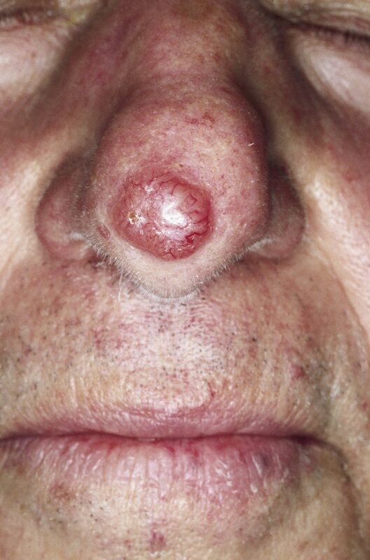
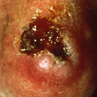
| Appearance | Pearly, waxy nodule | Scaly, ulcerated plaque | Asymmetric pigmented lesion |
| Location | Face (especially nose) | Hands, face, lips, ears | Anywhere (back in men, legs in women) |
| Precursor | None (de novo) | Actinic keratosis | Dysplastic nevus |
| Metastasis | Almost never | Can (2-5%) | Aggressive |
| Mortality | Very low | Low-moderate | Highest of all skin cancers |
| Histology | Palisading nuclei | Keratin pearls | S-100+, HMB-45+ |
| Treatment | Mohs surgery, excision | Excision, Mohs for high-risk | Wide excision (margins by depth) |
| Genetics | PTCH1 (Hedgehog) | p53 mutation | BRAF (V600E) |
A lesion walks into your exam. Work through it:
Patient has a pigmented lesion. Walk the algorithm.
Tap each card. Front = the buzzword clinical medicine hands you. Back = everything you need to close the case.
A lesion just walked into your clinic. Work through the branches.
5 patients just walked in with skin lesions. Don't miss the diagnosis.
4 of these from a pool of 14. Different every time. Let's go.