Three superior alveolar nerves carry the upper teeth, and the palate has its own wiring. Front teeth are ASA. Stop drifting to the molar nerve.
Tap a tooth. Watch which superior alveolar nerve lights up and trace the signal up to the maxillary nerve and the trigeminal ganglion. Then flip to palate mode to separate pulp from the gum on the roof of the mouth.
Three superior alveolar nerves, front to back. ASA for the incisors and canine. MSA for the premolars and the mesiobuccal root of the first molar. PSA for the molars. ASA and MSA branch off the infraorbital nerve inside the canal. PSA splits off V2 earlier, back in the pterygopalatine fossa. All of it is the maxillary nerve, V2. When a front tooth breaks, the pain is ASA. Do not drift to the molar nerve because it sounds familiar. Know your clues.
Three for the teeth, two for the palate, one for the face. Tap a tab.

Two questions split this whole topic. Is the problem the tooth or the gum? Tooth pulp is the superior alveolar nerves: ASA, MSA, PSA. Palatal soft tissue is the palatine nerves: nasopalatine in front, greater palatine in back. Then, which tooth? Front equals ASA, middle equals MSA, back equals PSA. Pulp versus palate, then front versus back. Answer those two and you never miss one of these.
The single distinction that defeats these questions. Each row shows the structure; the nerve, tissue, and exit start blurred. Predict each row, then tap to reveal.
read the structure, call the nerve, then tap that one row to check it
| Structure | Nerve | Tissue | Exit |
|---|---|---|---|
| Incisors and canine | Anterior superior alveolar (ASA) | Tooth pulp | Off infraorbital nerve in the canal |
| Premolars | Middle superior alveolar (MSA) | Tooth pulp | Off infraorbital nerve in the canal |
| Molars | Posterior superior alveolar (PSA) | Tooth pulp | Off V2 in the pterygopalatine fossa |
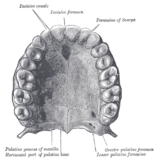
| Gum behind the incisors | Nasopalatine | Palatal soft tissue | Incisive foramen |
| Posterior palatal gum | Greater palatine | Palatal soft tissue | Greater palatine foramen |
| Lower eyelid, side of nose, upper lip | Infraorbital | Facial skin | Infraorbital foramen |

Look at the third column. Three rows of pulp, two rows of palatal gum, one row of facial skin. The superior alveolar nerves never touch the roof of the mouth, and the palatine nerves never feel a toothache. The infraorbital nerve is the parent trunk on the face, and after it leaves the foramen it is pure skin: lower eyelid, side of the nose, upper lip. Mix up the columns and you pick the wrong answer. Keep them straight and the question answers itself.
Every distractor has a reason it tempts you. Tap to flip and find it.
Predict first, then tap to unblur.
