>

Sarcoidosis

The granuloma that touches everything → lungs, skin, eyes, heart. How one mechanism creates a whole-body disease.

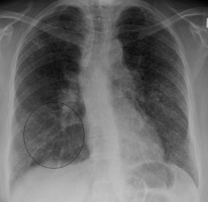
A 29-year-old African-American woman presents with 4 months of fever, progressive shortness of breath, and a dry cough. She's lost 5 kg. She visited Kenya 2 months ago. Exam: fine crackles and wheezing bilaterally. Labs: calcium 11.8 mg/dL (normal: 8.4-10.2). CXR: reticular opacities + bilateral hilar lymphadenopathy.

What's the underlying mechanism?

A. Granulomatous inflammation
B. Bronchial hyperresponsiveness
C. Neoplastic transformation
D. Monoclonal plasma cell production
E. Necrotizing inflammation
F. Viral infection

Break Down the Clues

Patient walks in. Three clues hit you immediately. What do you make of them?

Clue 01
29-year-old African-American woman
Demographics are the clue. Sarcoidosis peaks in 20-40 year-old African Americans, especially women. It's also common in Scandinavians. If boards give you a young Black woman with lung symptoms → sarcoidosis should already be on your radar.
Makes less likely: Lung cancer (rare under 40, especially without smoking history)
Clue 02
Dry cough + progressive SOB + fever × 4 months
Chronic and dry → this is an interstitial processInterstitial lung diseases affect the tissue between the air sacs (interstitium), not the airways themselves. That's why the cough is dry → there's no mucus production. The lung tissue is getting stiff and scarred., not an infection that produces sputum. The timeline (4 months) rules out acute processes. The fever says this is inflammatory, not just fibrotic.
Makes less likely: Typical pneumonia (too chronic), viral infection (self-limited)
Clue 03
Kenya travel → the red herring
They WANT you to think TB. Kenya is endemic for TB, and TB also causes granulomas + hilar lymphadenopathy. But here's the thing: TB granulomas are caseating (cheesy necrotic centers). Sarcoidosis granulomas are non-caseating. Plus, TB cough is usually productive (sputum, hemoptysis), and TB CAN cause hypercalcemia through the same 1-alpha-hydroxylase mechanism (any granulomatous disease can), but the dry cough and non-caseating granulomas here point away from TB.
Makes less likely: TB (caseating granulomas expected; TB also causes productive cough and hemoptysis, not dry cough)

Two Clues That Seal It

Lab + imaging. These two together lock the diagnosis.

Clue 04
Calcium: 11.8 mg/dL (high)
This is the sneaky clue. Two main causes of hypercalcemia you need to tell apart:

Sarcoidosis: Activated macrophages in granulomas express 1-alpha hydroxylaseThis enzyme normally lives in the kidney and converts 25-OH vitamin D (inactive storage form) → 1,25-(OH)2 vitamin D (calcitriol, the active form). In sarcoidosis, the granuloma macrophages hijack this enzyme, making calcitriol uncontrollably → calcium absorption goes up. → converts vitamin D to active calcitriol → increased calcium absorption from the gut.

Lung cancer: Squamous cell carcinoma secretes PTHrPParathyroid hormone-related peptide. Acts just like PTH (activates osteoclasts, increases calcium) but is made by the tumor, not the parathyroid glands. This is "humoral hypercalcemia of malignancy." → mimics PTH → pulls calcium from bones.

Both cause high calcium, but the mechanism is completely different.
Clue 05: The Lock
CXR: Bilateral hilar lymphadenopathy + reticular opacities
Bilateral hilar lymphadenopathy is THE classic CXR finding of sarcoidosis. It's so associated that it practically seals the diagnosis when combined with the right demographics. Lung cancer would show a solitary nodule or mass, not symmetric bilateral hilar nodes. Lymphoma can also cause bilateral hilar LAD, but the hypercalcemia mechanism + demographics point to sarcoidosis.
AP chest X-ray showing bilateral hilar lymphadenopathy in sarcoidosis
See the bulging hila on both sides? That's bilateral hilar lymphadenopathy → the hallmark CXR of sarcoidosis. AP chest X-ray, sarcoidosis. Credit: James Heilman, MD → CC BY-SA 3.0, Wikimedia Commons.
Diagnosis: Sarcoidosis
Young Black woman + dry cough + hypercalcemia + bilateral hilar LAD = sarcoidosis until proven otherwise. The mechanism is granulomatous inflammation.

Why Granulomas?

One immune reaction, systemic consequences

Sarcoidosis is a Type IV hypersensitivityDelayed-type hypersensitivity. T-cells (not antibodies) drive the reaction. Same category as TB skin test reactions, contact dermatitis, and transplant rejection. Takes 24-72 hours to develop, which is why sarcoidosis is chronic, not acute. reaction to an unknown antigen. Nobody knows what triggers it. The immune system just... decides to fight something, and can't stop.

Here's the chain:

1. Unknown antigen activates CD4+ T-helper cells (Th1 subtype)
2. Th1 cells release IL-2 and IFN-gamma → recruit and activate macrophages
3. Macrophages fuse into epithelioid cells and multinucleated giant cells
4. These form tight clusters → non-caseating granulomas (no central necrosis)
5. Granulomas deposit everywhere → lungs, lymph nodes, skin, eyes, liver, heart, kidneys, CNS

The granuloma macrophages also secrete ACE (angiotensin-converting enzyme) → which is why elevated serum ACE is a lab marker for sarcoidosis.🔑Sarcoidosis → Serum ACE ↑ (the granulomas are little ACE factories)

⚠️
Board Trap: Caseating vs Non-Caseating
Both TB and sarcoidosis form granulomas. The difference:
TB = caseating (central cheesy necrosis → the immune system is trying to wall off AND destroy the bacteria)
Sarcoidosis = non-caseating (no central necrosis → the immune system is walling off... nothing? The antigen is unknown)

If a question says "non-caseating granulomas" → think sarcoidosis.
If it says "caseating granulomas" → think TB or fungal infection. 🔑Sarcoid is clean inside (non-caseating). TB is cheesy inside (caseating). Clean vs Cheese.

Two Roads to Hypercalcemia

Same lab value, completely different mechanisms

Sarcoidosis Malignancy (SCC Lung)
Trace It Granuloma macrophages make 1-alpha hydroxylase → excess active vitamin D → ↑ gut Ca²⁺ absorption Tumor secretes PTHrP → mimics PTH → ↑ bone resorption
Vitamin D ↑ 1,25-(OH)₂D (calcitriol HIGH) Normal or low
PTH Suppressed (calcium is high, so PTH shuts down) Suppressed (PTHrP, not PTH, is driving it)
PTHrP Normal Elevated
CXR Bilateral hilar LAD, reticular opacities Solitary mass or nodule
Demographics Young (20-40), often African American Older (50+), smoker
Treatment Steroids (suppress the granulomas) Treat the cancer + IV fluids + bisphosphonates
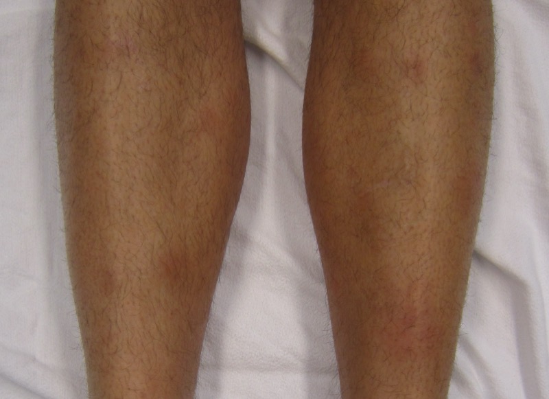
Where Sarcoidosis Strikes

Tap each organ to see what happens when granulomas show up there

🫁
Lungs
#1 site → >90% of patients
Dry cough, dyspnea, bilateral hilar LAD on CXR. Can progress to pulmonary fibrosis in severe cases. Staging is based on CXR:
Stage I: Bilateral hilar LAD only
Stage II: LAD + pulmonary infiltrates
Stage III: Pulmonary infiltrates only (LAD resolved)
Stage IV: Pulmonary fibrosis
Most patients (Stage I) resolve spontaneously.
👁️
Eyes
Most common extrapulmonary site
Anterior uveitis → eye pain, redness, blurred vision, photophobia. Can cause blindness if untreated. All sarcoidosis patients need an eye exam. Also: lacrimal gland swelling, keratoconjunctivitis sicca (dry eyes).
🩹
Skin
~30% of patients
Erythema nodosum → tender red nodules on the shins (actually a good prognostic sign).
Lupus pernio → violaceous (purple-blue) plaques on the nose, cheeks, ears. Looks like frostbite. Associated with chronic sarcoidosis.
Papules, plaques, and nodules that can appear anywhere.
Erythema nodosum → tender red nodules on the shins
Tender, red, raised nodules on the anterior shins → classic erythema nodosum. Credit: James Heilman, MD → CC BY-SA 3.0, Wikimedia Commons.
❤️
Heart
~5%, but major cause of death
Granulomas infiltrate the conduction system → arrhythmias, heart block, sudden cardiac death. Can also cause restrictive cardiomyopathy. Cardiac sarcoidosis is the #1 cause of sarcoidosis death in some populations. Diagnose with cardiac MRI or PET scan.
🧠
CNS
~5-10% → neurosarcoidosis
CN VII palsy (Bell's palsy) is the most common neurologic finding. Can also cause: cranial neuropathies, seizures, meningitis, hypothalamic/pituitary dysfunction (diabetes insipidus). If a young patient has Bell's palsy + any lung symptoms → think sarcoid.
🦴
Calcium / Kidneys
Hypercalcemia in ~10-15%
1-alpha hydroxylase from granuloma macrophages → excess calcitriol → hypercalcemia + hypercalciuria. Can lead to nephrocalcinosis and kidney stones. Chronic hypercalcemia → renal failure. This is why sarcoidosis patients need calcium monitoring.
Löfgren Syndrome → the "good sarcoidosis." Classic triad: bilateral hilar LAD + erythema nodosum + polyarthralgia + fever. Acute onset, excellent prognosis (usually self-resolves). If boards describe this combination → sarcoidosis with good prognosis.

Diagnosis & Treatment

What to order and what to give

Diagnosis:

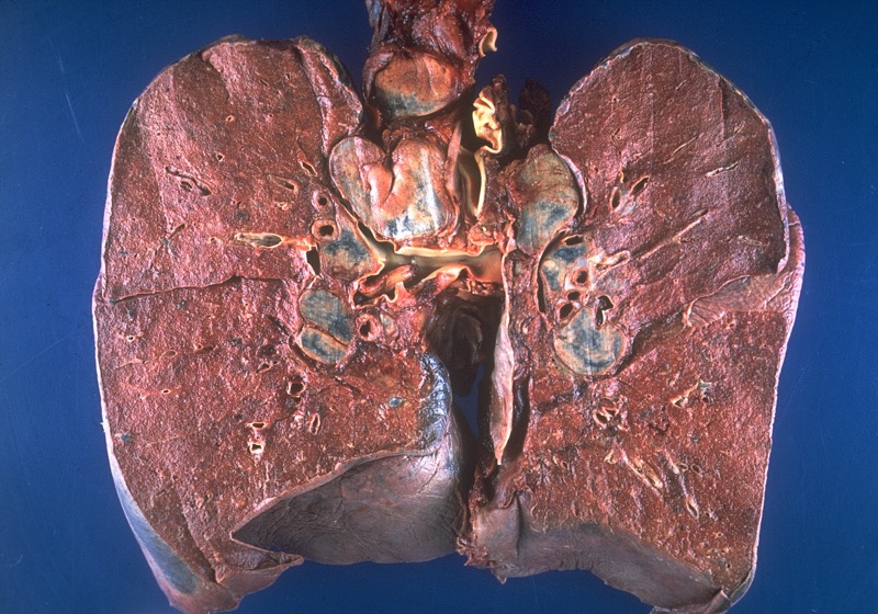
Gold standard is biopsy showing non-caseating granulomas. Transbronchial biopsy is most common approach. You'll also see:

TestFindingWhy
CXRBilateral hilar LADGranulomas in hilar lymph nodes
Serum ACE↑ ElevatedGranuloma macrophages produce ACE
Calcium↑ Hypercalcemia1-alpha hydroxylase → excess calcitriol
1,25-(OH)₂D↑ ElevatedSame mechanism → granulomas make active vit D
BiopsyNon-caseating granulomasConfirms diagnosis, rules out TB/fungal
CD4:CD8 ratio (BAL)↑ ElevatedTh1-predominant response in alveoli

Treatment:

Corticosteroids → suppress the granulomatous inflammation. Not everyone needs treatment though → many patients with Stage I disease self-resolve. Treat when there's: organ damage, hypercalcemia, eye involvement, cardiac or neuro involvement, or progressive lung disease.

Steroid-sparing agents for refractory cases: methotrexate, azathioprine, infliximab.

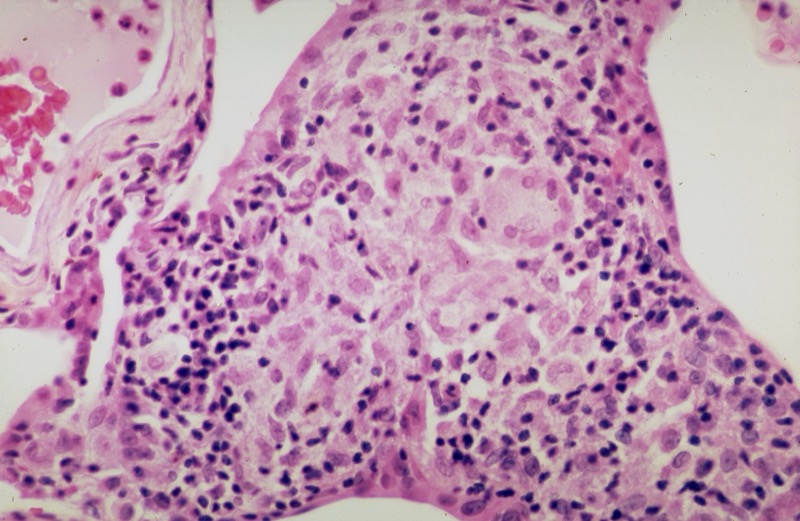
Histology buzzwords:

FindingWhat It Is
Non-caseating granulomasTight clusters of epithelioid macrophages, NO central necrosis
Schaumann bodiesCalcified, laminated concretions inside giant cells (calcium + protein deposits)
Asteroid bodiesStar-shaped inclusions inside giant cells (cytoskeletal remnants)
Multinucleated giant cellsFused macrophages → Langhans-type (nuclei arranged in horseshoe pattern)
Non-caseating granuloma histology in sarcoidosis
Tight cluster of epithelioid histiocytes + giant cells with NO central necrosis → that's the non-caseating granuloma. If you saw cheesy stuff in the center, it'd be TB. Sarcoidosis granuloma, H&E stain. Credit: Yale Rosen → CC BY-SA 2.0, Wikimedia Commons.
⚠️
Schaumann & Asteroid Bodies Are NOT Specific
These inclusion bodies are found in sarcoidosis but also in other granulomatous diseases. They're suggestive, not diagnostic. The diagnosis requires non-caseating granulomas + clinical picture + exclusion of other causes (TB, fungal, foreign body).

Mechanism Elimination

Four mechanisms. One matches sarcoidosis. Watch the clues knock them out.

Bronchial Hyperresponsiveness
Airway smooth muscle overreaction
Granulomatous Inflammation
Macrophages forming organized clusters
Neoplastic Transformation
Uncontrolled cell growth → tumor
Necrotizing Inflammation
Tissue death from inflammatory damage
Ready? Let's eliminate.

Clinical Vignettes

5 patients just showed up. Don't let the granulomas fool you.

Not bad for someone who just learned this.
Board-Style Walkthrough

Board-Style Walkthrough

Original board-style vignettes. Shuffled, never-repeat, full explanations for every choice.