The most common extracranial solid tumor in children. You diagnose it by watching the baby dance.
A 2-year-old boy is brought by his mother because his eyes keep darting around randomly, his feet jerk when he tries to stand, and he has become unusually irritable and clumsy over the past 3 weeks. On exam, you palpate a firm, irregular abdominal mass crossing the midline.
What is the most likely diagnosis?
THE HOOK
Dancing Eyes, Dancing Feet, Dancing Mind
This is the mnemonic that separates neuroblastoma from every other pediatric abdominal mass.
Opsoclonus-Myoclonus-Ataxia Syndrome
The tumor triggers an autoimmune responseThe immune system attacks the neuroblastoma cells but also cross-reacts with cerebellar neurons. The antibodies meant for the tumor hit the brain instead. that attacks the cerebellum. Three things happen:
Opsoclonus (dancing eyes): rapid, chaotic, multi-directional eye movements. Not nystagmus, which is rhythmic. This is random.
Myoclonus (dancing feet): sudden, involuntary muscle jerks, especially in the limbs
Ataxia (dancing mind): cerebellar dysfunction causing clumsiness and behavioral changes
★If you see a child with chaotic eye movements + jerky limbs + an abdominal mass, the answer is neuroblastoma. There is nothing else in the differential that looks like this. 🔑Dancing Eyes + Dancing Feet + Belly Mass = Neuroblastoma. All three together have only one answer.
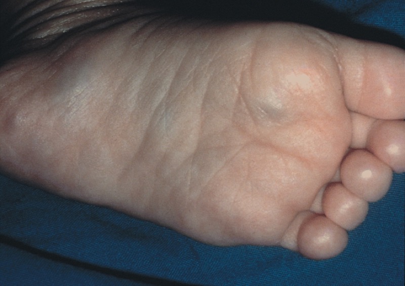
📷 BLUEBERRY MUFFIN BABY: Stage IV-S neuroblastoma with skin metastases · tap to expand
TUMOR PATH
Neural Crest to Tumor
Where it comes from, what it makes, how you find it.
Origin
Neuroblastoma arises from neural crest cellsThese are the embryonic cells that migrate out of the neural tube and become the sympathetic nervous system, adrenal medulla, and melanocytes. Tumors from this lineage include neuroblastoma, pheochromocytoma, and melanoma. of the sympathetic nervous system. The most common location is the adrenal medulla, but it can arise anywhere along the sympathetic chain (abdomen, chest, pelvis, neck).
Because it comes from sympathetic tissue, it produces catecholamines and their breakdown products:
🔬90% of neuroblastomas produce elevated urine HVA and VMA. These are the lab markers you order when you suspect the diagnosis. 💡HVA = dopamine metabolite. VMA = norepinephrine/epinephrine metabolite. Both elevated in neuroblastoma. Carcinoid uses 5-HIAA instead.
Match the Marker
Drag the correct lab test to each tumor. Tap the right match.
Neuroblastoma is diagnosed with:
Pheochromocytoma is diagnosed with:
Key Features at a Glance
Most common extracranial solid tumor in children
Most common malignancy in infants (under 1 year)
Median age at diagnosis: 18 months
Crosses the midline (unlike Wilms tumor, which stays on one side)
Can present with hypertensionFrom the catecholamines the tumor produces. Same mechanism as pheochromocytoma, just in a baby. from catecholamine release
Prognosis linked to N-myc amplification💡N-myc amplified = N for "Not good." More copies of the oncogene = worse prognosis.: more copies = worse outcome
THE DIFFERENTIAL
Three Masses. One Answer.
Tap each villain to see what makes it different. Know all three cold.
Your answer
Neuroblastoma
Origin: Adrenal medulla / sympathetic chain (neural crest) Age: Under 2 years (median 18 months) Location: Retroperitoneal, crosses midline freely Markers: Urine HVA + VMA (catecholamine metabolites) Imaging: MIBG scan (gold standard for staging/mets) Prognosis marker: N-myc amplification Paraneoplastic: Opsoclonus-myoclonus-ataxia (dancing eyes/feet/mind)
The other mass
Wilms Tumor
Origin: Intrarenal (metanephric mesoderm) Age: 3-4 years Location: Within the kidney, stays on ONE side (does not cross midline) Markers: None specific (no catecholamines) Imaging: Ultrasound first, then CT for staging Histology: Triphasic (blastemal + stromal + epithelial) Associations: WAGR syndrome, Beckwith-Wiedemann, aniridia, hemihypertrophy
Liver mass
Hepatoblastoma
Origin: Liver (endoderm) Age: Under 3 years (infants and toddlers) Location: Liver, does not cross midline Markers: Serum AFP (markedly elevated) Imaging: Ultrasound + CT/MRI liver Histology: Fetal or embryonal hepatic cells Associations: Beckwith-Wiedemann, familial adenomatous polyposis (APC)
🔑The one-line differentiator: Crosses midline = neuroblastoma. Stays in the kidney = Wilms. Stays in the liver + AFP elevation = hepatoblastoma. 💡NWH: Neuroblastoma crosses midline, Wilms stays in kidney, Hepatoblastoma in liver. Three pediatric masses, three locations.
THE ALGORITHM
Abdominal Mass in a Child
Tap each step to follow the clinical logic. One question at a time.
Child with an abdominal mass. Walk the decision tree to the diagnosis.
Biopsy confirms neuroblastoma. Check N-myc amplification for prognosis. Stage 4S in infants: may spontaneously regress.
VISUAL ANCHORS
See the Tumor
Tap to expand. The blueberry muffin and the rosettes are both tested.
Blueberry Muffin Baby
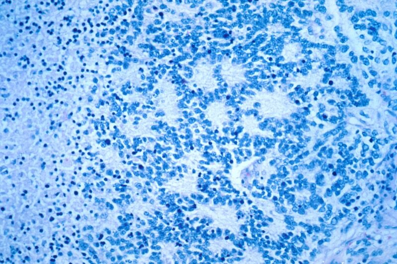
Homer-Wright Rosettes
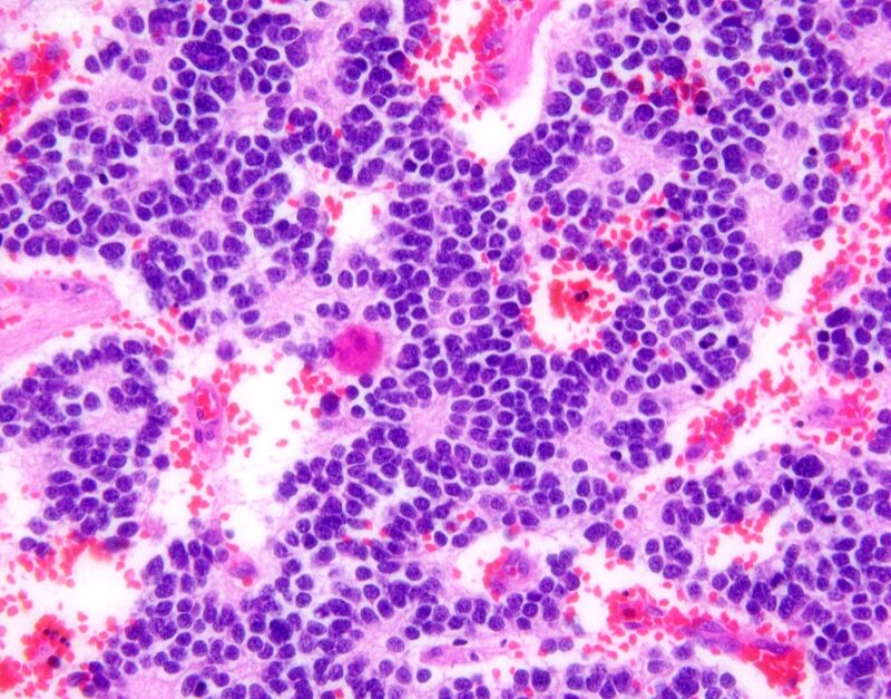
H&E Rosettes
THE PATTERN
Homer-Wright Rosettes
The histology finding that links neuroblastoma to its brain cousin.
📷 HOMER-WRIGHT ROSETTES: tumor cells circling neuropil core (40x) · tap to expand
📷 H&E STAIN: Homer-Wright pseudorosettes under higher magnification · tap to expand
Both neuroblastoma and medulloblastomaThe most common malignant brain tumor in children. Found in the posterior fossa (cerebellum). Same neural origin as neuroblastoma, but in the brain instead of the abdomen. show Homer-Wright rosettes on biopsy, because both tumors come from primitive neural tissue. The rosette is a ring of tumor cells arranged around a central core of neuropil (tangled nerve fibers).
Neuroblastoma
Tumor cells circle a core of tangled nerve fibers (neuropil). Found in the abdomen.
EXTRACRANIAL
Medulloblastoma
Same rosette pattern, same neural origin. Found in the posterior fossa (cerebellum).
INTRACRANIAL
💡Same rosette, different address. Homer-Wright rosettes in the abdomen = neuroblastoma. Homer-Wright rosettes in the brain = medulloblastoma. Both are neural-origin tumors.
⚠️
Board Trap: Rosette Confusion
Don't confuse Homer-Wright rosettes (neural tumors, cells around neuropil) with Flexner-Wintersteiner rosettes (retinoblastoma, cells around an empty lumen). Homer-Wright = nerve center. Flexner-Wintersteiner = empty center.
Feature
Neuroblastoma
Medulloblastoma
Location
Abdomen (adrenal medulla, sympathetic chain)
Posterior fossa (cerebellum)
Age
Usually < 2 years
Usually 4-8 years
Histology
Homer-Wright rosettes
Homer-Wright rosettes
Origin
Neural crest (sympathetic)
Cerebellar granule cell precursors
Lab marker
Urine HVA / VMA
None specific (imaging-based)
Classic clue
Dancing eyes/feet/mind
Truncal ataxia, morning vomiting
Prognosis marker
N-myc amplification (bad)
WNT subgroup (good)
Midline
Crosses midline
Arises at midline (vermis)
CLINICAL REASONING
Decision Tree: Pediatric Abdominal Mass Workup
Child with abdominal mass. Work through the branches.
Child with abdominal mass. Where does imaging point?
Neuroblastoma. Most common extracranial solid tumor in children under 5. Arises from adrenal medulla most often. N-MYC amplification = poor prognosis. Treat: surgery + chemo, plus radiation for high-risk disease.
Staging sub-branches:
Localized: surgical resection. Favorable biology (hyperdiploid, MYCN not amplified) = good prognosis.
Metastatic stage 4: high-dose chemo + autologous SCT + anti-GD2 immunotherapy (dinutuximab).
Stage 4S (special, under 18 months): regression often spontaneous. Watch and wait.
Wilms tumor (nephroblastoma). Most common renal tumor in children. Peak age 3-4 years. Associated with WAGR syndrome (Wilms + aniridia + GU abnormalities + intellectual disability). Treat: surgery + chemo, plus radiation for advanced disease. Prognosis generally good.
Hepatoblastoma. Hepatic mass with elevated alpha-fetoprotein. Treat: surgical excision + chemotherapy.
Appendicitis. Classic point tenderness at McBurney with fever. Treat: surgical appendectomy.
CHALLENGE
Clinical Questions
6 randomized questions from a pool of 10. Reload for a fresh set.
Medically reviewed by Kaitlyn Cocuzzo, MD and Fatima Ali, DO · Last updated July 1, 2026 at 10:03 PM ET
Bone Wizardry is an independent educational resource for visual learning in the medical sciences. It is not affiliated with, endorsed by, or sponsored by any licensing or examination board, contains no real or recalled examination questions, and does not guarantee any educational or examination outcome.