RSV virion: fusion (F) protein is the palivizumab target · tap to expand
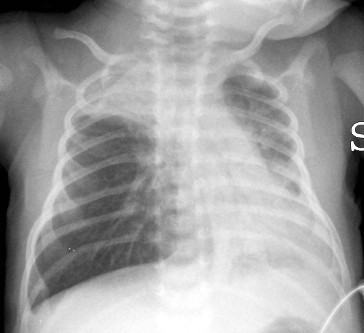
Hyperinflation and peribronchial cuffing, not antibiotics · tap to expand
The plugged bronchiole: sloughed cells, mucus, edema · tap to expand
🫁
Bronchiolitis and RSV
The winter virus that plugs an infant's smallest airways. The whole exam game is recognizing it, supporting it, and resisting the urge to treat it like asthma or pneumonia.
A 6-month-old boy is brought in during January after four days of runny nose and congestion that have now turned into noisy, wheezy breathing. He is breathing fast at 60 per minute with nasal flaring and pulling in between his ribs. On exam there are diffuse wheezes and fine crackles. He is alert, has wet diapers, and saturates 94 percent on room air.
What is the single most likely diagnosis?
Bacterial pneumonia
RSV bronchiolitis
Foreign body aspiration
Croup
The Mechanism
Why a Tiny Airway Makes a Big Wheeze
Tap each card. Front is the fact. Back is why the board cares.
🦠
RSV
tap
The bug
Respiratory syncytial virus, a paramyxovirus, infects the lining of the small airways. It is the number one cause of bronchiolitis and the number one cause of infant hospitalization in winter.
🔗
Syncytia
tap
Why the name
The viral fusion (F) protein melts neighboring cells into giant multinucleated syncytia. That same F protein is the exact target of palivizumab.
🚰
Tiny radius
tap
Resistance law
Airway resistance climbs with the fourth power of shrinking radius. A little edema and mucus that an adult shrugs off nearly chokes an infant bronchiole.
🎐
Ball valve
tap
Air trapping
The plug lets air in but not out. Air gets trapped behind it, the lungs hyperinflate, and the diaphragm flattens. That is the barrel chest and the hyperinflated X-ray.
The cause-and-effect chain
Virus lands in the bronchioles: RSV infects and kills the airway lining, which sloughs into the lumen.
The wall swells and mucus pours in: edema plus dead cells plus mucus equals a plug stuffed into a tube already the width of a coffee stirrer.
Resistance explodes: because resistance scales with the fourth power of radius, that small plug causes a huge jump in the effort needed to move air. You hear it as a wheeze.
Air gets trapped: the plug acts as a one-way valve, so the lungs hyperinflate and gas exchange suffers.
The baby works harder: tachypnea, nasal flaring, retractions, and grunting are the visible signs of that extra work.
💡Memory hook: bronchiolitis is a plumbing problem, not a muscle problem.🔑The airway is physically plugged with debris and swelling. There is no bronchial muscle spasm to relax, which is exactly why albuterol disappoints.
At the Bedside
The Story, the Red Flags, the High-Risk Babies
It starts as a cold and marches down into the chest. Your job is to spot the babies who cannot ride it out at home.
The classic course
Who: infant under 2, peak at 2 to 6 months, in winter to early spring.
Prodrome: 1 to 3 days of upper-airway symptoms, runny nose, congestion, cough, maybe a low-grade fever.
Descent: then it moves down: wheezing, crackles, tachypnea, retractions, nasal flaring, prolonged expiration.
Feeding drops: a baby breathing 60 times a minute cannot coordinate sucking, so intake falls and dehydration creeps in.
Time course: self-limited, worst around day 3 to 5, then better over 1 to 2 weeks. The cough can linger for weeks.
⚠
Apnea can be the first sign
In young infants (under about 2 months) and former premature babies, RSV can cause apnea before much wheezing appears. A reported pause-and-turn-dusky episode is an admission-level red flag, not a footnote.
Admit When You See
Apnea: observed or reported. Full stop, this one admits.
Why: the smallest airways and the highest apnea risk.
Also high risk: immunodeficiency and neuromuscular disease.
Action: lower the threshold to admit and observe.
Workup
Diagnose With Your Eyes, Not the Lab
Bronchiolitis is a clinical diagnosis. The traps here are tests that confirm what you already know and antibiotics for a virus.
History plus exam
Winter, a baby under 2, a cold that descended into wheezing and retractions. That pattern is the diagnosis.
Why: routine testing rarely changes what you do. The story and the exam carry the day.
Nasopharyngeal RSV testing (selective)
A rapid RSV antigen swab or PCR can confirm the virus. It is most useful for cohorting and isolation of admitted infants, not for deciding treatment.
Why: a positive swab does not change supportive management of a typical case. It groups infected babies and reassures against a bacterial hunt.
Chest X-ray (only if you truly suspect something else)
Not routine. When obtained it shows hyperinflation, flattened diaphragms, peribronchial cuffing, and patchy atelectasis.
Why: those viral findings get misread as pneumonia and trigger needless antibiotics. Order film only for a focal or atypical picture.
⚠
Peribronchial cuffing is not pus
Hyperinflation and peribronchial cuffing are the puffy outline of inflamed small airways, the signature of a virus. A dense focal consolidation is what a bacterial pneumonia looks like. Do not let a hazy viral film talk you into ceftriaxone.
🧪Atelectasis from a mucus plug can mimic an infiltrate. Reassess after suctioning before you call it pneumonia.
Treatment
Support It, Do Not Over-Treat It
The cure is time. Your job is to keep the airway clear, the baby fed, and the oxygen up while the virus runs its course.
What actually helps (supportive care)
Nasal suctioning: clear secretions, especially before feeds, so the baby can breathe and eat.
Hydration: oral if tolerated, IV or nasogastric if the baby cannot keep up.
Oxygen: supplemental oxygen when saturation stays below 90 percent.
Monitoring: watch the work of breathing, feeding, and oxygen. Most babies improve with this alone.
⚠
What NOT to give routinely
No albuterol (there is no muscle spasm to relax), no corticosteroids (this is not allergic asthma inflammation), no antibiotics (it is viral), and no routine chest physiotherapy. Nebulized hypertonic saline has at best a small, inconsistent effect and is not a substitute for the basics.
Palivizumab: prevention, not treatment
Palivizumab is a monoclonal antibody against the RSV fusion (F) protein. It is passive immunization given as monthly injections through RSV season (up to 5 doses) to the babies most likely to be hospitalized. It does nothing for an infant who is already sick.
Born under 29 weeks gestation: in the first year of life.
Chronic lung disease of prematurity: infants under 12 months who needed prolonged oxygen.
Hemodynamically significant congenital heart disease: infants under 12 months.
Newer agents (the long-acting monoclonal nirsevimab and the maternal RSV vaccine) now broaden protection, but palivizumab and its three classic indications remain the high-yield board answer.
Walk the Logic (Tap an Answer)
An 11-month-old with a first-ever wheeze that began with a runny nose, no atopy. Bronchiolitis or asthma?
A single virus-triggered wheeze under age 2, without atopy, is bronchiolitis. Asthma needs a recurrent, reversible pattern over time. One viral wheeze is not asthma.
Do you need a chest X-ray and antibiotics to manage a typical case?
Bronchiolitis is diagnosed clinically. Routine film leads to misread viral findings and needless antibiotics. No routine X-ray, no antibiotics for a virus.
What is first-line treatment for the wheezing, tachypneic but well-hydrated infant?
Supportive care is the whole treatment. Albuterol and steroids do not change the course of a first bronchiolitis episode. Suction, hydrate, oxygenate.
Which finding sends this baby to the hospital?
Rhinorrhea and cough are the disease being the disease. Apnea, oxygen below 90 percent, and failing feeds are the tripwires that admit. Admit for apnea, hypoxia, or dehydration.
Prove It
Board Walkthrough
Original clinical vignettes, dealt a few per round, answer choices shuffled. Tap a wrong answer first to see why it almost works, then read the glowing clues.
5 of 5
Medically reviewed by Kaitlyn Cocuzzo, MD and Fatima Ali, DO · Last updated July 1, 2026 at 10:03 PM ET
Bone Wizardry is an independent educational resource for visual learning in the medical sciences. It is not affiliated with, endorsed by, or sponsored by any licensing or examination board, contains no real or recalled examination questions, and does not guarantee any educational or examination outcome.