A psammoma body is a tiny ring of calcium that builds layer by layer, like an onion of dead cell + lime, inside a tumor. Four cancers leave this calling card. Know the club roster and the clinical medicine stop being scary.
Pathology Rounds · Pick One
A pathology resident is reviewing a thyroid biopsy. Under high-power magnification she sees concentric calcified rings nestled inside a papillary cluster, alongside nuclei that appear cleared out, ground-glass, almost hollow, like someone erased the inside of each cell. What is the most likely diagnosis?
Follicular thyroid carcinoma
Papillary thyroid carcinoma
Medullary thyroid carcinoma
Anaplastic thyroid carcinoma
Good instinct if you hesitated on follicular, it is also a thyroid cancer. But follicular doesn't give you psammoma bodies or those hollow nuclei. Think of those cleared-out nuclei like someone took Photoshop's eraser to the nucleus. That is Orphan Annie eye nucleus, and it belongs to one tumor only: papillary thyroid carcinoma. Psammoma bodies plus Orphan Annie = you are done. Bottom line: Cleared-out nuclei + psammoma bodies in a thyroid biopsy = Papillary Thyroid Carcinoma every time.
Section 1 of 7
Build the PSaMMoMa
Four letters, four tumors. Tap each letter in order to light it up and meet the member.
From the Attending
PSaMMoMa is the mnemonic. Each letter = one tumor that makes psammoma bodies: P · Papillary thyroid carcinoma (also has Orphan Annie eye nuclei + nuclear grooves). S · Serous papillary cystadenocarcinoma of ovary (high-grade serous, BRCA-associated). M · Meningioma (spindle cells in whorls + psammoma). M · Mesothelioma (asbestos history, pleural rind). The bodies themselves are laminated calcium concretions · layers built around dying papilla tips. If a stem describes "concentric calcified bodies on biopsy," you're picking from those four.
Psammoma comes from the Greek word for sand. Picture a single dying cell at the tip of a finger-like tumor growth. Calcium drifts in, settles around it, then layer by layer wraps it like a pearl. Now picture that happening hundreds of times in one tumor. That is psammoma. Four tumors do this. Tap each tile in order to recruit the club.
Recruited 0 of 4
P is for Papillary Thyroid
Most common thyroid CASpreads via lymphExcellent prognosis
The finger-like (papillary) projections of this thyroid tumor pile up calcium at their tips. Risk factor in clinical practice: prior neck irradiation as a kid. The microscope shows two things together: psammoma bodies plus eerily pale, hollow-looking nuclei called Orphan Annie eyes.
Bottom line: psammoma bodies + cleared (Orphan Annie) nuclei in a thyroid biopsy = papillary thyroid carcinoma.
S is for Serous Ovarian
Most common ovarian CAOften bilateralBRCA1/2
High-grade serous adenocarcinoma actually starts in the fallopian tube and seeds the ovary, which is why it is so often bilateral. Family history of breast or ovarian cancer in mom or sister = think BRCA1 first. The tumor builds papillary fronds just like the thyroid version, so it lays down the same calcium rings.
Bottom line: bilateral ovarian mass + papillary tumor with psammoma bodies + family BRCA history = high-grade serous.
M is for Meningioma
Most common intracranial tumorBenign (WHO I)Best prognosis
A tumor of the brain's wrapping, not the brain itself. Arises from arachnoid cap cells and grows in concentric whorls, like the inside of a cinnamon roll. Those whorls calcify into psammoma bodies. Classic clinical medicine setup: middle-aged woman, parasagittal mass, dural tail on MRI.
The lining of the lung, not the lung itself. Shipyard, plumbing, insulation, or brake-pad worker decades ago. The clinical medicine stem always tells you the job. The biopsy has psammoma bodies PLUS golden-brown dumbbell-shaped fibers called ferruginous bodies, which are asbestos needles wrapped in iron-protein rust.
PSaMMoMa
Roster complete. Four tumors, one calcium calling card. The two M's get mixed up the most. We will untangle them in Section 3. First, flip the cards below to see the full villain dossier on each one.
The Villain Cards
Same four. Now with full stats and a deep dive on the back of each card.
Papillary clusters · tap to expand
P · Thyroid
Papillary Thyroid CA
Most common thyroid cancer
🔍 Orphan Annie eye nuclei + psammoma bodies
MetastasisLymph nodes (local)
Risk factorPrior neck irradiation
PrognosisExcellent
tap to flip →
Why This Happens
Psammoma Connection
Papillary projections develop areas of dystrophic calcificationDead cells calcify at their core when calcium precipitates around necrotic debris. In papillary thyroid CA, this happens inside the papillary stalks themselves., building up layer by layer into those concentric rings. Psammoma bodies here are inside the actual papillary fronds.
The Orphan Annie Sign
The nuclei have an optically clear appearance because the chromatin is pushed to the periphery, leaving the center appearing empty. On H&E stain they look like the eyes of the comic strip character Annie: wide, pale, and haunting. This is pathognomonic for papillary thyroid CA.
Board Trap
Most common thyroid cancer = papillary, not follicular. Follicular metastasizes via blood (hematogenous). Papillary goes lymph nodes first. Two completely different spread patterns.
Serous carcinoma · tap to expand
S · Ovary
Serous Adenocarcinoma
Most common ovarian cancer (high-grade)
🔍 Psammoma bodies on histology
MutationBRCA1/2
LateralityOften bilateral
Staging concernOften advanced at dx
tap to flip →
Why This Happens
Psammoma Connection
High-grade serous carcinoma of the ovary arises from the fallopian tube epitheliumCurrent evidence shows most "ovarian" serous carcinomas actually start in the distal fallopian tube epithelium (fimbrial end), then implant on the ovary. This has changed how prophylactic surgeries are planned.. As the tumor invades and necroses, calcium deposits form laminated rings within the papillary projections. Same mechanism as thyroid, different organ.
BRCA Connection
BRCA1 and BRCA2 are DNA repair genes. When mutated, cells accumulate DNA errors faster and undergo malignant transformation. BRCA1 mutation is a stronger driver for ovarian cancer; BRCA2 is more strongly tied to male breast cancer. Both massively increase high-grade serous risk.
Board Note
The question stem will say bilateral ovarian masses + psammoma bodies on biopsy. That locks in serous adenocarcinoma. Bilateral is a huge clue. Mucinous carcinomas are almost always unilateral.
Meningioma whorls · tap to expand
M · Brain
Meningioma
Most common intracranial tumor (overall)
🔍 Whorling pattern + psammoma bodies
LocationParasagittal (falx)
OriginArachnoid cap cells
PrognosisBest of all brain tumors
tap to flip →
Why This Happens
Psammoma Connection
Meningiomas grow from arachnoid cap cellsThe arachnoid layer is the middle meningeal layer. Cap cells are specialized cells at the tips of arachnoid villi. They're the source of meningioma growth and the reason meningiomas are slow-growing and well-demarcated. and characteristically form concentric whorls of cells. Inside these whorls, over time, the cells calcify into psammoma bodies. So the psammoma bodies sit WITHIN the whorls on histology. A two-feature duo the clinical medicine love.
Why Best Prognosis
Meningiomas are benign (WHO grade I) in most cases. They compress brain rather than invade it. Because they're extra-axial (outside brain tissue), complete surgical removal is possible. Malignant meningiomas exist but are rare. Contrast this with glioblastoma (GBM), which is the most common primary brain tumor in adults. Meningioma is most common overall counting all types.
clinical medicine Trick
Question will say "parasagittal location" or "along the falx" + dural tail on MRI + psammoma bodies on biopsy. That combo = meningioma, not GBM, not metastasis.
Ferruginous body · tap to expand
M · Pleura
Mesothelioma
Pleural cavity malignancy
🔍 BOTH psammoma bodies AND ferruginous bodies
CauseAsbestos exposure
Job clueShipyard / construction
ImagingPleural thickening
tap to flip →
Why This Happens
Asbestos Mechanism
Inhaled asbestos fibers are too long for macrophages to fully engulf. The macrophages try anyway, get frustrated, and die, releasing reactive oxygen species that damage mesothelial DNA over 20-40 years. This long latency periodTime from first asbestos exposure to mesothelioma diagnosis is typically 20-50 years. The patient worked in a shipyard decades ago and just now develops symptoms. Always ask about occupation history in a pleural mass question. means the patient worked in shipyards in the 1970s and presents today.
Ferruginous Bodies
Ferruginous bodies = asbestos fibers coated in iron-proteinMacrophages coat the asbestos fiber with hemosiderin (an iron-containing protein). The result looks golden-brown and dumbbell-shaped under the microscope. "Ferruginous" comes from the Latin for iron (ferrum).. Golden-brown, dumbbell-shaped on microscopy. These are EXCLUSIVE to asbestos exposure. No other tumor in the PSaMMoMa group has them. Mesothelioma is the only tumor in this group with both psammoma bodies AND ferruginous bodies.
The Test Trap
Asbestos also causes bronchogenic carcinoma (lung cancer, specifically small cell and squamous), actually more commonly than mesothelioma. But psammoma + ferruginous bodies = mesothelioma. Bronchogenic carcinoma does NOT have psammoma bodies.
⚠
Board Trap: Most Common vs. What Has Psammoma Bodies
Most common thyroid cancer: papillary (YES, has psammoma bodies). Most common ovarian cancer: serous (YES). Most common intracranial tumor overall: meningioma (YES). The board loves to ask the most common one, and it's always the psammoma one. Don't get confused: GBM is most common primary malignant brain tumor; meningioma is most common intracranial tumor overall (because it's benign and very common).
Section 2 of 7
Through the Microscope
Two slides. One detective story. Walk a thyroid biopsy from normal to malignant, then sit with the asbestos slide that fed mesothelioma for forty years.
Case file · Thyroid FNA
The Biopsy
34F · new thyroid nodule
"The FNA biopsy hit the slide twenty minutes ago. You're at the scope. Two slides. Two verdicts. One diagnosis."
Slide 1 · Low power FNA cytology
First field. What's the verdict?
Round purple clusters of cells around a pale pink center. Single layer. No piling up.
Tap to zoom
Benign thyroid follicles. The nuclei are small, dark, packed dense, and lined up in a single layer around pink colloid. This is healthy thyroid tissue. No psammoma bodies, no cleared-out nuclei. Now the pathologist swaps slides.
Slide 2 · High power H&E
Same patient. Same biopsy. Verdict?
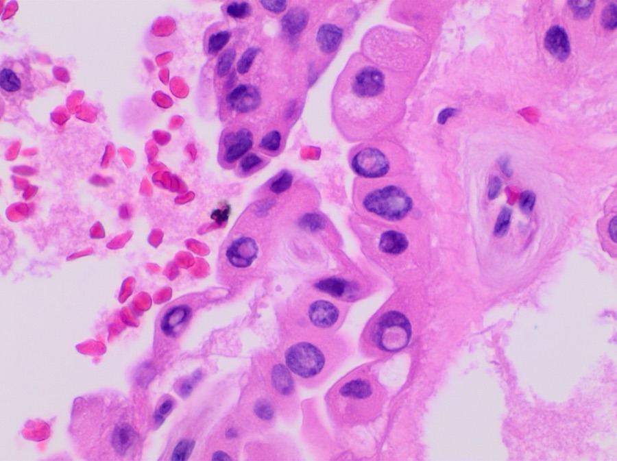
Nuclei look hollow, almost ghost-like. Three small concentric rings sit between the cells.
Tap to zoom
What you're hunting for
Orphan Annie eye
Chromatin shoved to the rim. The center reads hollow.
Psammoma body
Laminated calcium rings, stacked layer by layer.
Papillary thyroid carcinoma. Two findings light up the slide:
Orphan Annie eye the cell's chromatin gets shoved to the rim, leaving the nucleus optically clear. Wide, blank, hollow.
Psammoma body laminated calcium rings stacked layer by layer at the tips of necrotic papillae.
Verdict
Papillary Thyroid Carcinoma
RoutePapillae pile calcium at the tips → psammoma bodies form. Nuclei lose their chromatin to the rim → cells look hollow.
PatternOrphan Annie eye nuclei + psammoma bodies on a thyroid FNA.
PearlThese two together = papillary CA. No other thyroid tumor does this.
Orphan Annie
Chromatin pushed to the rim. Nucleus reads hollow on H&E.
Psammoma body
Laminated calcium rings. Built layer by layer at papillary tips.
Ferruginous Body: needle wrapped in rust
Tap the slide to coat the asbestos fiber in iron-protein and watch a ferruginous body form.
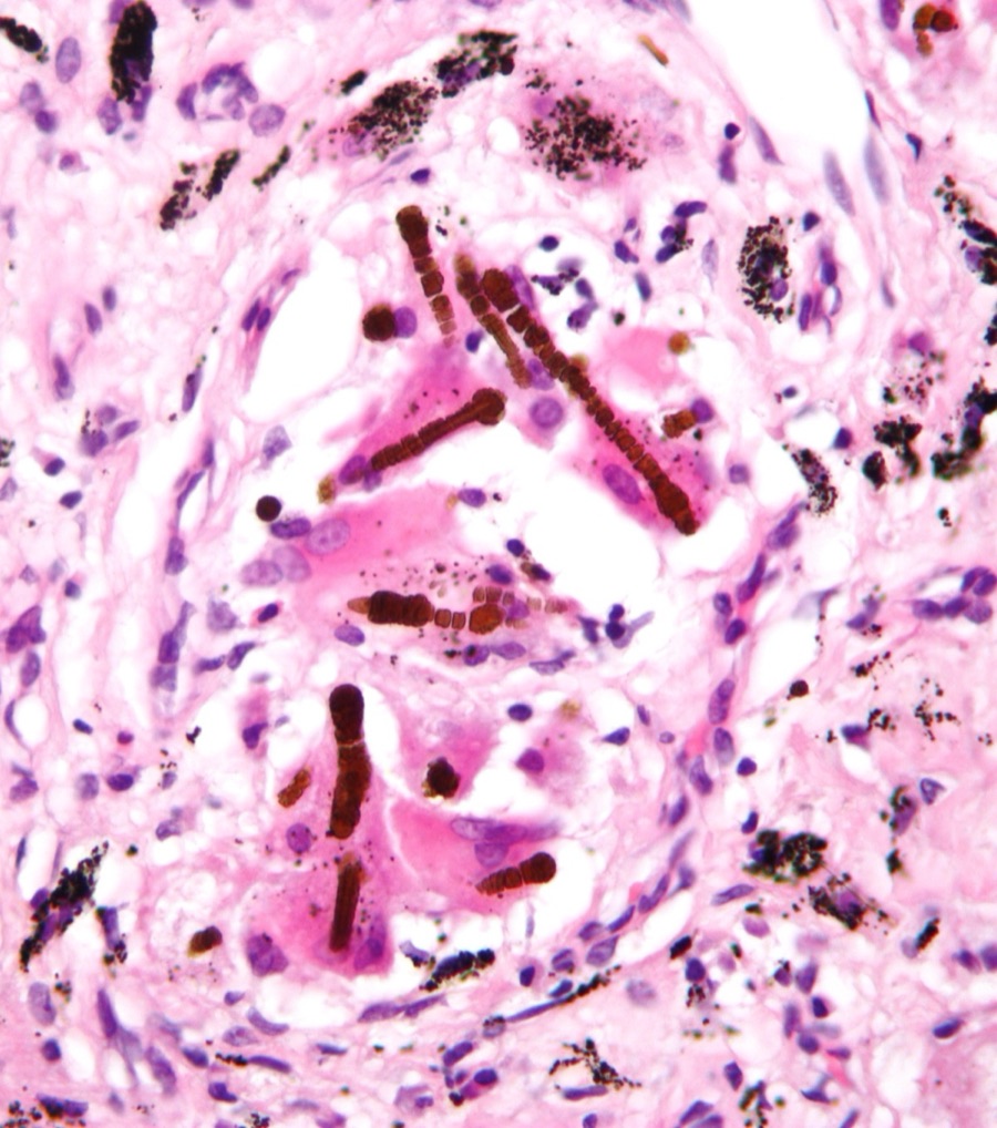
Under the microscope
Tap to zoom
The schematic above explains the mechanism. This is what one looks like. Golden-brown, dumbbell-tipped, sitting in lung tissue. Once a pathologist sees this color and shape together, asbestos exposure is on the chart.
Why this matters: the body cannot digest asbestos. A macrophage tries to eat the fiber, fails, dies, and the next macrophage in line wraps the fiber in hemosiderin, an iron-storage protein. After years of this, the naked needle becomes a rusty dumbbell. That dumbbell shape, golden-brown on H&E stain, is the ferruginous body. It only shows up with asbestos exposure. In the psammoma club, only one tumor carries it: mesothelioma.
The Shipyard Story
Why does the clinical medicine stem always say "retired shipyard worker"? Because asbestos has a 30-50 year fuse. Follow the timeline.
1970s · Age 25
Day one in the shipyard. He cuts insulation, sprays asbestos lining, breathes it in. The fibers are too long and too sharp for the lungs to clear. They lodge in the pleura, the thin slippery sheet wrapping the lung.
Weeks later
Frustrated macrophages. Lung macrophages, the cleanup crew, try to engulf the fibers. They cannot. The fibers are like swallowing a glass needle. The macrophages die and release reactive oxygen species, which damage the DNA of nearby mesothelial cells. No symptoms.
Years later
The fibers go rusty. Surviving macrophages keep coating the fibers with iron-protein (hemosiderin). The naked needles slowly turn into golden-brown dumbbells, the ferruginous bodies. Still no symptoms.
30-50 year fuse
DNA damage stacks. Decades of low-level reactive oxygen species mutate the mesothelial cells. Tumor suppressors fail. He retires. He has no clue any of this is happening. The mesothelial cells start growing where they should not.
2020s · Age 70
Breathlessness and pleural effusion. He shows up short of breath. CT shows diffuse pleural thickening that wraps the lung like a rind. Biopsy: malignant mesothelioma. The slide shows both psammoma bodies AND ferruginous bodies. The shipyard from 45 years ago wrote his diagnosis today.
Section 3 of 7
The Two M's, Untangled
Same letter, completely different tumor. This is the trap the clinical medicine loves.
Both end in -oma. Both are in the PSaMMoMa club. After that, they share almost nothing. Compare them side by side, then take the snap quiz.
Meningioma
Brain wrapping tumor
MRI · Dural tail · tap to expand
Lives inSkull, on the surface of the brain (extra-axial). Most often along the falx (parasagittal).
Comes fromArachnoid cap cells (the middle meningeal layer).
HistologyConcentric whorls of spindle cells, calcium rings (psammoma) tucked inside the whorls.
Imaging clueDural tail on contrast MRI. Round, well-circumscribed.
Risk factorMost cases sporadic. NF2 mutation in some.
BehaviorUsually benign (WHO grade I). Compresses brain, does not invade. Best prognosis of all intracranial tumors.
Mesothelioma
Lung-lining tumor
Ferruginous body · tap to expand
Lives inPleural cavity, the space wrapping the lung. Sometimes peritoneal.
Comes fromMesothelial cells of the pleura, damaged by asbestos.
HistologyPsammoma bodies PLUS golden-brown ferruginous (dumbbell) bodies.
Imaging clueDiffuse pleural thickening, often unilateral effusion. Lung wrapped in a rind.
Risk factorAsbestos exposure, 20-50 year latency. Shipyard, insulation, brake-pad work.
BehaviorAggressive malignancy. Poor prognosis, often months from diagnosis.
Question 1: A stem says "55-year-old woman, parasagittal mass, dural tail on MRI, psammoma bodies on biopsy." Which M is this?
Question 2: A stem says "65-year-old retired plumber, pleural thickening, dyspnea, golden-brown dumbbell bodies on biopsy." Which M is this?
Question 3: Which M sits in the same anatomic ZIP code as the lung, not the brain?
⚠
Quick rescue if you blank
Mening sounds like meninges = brain wrap. Meso like mesoTHELial = the lining cells of the pleura. If the stem says any occupation that involves old buildings, ships, or pipes, you are looking at the lung-lining version. If the stem says skull or brain or dural tail, you are looking at the brain-wrap version.
Section 4 of 7
Pathology Rounds
Four biopsies came back. Use the clues to eliminate tumors one by one. Last one standing is your diagnosis.
Four mystery tumors. Each clue rules one out. Click the card you're eliminating. Wrong guess? You'll get corrected. Right guess? It fades. Final survivor gets the crown.
Tumor A
Thyroid biopsy
Tumor B
Ovarian mass
Tumor C
Intracranial tumor
Tumor D
Pleural mass
Clue 1 of 4Loading first clue...
Section 5 of 7
What's On This Slide?
Work through the clues before you commit. Answer first, then the path reveals.
1
A slide shows psammoma bodies plus whorling concentric cell clusters in an intracranial location. The patient is a 55-year-old woman. What structure do these whorls arise from?
Arachnoid cap cells
Oligodendrocytes
Astrocytes
Exactly. Meningiomas come from arachnoid cap cells. That's why they form concentric whorls: the cells naturally spiral in this pattern as the tumor grows. The calcified version of those whorls are the psammoma bodies.
Oligodendrocytes give rise to oligodendrogliomas, which have "fried egg" cells, not whorls. The whorling pattern is unique to meningiomas and their arachnoid cap cell origin.
Astrocytes are the source of glioblastoma (GBM) and other gliomas. GBM has pseudopalisading necrosis, not whorls. Whorls = arachnoid cap cells = meningioma.
Arachnoid cap cells spiral into whorls as meningiomas grow. The whorls calcify into psammoma bodies over time. This duo is the histologic signature of meningioma. Parasagittal location along the falx cerebri is the most common site in clinical practice.
Meningioma histology showing the concentric whorls that calcify into psammoma bodies. Tap to expand.
2
Next slide. A 62-year-old shipyard worker presents with progressive dyspnea and pleural effusion. Biopsy shows golden-brown, dumbbell-shaped fibers in addition to psammoma bodies. Which structure are those dumbbell fibers?
Ferruginous bodies (iron-coated asbestos)
Amyloid deposits
Free hemosiderin granules
Correct. Ferruginous bodies are asbestos fibers that macrophages tried and failed to digest. They coat them in iron-containing protein (hemosiderin), creating those golden-brown, dumbbell shapes under H&E. Exclusively seen with asbestos exposure.
Amyloid looks like homogeneous pink deposits under H&E and has apple-green birefringence under Congo red staining. It doesn't look dumbbell-shaped and isn't associated with asbestos exposure.
Free hemosiderin does appear golden-brown but it's granular, not dumbbell-shaped. Ferruginous bodies are specifically the iron-coated asbestos fibers. A distinct structure, not free granules.
Mesothelioma is the only PSaMMoMa tumor with BOTH psammoma bodies AND ferruginous bodies. The asbestos occupational history (shipyard, construction, insulation) is always in the stem. Ferruginous bodies = asbestos exposure = mesothelioma. No other tumor in this group has them.
Ferruginous bodies are golden-brown, dumbbell-shaped iron-coated asbestos fibers seen in mesothelioma. Tap to expand.
3
Final slide. A thyroid FNA shows papillary clusters. The nuclei appear pale, empty-centered, and haunting, as if the nuclear contents are pushed to the rim. What is this finding called?
Orphan Annie eye nuclei
Ground-glass cytoplasm
Koilocytic change
Nailed it. Orphan Annie eye nuclei: chromatin pushed peripherally, leaving the center optically clear. Looks like the wide-eyed, hollow pupils of the comic strip character. Pathognomonic for papillary thyroid carcinoma.
Ground-glass cytoplasm refers to the cytoplasm, not the nucleus. In hepatitis B, hepatocytes can have ground-glass cytoplasm from HBsAg accumulation. Orphan Annie eye is a nuclear change, not cytoplasmic.
Koilocytes are HPV-infected cells with nuclear enlargement, irregular borders, and a cytoplasmic halo. That's a cervical dysplasia finding, not thyroid. The thyroid finding with cleared nuclei is Orphan Annie.
Orphan Annie eye nuclei + psammoma bodies in a thyroid FNA = papillary thyroid carcinoma. These two findings together are considered diagnostic. No other thyroid tumor has this nuclear pattern. Prognosis is excellent. Most patients are cured with surgical resection.
Papillary thyroid carcinoma with optically clear Orphan Annie eye nuclei. Tap to expand.
Section 6 of 7
Lock It In
Tap each card to unblur the hook. Test yourself before peeking.
🧴
The PSaMMoMa Mnemonic
Papillary thyroid · Serous ovarian · Meningioma · Mesothelioma. The word psammoma literally comes from Greek for "sand" (psammos). These four tumors grow sand-like calcified rings in their stroma. Every time you see "psammoma" think: which of these four is the question pointing to?
tap to reveal
👀
Orphan Annie Nuclei
Only one tumor gets Orphan Annie: papillary thyroid carcinoma. Think of Annie's wide, pale, expressionless eyes: the nucleus looks hollow because the chromatin is pushed to the rim. No other tumor in this group has cleared nuclei. If you see psammoma bodies plus hollow nuclei in a thyroid biopsy, you are done.
tap to reveal
🔨
Ferruginous Bodies
Only one tumor has ferruginous bodies: mesothelioma. "Ferruginous" = iron-coated. Macrophages tried to eat asbestos fibers, got stuck, and coated them in iron protein. Golden-brown, dumbbell-shaped. If the question mentions a shipyard worker or construction worker and shows dumbbell structures on the slide, the answer is mesothelioma.
tap to reveal
🧠
Best Brain Tumor Prognosis
Meningioma = best prognosis of all brain tumors. It sits outside the brain (extra-axial), compresses but doesn't invade, and can usually be fully removed. Compare: GBM is the most common primary malignant brain tumor in adults. Worst prognosis. Meningioma is most common overall (counting benign) and best prognosis. The board tests this distinction constantly.
tap to reveal
📈
Bilateral Ovarian Mass
Bilateral ovarian mass + psammoma bodies = serous adenocarcinoma. The word "serous" is the tell. Serous = the tumor makes fluid resembling serous fluid. Most common ovarian cancer. Associated with BRCA1/2. Often bilateral, often diagnosed at advanced stage because symptoms come late.
tap to reveal
🔫
Mesothelioma vs. Lung Cancer (Asbestos)
Asbestos causes BOTH lung cancer (bronchogenic carcinoma) AND mesothelioma, but mesothelioma is the famous one. If the question says "pleural thickening + psammoma bodies + ferruginous bodies + shipyard worker" = mesothelioma. If it says "lung mass + smoking + asbestos" = bronchogenic CA. Psammoma and ferruginous bodies together = mesothelioma every time.
tap to reveal
Section 7 of 7
Prove It
Five stems drawn at random. Eight in the pool. Answers shuffle every load.
From the Attending
Disambiguate by clinical setting around the calcifications: Thyroid nodule + Orphan Annie eye nuclei + nuclear grooves · papillary thyroid. Postmenopausal woman + pelvic mass + elevated CA-125 + BRCA family · serous ovarian. Adult with seizures or focal neurologic deficit + dural-based mass + spindle cell whorls · meningioma. Shipyard / plumber / pipefitter + pleural rind + asbestos exposure 30 yrs ago · mesothelioma. Calcifications don't pick the tumor · the clinical setting does. Same body, four homes.
Medically reviewed by Kaitlyn Cocuzzo, MD and Fatima Ali, DO · Last updated July 5, 2026 at 8:04 PM ET
Bone Wizardry is an independent educational resource for visual learning in the medical sciences. It is not affiliated with, endorsed by, or sponsored by any licensing or examination board, contains no real or recalled examination questions, and does not guarantee any educational or examination outcome.