When organs scream and the spine listens. The reflex arc that turns visceral disease into somatic dysfunction -- and why DO physicians think differently.
A 52-year-old man presents with left shoulder pain and has no musculoskeletal history. OMM exam reveals T1-T5 paravertebral tissue texture changes on the left. His ECG shows ST elevations. The spinal finding is best explained by:
Cardiac sympathetic afferents travel with T1-T5 sympathetic fibers. When the heart is ischemic, these afferents converge on the same spinal segments as somatic fibers from the chest wall and left arm (Rami communicantes). The brain cannot distinguish -- it interprets the signal as somatic pain AND produces reflex facilitation of the T1-T5 spinal segments, creating palpable tissue texture changes. This is the viscerosomatic reflex in action.
A patient with epigastric pain has new T5-T9 paraspinal hypertonicity. Which signal path explains that finding?
Correct. Organ pain rides back to the matching spinal level. That facilitated segment then produces tissue texture change, tenderness, and hypertonicity at the segment you can palpate.
VISCEROSOMATIC (organ pain → spine tightens)
Visceral organ
injury / disease
→
Afferent nerve
travels with sympathetics
→
Dorsal horn
spinal segment
→
Somatic muscle
paraspinal hypertonicity
SOMATOVISCERAL (spine problem → organ affected)
Somatic dysfunction
restricted segment
→
Facilitated segment
spinal cord
→
Efferent nerve
travels with sympathetics
→
Visceral organ
altered function
Key Concept
Visceral and somatic afferents from the same spinal segment converge in the dorsal horn. The brain reads the signal as somatic because the body maps skin and muscle better than deep organs.
+
Viscerosomatic Reflex
Organ → Spine
Visceral disease activates afferents that share a spinal segment with somatic structures. The dorsal horn becomes facilitated, lowering the firing threshold. Result: predictable paraspinal hypertonicity, skin texture change, and tenderness.
+
Somatovisceral Reflex
Spine → Organ
Somatic dysfunction can drive efferent output back toward viscera at that level. A facilitated segment can increase sympathetic tone to the matching organ, so treating the segment may quiet the loop.
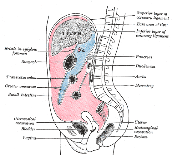
High-Yield Spinal Levels by Organ
Organ
Spinal Levels
Innervation Type
Clinical Clue
Heart
T1-T5 (L)
Sympathetic
Left arm + chest pain = T1-T5 somatic dysfunction
Lungs
T2-T7
Sympathetic
Pneumonia can produce upper thoracic somatic dysfunction
Esophagus
T2-T8
Sympathetic
GERD: mid-thoracic restriction common finding
Stomach
T5-T9
Sympathetic
PUD: T6-T8 paraspinal tenderness
Liver / Gallbladder
T7-T9 (R)
Sympathetic
Cholecystitis: right T7-T9 tissue texture changes; C3-C5 for diaphragmatic referral
Appendicitis: periumbilical pain (T10) then McBurney's
Kidney / Ureter
T10-L1
Sympathetic
Renal colic: flank to groin; CVA tenderness maps to T11-L1
Bladder / Uterus
T10-L2 + S2-S4
Sympathetic + Parasympathetic
Dual innervation: lower thoracic AND sacral dysfunction both possible
Ovary / Testis
T10-T11
Sympathetic
Ovarian torsion: periumbilical referral (T10)
Diaphragm
C3-C5
Phrenic nerve
Subdiaphragmatic irritation (blood, pus, gallbladder) = shoulder tip pain
Memory Trick
Think in terms of embryological gut: Foregut (T5-T9), Midgut (T10-T11), Hindgut (T11-L1). The spinal levels follow embryological origin, not anatomical position.
Viscerosomatic vs Somatovisceral
Direction
Clinical Use
Trace It
Viscerosomatic
Organ → Spine
Organ pathology creates somatic dysfunction at the corresponding spinal segments. The spine becomes a "read-out" of visceral disease.
Somatovisceral
Spine → Organ
Spinal somatic dysfunction sends abnormal efferent signals to the viscera, potentially altering organ function. Treatment of the spine can improve organ function.
Viscerosomatic Use
Diagnostic:
Finding tissue texture changes at predictable spinal levels guides workup. T1-T5 changes in chest pain patient = rule out MI first. T10-T11 changes + periumbilical pain = consider appendicitis.
Somatovisceral Use
Therapeutic:
Treating somatic dysfunction at T5-T9 may improve gastric function. Sacral OMM (S2-S4) can modulate bladder/bowel motility. The rationale for OMM in systemic disease is largely somatovisceral.
Viscerosomatic Mechanism
Visceral afferents (traveling with sympathetics) converge on dorsal horn neurons that also receive somatic input. Repetitive visceral firing lowers the threshold (facilitates) the spinal segment. Result: tissue texture changes, restricted ROM, tenderness.
Somatovisceral Mechanism
Somatic dysfunction facilitates the same spinal segment. Facilitated neurons fire more easily and more frequently. The efferent limb (sympathetic fibers to viscera) is activated, increasing organ tone, vasospasm, or altering secretion.
The Facilitated Segment
Irvin Korr's Concept
A facilitated segment is a spinal cord level with a chronically lowered threshold for neural activity. Inputs that would normally be sub-threshold now cause full firing. Both viscerosomatic (organ signals in) and somatovisceral (organ signals out) are amplified at a facilitated segment.
+
How Facilitation Develops
Chronic input
Repeated afferent bombardment (from either soma or viscera) causes increased synaptic efficiency at that cord level. Fewer incoming signals are needed to trigger output. Analogy: a hair-trigger after prolonged stress.
+
Exam Findings at Facilitated Segment
What you feel on palpation
Tissue texture changes (boggy, ropey, stringy), paraspinal hypertonicity, skin temperature changes (sympathetic vasomotor changes), tenderness to palpation, restricted segmental motion. These are the TART criteria.
+
TART Criteria
Somatic dysfunction findings
Tissue texture changes, Asymmetry, Range of motion restriction, Tenderness. Any combination qualifies as somatic dysfunction. At a facilitated segment, all four are often present.
+
Clinical Implication
Why it matters
A chronic facilitated segment at T4-T5 in a patient with recurrent "chest tightness" should prompt cardiac evaluation -- even without classic ECG changes. Conversely, treating the facilitated segment may reduce visceral symptoms by normalizing efferent sympathetic tone.
Thigh Chapman: The Flip Rule
Recca emphasis
The colon Chapman points on the thigh do not lie where you instinctively place a colon segment in space. They wrap around the thigh like a tube. Cecum and ascending colon sit on the lateral thigh, transverse colon on the anterior thigh, descending colon on the medial thigh, and sigmoid on the posterior thigh. The flip is the test trap: students place ascending colon medially because they picture the colon laid flat on a table. On the thigh, the layout wraps.
Right thigh, axial cross-section (looking down) · tap a segment
Tap a colored segment to see the matching colon part, the SNS spinal level, and the abdominal cross-reference Chapman point.
The mechanism
Tenderness on the thigh maps colon dysfunction through visceral afferents that converge at lumbar segments and travel via the lumbar splanchnic nerves and the inferior mesenteric ganglion to the hindgut. The thigh point is fascial and neurologic, not anatomic: the colon segment is not under your hand. The afferent shares the spinal level, and the spinal level facilitates the IT band region.
Why it matters clinically
When the abdomen is unavailable (postoperative dressing, open wound, severe guarding, modesty during exam, or a patient who cannot lie supine), the thigh Chapman points give a quiet backup palpation site for colon dysfunction. The test is the same: firm, discrete, pea-sized tenderness. Apply rotary friction at the posterior point if treating, or use the thigh tenderness as a viscerosomatic flag to push the workup toward the colon.
Thigh surface
Colon segment
SNS spinal level
Abdominal cross-reference
Lateral (IT band)
Cecum / Ascending
T10-T11
Tip of right 12th rib (cecum/appendix anterior)
Anterior (mid)
Transverse
T10-T11
Periumbilical region (midgut afferent zone)
Medial (IT region, L thigh)
Descending
T12-L2
Lateral to rectus, mid-abdomen on the left
Posterior
Sigmoid
L1-L2
Lower left quadrant near anterior superior iliac spine
The "second hand on the same clock" hook
Descending colon tenderness on the medial thigh corresponds to the same T12-L2 sympathetic level as descending colon tenderness in the lumbar paraspinals. The thigh point is not a different reflex, it is the same viscerosomatic clock face read at a second location. If the abdominal Chapman is positive AND the thigh Chapman is positive, that is two confirmations of the same colon segment, not two separate findings.
A 58-year-old woman with chronic constipation and intermittent left lower quadrant pain is examined osteopathically. Tenderness is palpated on the medial mid-thigh. Which colon segment is most likely dysfunctional?
Option A (cecum) fails because the cecum sits on the lateral thigh in the IT band region, not the medial thigh. Cecum is the start of the wraparound on the lateral surface. If you placed the cecum medially, you flipped the rule incorrectly: the rule already does the flipping for you.
Option B (ascending colon) fails for the same reason as cecum. Ascending colon shares the lateral thigh location with the cecum because both are right-sided midgut structures innervated at T10-T11. Medial thigh is left-sided territory, not right-sided.
Option C (transverse colon) fails because the transverse colon sits on the anterior thigh, not the medial thigh. Mid-anterior is the transverse colon's surface. The patient's clinical history (LLQ pain plus chronic constipation) also fits descending or sigmoid much better than transverse: transverse colon dysfunction would refer more centrally, around the umbilicus.
Option D (descending colon) is correct. The descending colon's thigh Chapman point is on the medial thigh. The clinical picture fits, and the location fits. Think of the four thigh surfaces as a compass: lateral = right-sided colon (cecum and ascending), anterior = transverse (the colon's "top" laid across the abdomen), medial = left-sided descending, posterior = sigmoid where the colon turns toward the rectum. Medial thigh tenderness in a patient with LLQ pain is descending colon until proven otherwise.
Break it down: Lateral thigh = cecum/ascending (right). Anterior thigh = transverse (top). Medial thigh = descending (left). Posterior thigh = sigmoid (back/down). Match the surface to the segment, then check the spinal level for confirmation.
During an osteopathic exam, a tender, firm, pea-sized nodule is palpated on the iliotibial band region of the right lateral thigh. Which colon segment and which sympathetic spinal level does this finding correspond to?
Option A (descending colon, T12-L2) fails on both counts. Descending colon's thigh point is on the medial thigh, not lateral, and the spinal level is T12-L2, which is hindgut territory. The lateral thigh point belongs to the right-sided midgut (cecum and ascending), not to the left-sided hindgut.
Option C (sigmoid, L1-L2) fails because the sigmoid colon's thigh point is on the posterior thigh, not lateral. Sigmoid is hindgut and sits at L1-L2 sympathetically, which is the right level for sigmoid but the wrong location for the IT band finding.
Option D (transverse, T5-T9) fails on both counts. Transverse colon sits on the anterior thigh, not lateral, and T5-T9 is foregut sympathetic territory (stomach, liver, gallbladder, pancreas), not midgut. Anyone choosing T5-T9 for a colon segment has confused embryologic divisions.
Option B (cecum/ascending, T10-T11) is correct. Lateral thigh (IT band) = right-sided midgut colon (cecum and ascending). Sympathetic supply to the midgut is T10-T11 via the lesser and least splanchnic nerves to the superior mesenteric ganglion. The flip rule places the right-sided colon segments on the lateral thigh because the colon "U" wraps around the thigh tube starting laterally with the cecum.
A 47-year-old man with diverticulitis has guarding and a postoperative abdominal dressing that prevents anterior abdominal palpation. The osteopathic physician wants to assess for colon viscerosomatic involvement using the lower-extremity Chapman points. Tenderness is found on the posterior thigh. Which colon segment does this most likely indicate?
Option A (transverse) fails because transverse colon's thigh Chapman point is on the anterior thigh. Anterior is the colon's "top" surface in the wraparound. Posterior thigh is reserved for the segment that finishes the colon's path: sigmoid.
Option B (cecum) fails because cecum sits on the lateral thigh in the IT band region. Cecum is the right-sided start of the midgut colon. It is also clinically less likely here: diverticulitis classically involves the sigmoid in adults, not the cecum.
Option D (ascending colon) fails for the same surface reason. Ascending colon also lives on the lateral thigh. And ascending colon is not the typical site of diverticulitis in adults (sigmoid is the heavy hitter).
Option C (sigmoid) is correct. Posterior thigh = sigmoid. The flip rule wraps the colon onto the thigh like a tube: starting lateral (cecum/ascending), going across the top (anterior, transverse), down the other side (medial, descending), and finishing at the back (posterior, sigmoid). The clinical context closes the loop: adult diverticulitis is overwhelmingly sigmoid, and the LE Chapman point matches.
Break it down: Posterior thigh = sigmoid. Memory check: the colon's "exit" segment (sigmoid heading toward the rectum) maps to the back of the thigh, mirroring the colon's path from front of the abdomen around to the back where the rectum exits. The wraparound visual makes posterior = sigmoid intuitive once you commit to it.
A student about to begin OMM rotations is taught the LE Chapman flip rule for the colon. She asks why the cecum (a right-sided abdominal organ) maps to the lateral thigh rather than the medial thigh. Which explanation is most accurate?
Option A fails because dermatomes are sensory body-surface maps from somatic nerve roots, and the cecum (a visceral organ) does not have a dermatome. Visceral structures map through visceral afferents that converge on dorsal horn segments shared with somatic territories, but the resulting Chapman point is fascial and lymphatic, not a "dermatome on the opposite side." This explanation invents a contralateral mechanism that does not exist in osteopathic theory.
Option C fails because deep inguinal lymphatic drainage of the cecum does not produce a contralateral reflex. Chapman points are not located based on lymphatic drainage routes to a specific node group: they are predictable fascial points tied to viscerosomatic reflex arcs at specific spinal levels. The "drainage causes a reflex on the opposite side" reasoning is a fabrication that sounds anatomic but has no basis in the literature.
Option D fails because the iliotibial band exists only on the lateral thigh, true, but that does not explain why ALL the colon segments map to specific thigh surfaces (anterior for transverse, medial for descending, posterior for sigmoid). If "lateral by default" were the rule, every colon Chapman would be on the IT band. The flip rule's structure is the wraparound, not the IT band's location.
Option B is correct. The colon Chapman points wrap the thigh like a tube. Lateral surface of the right thigh = right-sided midgut colon (cecum and ascending). Anterior thigh = transverse colon's "top" surface. Medial thigh = left-sided descending colon. Posterior thigh = sigmoid where the colon turns toward the rectum. The point's surface matches the colon segment's anatomic position when you imagine the colon laid on the thigh as a U-shaped tube wrapping around. The flip is the wrap.
Break it down: Chapman points are fascial reflex points, not dermatomes or lymphatic drainage targets. The colon LE Chapman map is a wraparound (lateral, anterior, medial, posterior = cecum/ascending, transverse, descending, sigmoid). Lateral matches right-sided colon because the thigh tube is wrapped from the right.
Identify the Organ
A patient has right-sided T7-T9 somatic dysfunction with tissue texture changes. Clues appear one at a time. Eliminate organs that no longer fit.
Clue 1: The dysfunction is right-sided and reproducible with deep inspiration.
Clue 2: Blood work shows elevated alkaline phosphatase and direct bilirubin.
Clue 3: Ultrasound shows wall thickening and a positive Murphy's sign.
Clue 4: The patient also reports shoulder tip pain on the right side.
Stomach
Gallbladder
Kidney
Pancreas
Press "Next Clue" to begin.
Viscerosomatic Reflexes Quiz
A 45-year-old woman with known cholelithiasis develops T7-T9 right paravertebral tissue texture changes and restricted motion at those segments. This finding is best characterized as:
Option A fails because somatovisceral runs spine-to-organ, not organ-to-spine. A spine problem can alter gallbladder function via efferent sympathetics, but it cannot create gallstones. The pathology started in the gallbladder, and the spinal findings are the downstream reaction.
Option C fails because C3-C5 (phrenic nerve) is the referral route for shoulder tip pain from diaphragmatic irritation, not paraspinal tissue texture changes. C3-C5 produces shoulder pain; T7-T9 produces somatic dysfunction in the mid-back. This question asks about the spinal palpatory finding, not shoulder pain.
Option D fails because a facilitated spinal segment can alter organ tone and blood flow, but it cannot manufacture gallstones. Gallstones form from bile composition changes like cholesterol supersaturation, not from nerve signals. Think of it like a thermostat: the spine controls the temperature in the room, but it cannot build furniture.
Option B is the mechanism: gallbladder afferents travel with T7-T9 sympathetic fibers. Gallbladder inflammation fires those afferents, which converge on the T7-T9 dorsal horn, facilitating that segment and producing the palpable tissue texture changes found on OMM exam. Organ fires first, spine reacts. That is the viscerosomatic direction.
Break it down: Viscerosomatic = organ fires first, spine reacts (diagnostic finding). Somatovisceral = spine fires first, organ reacts (therapeutic target). C3-C5 = shoulder tip only (phrenic nerve/diaphragm). Facilitated segments alter function; they do not cause structural pathology like stones.
The embryological basis for why appendicitis classically presents as periumbilical pain before localizing to the right lower quadrant is best explained by:
Option A fails because the phrenic nerve (C3-C5) is the diaphragm's nerve, not the appendix's. Phrenic nerve referral produces shoulder tip pain, not periumbilical pain. The appendix has no phrenic nerve connection, embryologically or anatomically.
Option B fails because L1-L2 somatic innervation covers the groin and inner thigh (ilioinguinal and genitofemoral nerve territory), not the periumbilical region. Somatic localization also only appears later in appendicitis, when the parietal peritoneum (the body-wall lining of the abdomen, which carries sharp somatic nerve supply) becomes inflamed. L1-L2 explains the eventual RLQ localization, not the initial periumbilical pattern.
Option D fails on two counts: T4-T5 is cardiac territory, not midgut. And somatovisceral runs spine-to-organ, meaning a T4-T5 lesion would alter upper thoracic organ function, not cause the appendix to refer pain to the umbilicus. Somatovisceral does not produce a pain referral pattern in the patient; it alters organ function.
Option C is the mechanism: the appendix is midgut-derived. Midgut sympathetic afferents converge at T10, which maps to the periumbilical dermatome. Early appendicitis fires visceral afferents at T10, producing the poorly localized periumbilical pain. It works like a fire alarm: the panel rings at the main board (T10, periumbilical) before the sprinklers activate at the actual fire location (McBurney's point, once the parietal peritoneum inflames).
Break it down: Match embryological origin to spinal level. Foregut = T5-T9. Midgut = T10-T11. Hindgut = T11-L1. Phrenic = C3-C5 (diaphragm only, always shoulder tip). Somatic localization always comes second, after the parietal peritoneum gets involved.
A patient with chronic T5-T9 somatic dysfunction is noted to have recurrent peptic ulcer disease. An osteopathic physician treating the thoracic dysfunction is utilizing which principle?
Option A fails because viscerosomatic runs organ-to-spine. If the ulcer were creating the spinal findings, that would be viscerosomatic. But the question asks what principle the physician is leveraging to treat the organ by working on the spine, which requires the somatovisceral direction (spine-to-organ).
Option B fails because Chapman reflexes are specific anterior and posterior fascial points thought to reflect lymphatic congestion in a given organ. They are a separate mechanism from facilitated spinal segments and sympathetic efferent outflow. Chapman's does not explain the link between T5-T9 somatic dysfunction and gastric acid secretion.
Option D fails because counterstrain is a treatment technique (indirect positional release of tender points), not a reflex mechanism. The question asks which principle explains WHY treating T5-T9 somatic dysfunction would affect peptic ulcer disease. Counterstrain names a method, not the neurological pathway connecting the spine to the stomach.
Option C is the mechanism: somatovisceral runs spine-to-organ. A facilitated T5-T9 segment (the stomach's sympathetic territory) drives increased sympathetic efferent firing to the stomach wall, increasing acid secretion and decreasing mucosal protective blood flow. Treating the somatic dysfunction with OMM reduces the facilitation, normalizing that efferent output. The physician is interrupting the spine-to-organ pathway.
Break it down: Somatovisceral = spine sends the problem downstream to the organ. If a physician treats the spine to fix the organ, that is somatovisceral logic. Viscerosomatic = organ sends the signal up to the spine (you read it as a diagnostic finding). Chapman and counterstrain are separate mechanisms and techniques, not this reflex arc.
During OMM examination of a 60-year-old man presenting with vague low back pain, you find bilateral T10-L1 tissue texture changes and tenderness. Urinalysis shows microscopic hematuria. The somatic findings at T10-L1 most directly reflect:
Option A fails because the ilioinguinal nerve (L1) is a somatic sensory nerve supplying the groin and inner thigh. It cannot produce bilateral paraspinal tissue texture changes, which require visceral afferent convergence at the dorsal horn. The ilioinguinal nerve also has no connection to the kidney or renal tract: it routes through the inguinal canal and stops there.
Option B fails because lumbar degenerative disc disease produces focal somatic findings tied to a single disc level, typically unilateral, and has no mechanism to cause hematuria. The combination of bilateral paraspinal texture changes plus a visceral sign (blood in urine) cannot be explained by a structural spinal problem alone. When a spinal finding and a visceral symptom map to the same territory, viscerosomatic should come first.
Option D fails because somatovisceral reflexes can alter organ tone, blood flow, and secretion, but they cannot cause hematuria. Hematuria requires physical disruption of the urinary tract lining, which only happens from pathology inside the kidney or ureter itself. The direction of causality here is reversed: the kidney fired first, and the spinal findings are the reaction.
Option C is the mechanism: renal and ureteral afferents travel with T10-L1 sympathetic fibers. Kidney or ureter pathology fires those afferents, facilitating the T10-L1 dorsal horn, and producing bilateral paraspinal somatic findings on OMM exam. The spinal findings are a read-out of the visceral problem. Hematuria is the flag that pushes the workup toward the kidney, not the spine.
Break it down: Bilateral paraspinal findings plus a visceral symptom = viscerosomatic until proven otherwise. Match the symptom to the sympathetic territory: kidney and ureter = T10-L1. Hematuria cannot be caused by a somatovisceral reflex. Somatic-only diagnoses (disc disease, nerve entrapment) do not produce visceral signs.
A 48-year-old woman with a history of recurrent cystitis and endometriosis is found on OMM examination to have bilateral T10-L2 somatic dysfunction and sacral restriction at S2-S4. This pattern of dual spinal-level involvement reflects the innervation of which organ?
Option A fails because the kidney has sympathetic innervation only at T10-L1. There is no sacral component to renal innervation. If only T10-L1 somatic dysfunction were present, the kidney would stay on the differential. But sacral restriction at S2-S4 means a second autonomic territory is involved, and the kidney sends no afferents through S2-S4.
Option B fails because the ovary has sympathetic innervation at T10-T11 only, matching its embryological origin as a gonadal structure derived from the same segment as the kidney. There is no sacral parasympathetic supply to the ovary. Ovarian pathology plus T10-T11 somatic dysfunction, yes. Dual-level findings including sacral restriction, no.
Option D fails because the appendix is midgut-derived with visceral referral at T10 only. It has no sacral innervation. Appendicitis never produces S2-S4 somatic findings.
Option C is the mechanism: the uterus and bladder receive both sympathetic input (T10-L2, via the hypogastric nerve) and parasympathetic input (S2-S4, via the pelvic splanchnic nerves). Viscerosomatic reflexes from either pathway show up on OMM exam, so pelvic organ pathology can produce findings at two separate spinal regions at the same time. Think of it like a building with two independent power supplies: a fault in either line shows up at both electrical panels simultaneously.
Break it down: Dual-level somatic dysfunction (lower thoracic/lumbar AND sacral) = pelvic organ with dual innervation (uterus, bladder). Single-territory only = kidney (T10-L1), ovary (T10-T11), appendix (T10). Sacral findings point to parasympathetic S2-S4 territory: bladder, uterus, rectum.
A facilitated segment at T4-T5 is identified on OMM examination of a 55-year-old male with chronic angina. Which of the following best describes why this segment remains persistently hyperactive even after the cardiac ischemia is managed medically?
Option A fails because the scalene muscles originate at C4-C6 vertebrae and insert on the first and second ribs, not at T4-T5. Scalene tension would produce cervical or upper rib somatic dysfunction, not persistent T4-T5 facilitation. Muscle anatomy here is simply wrong for the location.
Option C fails for two reasons: first, medications like beta-blockers and vasodilators do reduce sympathetic efferent tone significantly. Second, the question asks why the segment PERSISTS after ischemia is already managed, not whether efferents can be pharmacologically reduced. The mechanism of persistence lives in the spinal cord itself, not in the heart or the medications.
Option D fails because viscerosomatic reflexes do not cause structural spinal cord damage. Facilitation is a functional neural change (lowered firing threshold at the dorsal horn synapse), not anatomical injury. The spinal cord neurons are intact but primed to fire more easily. This is reversible with appropriate OMM treatment, unlike true hyperreflexia from actual cord damage.
Option B is the mechanism: Irvin Korr's research showed that repeated afferent input from an ailing organ drives down the firing threshold at the dorsal horn. Over time, this lowered threshold becomes the new baseline for that spinal segment. Like a violin string tuned so tight it vibrates from ambient room noise, the T4-T5 segment stays hyperexcitable because the neurophysiological change outlasts the original visceral stimulus. The segment is now self-sustaining without the cardiac input.
Break it down: Facilitation persists because the spinal cord threshold is permanently lowered, not because the visceral disease is still active. Scalene muscles do not maintain spinal segment facilitation. Medication controls the organ, not the dorsal horn threshold. Only direct treatment of the facilitated segment (OMM) addresses the threshold itself.
A 50-year-old man presents with new-onset chronic prostatitis. OMM examination reveals bilateral L1-L3 paraspinal tissue texture changes and tenderness. The treating physician uses OMM targeted at L1-L3 to reduce sympathetic efferent tone. This represents which reflex direction?
Option A fails on a subtle but important distinction: while viscerosomatic IS how the L1-L3 findings got there (prostate afferents firing at L1-L3 created the somatic dysfunction), the question asks about the treatment direction. When the physician treats L1-L3 to change what the prostate receives, that is the somatovisceral direction: spine-to-organ. Identifying the finding is viscerosomatic thinking; treating the spine to change organ function is somatovisceral thinking.
Option C fails because Chapman reflexes are specific anterior and posterior fascial points thought to reflect lymphatic congestion in a given organ. They are a separate diagnostic and treatment system from facilitated spinal segments and sympathetic efferent outflow. The prostate's Chapman points are not located at L1-L3 paraspinal tissue.
Option D fails because dermatomal referral describes a sensory pain distribution pattern from a somatic nerve root, not bilateral paraspinal tissue texture changes from visceral afferent convergence. The patient's diagnosis of chronic prostatitis is already established; this question is asking about which reflex mechanism the physician is using, not how to diagnose the condition.
Option B is the mechanism: the prostate receives sympathetic innervation from L1-L3. A facilitated L1-L3 segment bombards the prostate with increased sympathetic efferent tone, potentially worsening inflammation and pain. Treating the segment normalizes that efferent output, reducing the sympathetic burden on the prostate. The physician is using the spine as a lever to change what the organ receives.
Break it down: Treating the spine to improve organ function = somatovisceral. Organ pathology creating spinal findings = viscerosomatic (diagnostic direction). Chapman = lymphatic fascial points (separate system entirely). Dermatome = sensory distribution, not tissue texture change.
An OMM student asks how to distinguish a viscerosomatic reflex finding from a primary somatic dysfunction on physical examination. Which feature most strongly suggests the finding is due to an underlying visceral process rather than a primary musculoskeletal problem?
Option A fails because both primary somatic dysfunction and viscerosomatic reflex findings produce localized paraspinal pain and tenderness. A strained facet joint and cholecystitis can both produce the same T7-T9 back pain with the same quality of complaint. Subjective pain location alone cannot distinguish the source.
Option B fails because unilateral restriction following a dermatomal pattern is more consistent with primary somatic dysfunction (single nerve root involvement) than viscerosomatic reflex. Dermatomal patterns describe sensory distribution from a nerve root; viscerosomatic findings follow sympathetic territory maps, which do not align with dermatomes. Viscerosomatic findings can be unilateral, but dermatome distribution is not the mechanism.
Option D fails because TART criteria (Tissue texture change, Asymmetry, Range of motion restriction, Tenderness) are the definition of somatic dysfunction, present in both primary musculoskeletal and viscerosomatic causes. Single-level TART is actually more common in primary musculoskeletal problems, not a marker of visceral origin. TART alone cannot distinguish the source.
Option C is the mechanism: the key differentiator is behavior over time plus clinical context. Primary somatic dysfunction responds to appropriate OMM and holds. A viscerosomatic finding keeps coming back because the organ keeps firing afferents that re-facilitate the spinal segment. Like tightening a knot in a rope that is being pulled from the other end, you can loosen it temporarily, but the tension re-creates it. Constitutional symptoms, organ-specific history, and recurrence after competent OMM all point toward a visceral driver.
Break it down: Primary somatic dysfunction responds to OMM and stays better. Viscerosomatic recurs because the organ keeps reactivating the segment. TART is present in both; it does not distinguish the source. Look for the combination: recurrence + visceral symptoms + the right sympathetic territory.
0/8
Quiz complete
Board Practice
Board-Style Walkthrough
Five board-style vignettes. Answer, then read the full reasoning chain.
A patient has paraspinal tissue texture changes at T1-T5 left. Which visceral territory does this implicate first?
Cardiac/pulmonary sympathetics (T1-T5)
Renal sympathetics (T10-L1)
Pelvic parasympathetics (S2-S4)
Correct. T1-T5 left carries cardiac sympathetic afferents. Left-sided T1-T5 tissue texture changes are the viscerosomatic signature of myocardial ischemia. Rule out cardiac pathology before attributing these findings to musculoskeletal cause.
Renal afferents run with T10-L1 sympathetics, not T1-T5. T1-T5 is upper thoracic territory: heart and lungs. Flank findings and hematuria map to T10-L1.
S2-S4 is sacral parasympathetic territory for pelvic organs (bladder, uterus, rectum). Upper thoracic T1-T5 changes have no sacral mechanism. Think of the spine as a zip code: the address tells you which organ the signal is from.
The patient's EKG shows new ST elevations. The T1-T5 changes are a viscerosomatic reflex. What does this mean for treatment priority?
Treat the somatic dysfunction first, then reassess cardiac status
Cardiac workup and stabilization before any OMT
Viscerosomatic reflexes mean the organ fires first and the spine reacts. The T1-T5 findings are a diagnostic readout, not the primary problem. OMT on an actively ischemic heart is not the first move.
Correct. The viscerosomatic finding is a diagnostic flag pointing to the real problem. The spine is reading "cardiac distress." The cardiac emergency is treated first. OMT may support recovery after stabilization.
Walk the chain: A gallbladder inflames. What happens at the spinal cord?
1
Which nerve fibers carry pain signals from the gallbladder?
Tap to reveal
2
Where do those afferents converge in the spinal cord?
Tap to reveal
3
Why does the physician feel tissue texture changes at T7-T9 right?
Tap to reveal
4
What makes this a "facilitated" segment?
Tap to reveal
Viscerosomatic rule: organ fires the afferent, dorsal horn gets lowered threshold, paraspinals go ropey. The spine is just taking notes.
Memory Hook
How do I remember that T5-T9 is the stomach/gallbladder/liver territory?
Think Foregut Five-Nine: the embryological foregut becomes the stomach, duodenum, liver, and gallbladder. Foregut = T5-T9 sympathetics (greater splanchnic nerve). If a patient has epigastric pain and right-sided T7-T9 changes, think gallbladder. If left-sided T5-T9, think stomach or pancreas.
Memory Hook
How do I remember viscerosomatic vs somatovisceral direction?
Viscerosomatic = Organ Out. The organ sends the signal out to the spine (diagnostic). Somatovisceral = Spine In. The spine sends the signal in to the organ (therapeutic rationale). When an OMM physician treats the spine to improve organ function, that is somatovisceral thinking. When the OMM physician reads the spine to diagnose organ disease, that is viscerosomatic thinking.
Memory Hook
Why does a facilitated segment keep firing even after the organ heals?
Think of a hair-trigger gun. Repeated afferent bombardment lowers the dorsal horn firing threshold permanently. The segment now fires from inputs that would not trigger a normal segment. Irvin Korr described this as the spinal cord learning to be hypersensitive. OMM treatment is the only intervention targeting the threshold itself.