OMM
Anatomy

Lower Extremity · Nerve Injuries

Nerve Map

Six nerves. Six patterns of injury. Master the anatomy and you will never confuse foot drop with Trendelenburg again.

The Lower Extremity Nerve Map

The lumbosacral plexus (L2-S3) generates six nerves. Each has a unique anatomic pinch point and a unique injury signature. Tap a nerve to see its territory.

PELVIS KNEE ANKLE Femoral Sciatic Com. Peroneal Tibial Sup. Gluteal Fibular neck Med. malleolus
L2-L4
Femoral Nerve
Motor: Quadriceps (knee extension), iliopsoas (hip flexion)
Sensory: Anterior thigh + medial leg (saphenous)
Reflex: Patellar
Injury cause: Retroperitoneal hematoma, pelvic fracture, psoas abscess
Presentation: Quad weakness, absent patellar reflex, medial leg numbness
L2-L4
Obturator Nerve
Motor: Adductors (longus, brevis, gracilis, magnus)
Sensory: Medial thigh
Injury cause: Pelvic operations, obturator hernia
Presentation: Weak hip adduction, medial thigh numbness, normal reflexes
L4-S3
Sciatic Nerve
Motor: Hamstrings + all below knee (splits into peroneal + tibial)
Sensory: Posterior knee + all below knee except medial leg
Injury cause: Herniated disc, posterior hip dislocation, piriformis syndrome
Presentation: Foot drop + lost plantarflexion + posterior thigh weakness
L4-S2
Common Peroneal
Motor: Dorsiflexion (deep) + eversion (superficial)
Sensory: Dorsum of foot (except 1st webspace)
Injury cause: Fibular neck fracture, tight cast
Presentation: Foot dropPED - inverted + plantarflexed at rest; steppage gait
L4-S3
Tibial Nerve
Motor: Plantarflexion, toe flexion, foot invertors
Sensory: Sole of foot
Injury cause: Baker cyst (proximal), tarsal tunnel syndrome (distal)
Presentation: Can't stand on TIPtoes; sole numbness; Tinel at medial malleolus
L4-S1
Superior Gluteal
Motor: Gluteus medius + minimus + tensor fasciae latae
Sensory: None
Injury cause: IM injection in superomedial gluteal region, hip arthroplasty
Presentation: Trendelenburg gait - contralateral pelvis drops during stance
Three mnemonics you cannot afford to forget:
PED = Peroneal Everts and Dorsiflexes. If injured, foot dropPED.
TIP = Tibial Inverts and Plantarflexes. If injured, can't stand on TIPtoes.
Trendelenburg = ipsilateral superior gluteal nerve; contralateral hip drops.

Nerve Locator

Read the clinical scenario. Identify the nerve before choosing. The anatomy is the answer.
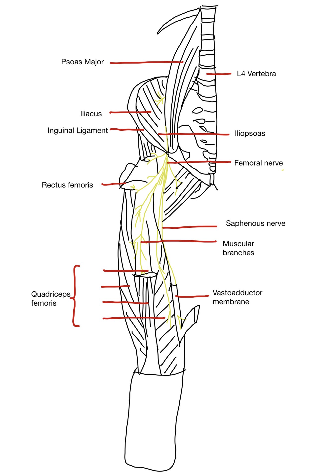
Patient cannot dorsiflex the foot. Sole sensation is normal.
Tap to reveal nerve
Common peroneal nerve. Dorsiflexion is the deep peroneal branch. Normal sole = tibial territory is intact. Foot is inverted and plantarflexed at rest.
Patient cannot adduct the hip. Medial thigh is numb. Patellar reflex is normal.
Tap to reveal nerve
Obturator nerve (L2-L4). Adductors = obturator. Normal patellar reflex excludes femoral. Medial thigh = obturator sensory territory. Pelvic surgery risk.
Pelvis drops on the left when the patient stands on the right leg.
Tap to reveal nerve
Right superior gluteal nerve. The lesion is on the WEIGHT-BEARING side. The right abductors cannot hold the pelvis level when the left foot lifts off.
Burning anterolateral thigh pain. No motor deficit. No reflex changes.
Tap to reveal nerve
Lateral femoral cutaneous nerve (L2-L3). Pure sensory. No motor fibers. Meralgia paresthetica from entrapment at the ASIS/inguinal ligament. Obesity and tight belts.
Nerve Locator · Question 1 of 3
A patient has foot drop - the foot is inverted and plantarflexed at rest. Plantarflexion strength is 4/5. The sole of the foot has normal sensation. Which of the following is the most likely injured nerve?
Common peroneal nerve. The key: plantarflexion is INTACT (tibial nerve is fine) and sole sensation is INTACT (also tibial). Only dorsiflexion and eversion are lost - that is precisely the common peroneal territory (PED). The sciatic nerve injury would have knocked out plantarflexion too.
Nerve Locator · Question 2 of 3
A patient has absent patellar reflex, 2/5 knee extension, and numbness over the anterior thigh and medial leg. Hip extension, plantar flexion, and Achilles reflex are all intact. Which nerve is most likely affected?
Femoral nerve (L2-L4). The triad: quadriceps weakness (knee extension) + absent patellar reflex (L4 arc via femoral) + sensory loss over anterior thigh and medial leg (saphenous branch). Intact Achilles rules out sciatic and S1. The obturator innervates adductors, not extensors. The lateral femoral cutaneous nerve is pure sensory with no reflex arc.
Nerve Locator · Question 3 of 3
A patient cannot flex the knee OR plantar flex the foot. Knee extension is 5/5. Sensation is absent over the sole and posterior calf. What is the most likely injured nerve?
Sciatic nerve (L4-S3). The sciatic is the trunk BEFORE it splits. Lost knee flexion (hamstrings) + lost plantarflexion (tibial division) + lost dorsiflexion (peroneal division) = all of these together point proximal, to the main sciatic trunk. If this were purely the peroneal nerve, plantarflexion would be intact.
All three localized correctly. The anatomy is the answer every time.

Clinical Anatomy Images

Tap any image to view full size. Source: Wikimedia Commons public domain anatomical plates.

Gray's Anatomy lower extremity nerves anterior view
LE nerves - anterior view
Sciatic nerve anatomy illustration
Sciatic nerve anatomy
Tibial nerve path to tarsal tunnel
Tibial nerve to tarsal tunnel
Anatomy reference

How the Board Tests This

From the Attending
The board has one move with lower extremity nerve injuries: they give you a mechanism, then show you the wrong territory.

Fibular neck fracture? They draw you toward L4-L5 disc herniation because dorsiflexion fails in both. The separator: disc herniation gives you back pain, a positive straight leg raise, and medial foot numbness. Peroneal nerve injury gives you dorsal foot numbness, no back pain, no reflex change at the knee.

Hip arthroplasty with Trendelenburg? They draw you toward femoral nerve (they both involve the hip). The separator: femoral nerve injury drops the patellar reflex and weakens the quad. Superior gluteal injury leaves the quad strong - the pelvis just can't stay level.

Medial malleolus Tinel sign? The board wants you to say S1 radiculopathy because the sole is numb. The separator: S1 radiculopathy kills the Achilles reflex. Tarsal tunnel syndrome leaves it intact. The Tinel sign is never present in radiculopathy.
Know the separator. That is the only question they can ask.
RootSensory territoryMotor lossReflex
L2-L3Anterior/medial thighHip flexion (partial)None
L4Medial leg, medial malleolusDorsiflexion weaknessPatellar
L5Lateral leg, dorsal foot, great toeDorsiflexion, hip abductionNone (or medial hamstring)
S1Lateral malleolus, lateral foot, solePlantarflexion weaknessAchilles
S2-S4Posterior thigh, perineum, perianalSphincter (S2-S4)Bulbocavernosus

Five Original Vignettes

Each vignette contains exactly one final question and a tap-to-reveal teaching chain after answering. Shuffle with the remix button.

VIGNETTE 1 OF 5
VIGNETTE 1 OF 5
All five vignettes complete. The nerve map is yours.
Bone Wizardry is an independent educational resource for visual learning in the medical sciences. It is not affiliated with, endorsed by, or sponsored by any licensing or examination board, contains no real or recalled examination questions, and does not guarantee any educational or examination outcome.