The #1 reason patients seek osteopathic care. The spondy triad, scoliosis grading, and the biomechanics of leg length discrepancy.
A fracture through the pars interarticularis visible as a "collar" on the Scotty Dog in an oblique X-ray describes which condition?




Three conditions, three different problems. The names sound alike but the pathology is completely different.
General degenerative changes of the spine: disc dehydration, osteophyte formation, facet joint arthropathy. This is wear and tear. Think of it as the "arthritis" of the spine.
A fracture of the pars interarticularis. This is a stress fracture, typically from repetitive hyperextension. Classic patient: young athlete (gymnast, football lineman).
Anterior slippage of one vertebra over the one below it. The vertebral body slides forward, creating a visible "step-off" on lateral X-ray.
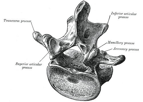
On an oblique X-ray, the lumbar vertebra looks like a Scottish Terrier in profile. Tap each part to reveal the anatomy.
Lateral curvature of the spine. Tap through each stage to see the mechanism happen.
Psoas involvement and L5 sacral torsion mechanics in low back pain.
Psoas spasm is a major contributor to low back pain. The psoas originates from the lumbar transverse processes and vertebral bodies (T12 to L5), crosses the pelvis, and inserts on the lesser trochanter of the femur.
When in spasm, it pulls the lumbar spine into flexion and the hip into flexion. The patient has difficulty standing upright and walks with a forward lean.
L5 somatic dysfunction is named by the sacral torsion it creates. Understanding the naming convention is the key to diagnosis.
Work through these clinical vignettes. Tap an answer, then tap through the teaching chain.
