The #1 reason patients seek osteopathic care. The spondy triad, scoliosis grading, and the biomechanics of leg length discrepancy.
Quick Challenge
A fracture through the pars interarticularis visible as a "collar" on the Scotty Dog in an oblique X-ray describes which condition?
Spondylolysis = fracture of the pars interarticularis. On an oblique X-ray, the fracture line appears as a "collar" around the Scotty Dog's neck. Spondylosis is general degeneration. Spondylolisthesis is anterior vertebral slippage, which can result FROM bilateral spondylolysis.
Three conditions, three different problems. The names sound alike but the pathology is completely different.
Degeneration
Spondylosis
General degenerative changes of the spine: disc dehydration, osteophyte formation, facet joint arthropathy. This is wear and tear. Think of it as the "arthritis" of the spine.
Key Imaging
Osteophytes, disc space narrowing, and sclerotic changes on plain films. No fracture, no slippage.
Clinical Significance
Most common cause of chronic low back pain in older adults. Can lead to spinal stenosis if osteophytes compress the canal.
SpondylOSIS = Old Spine, Osteoarthritis Situation. The "osis" ending screams chronic process, not acute injury.
Fracture
Spondylolysis
A fracture of the pars interarticularis. This is a stress fracture, typically from repetitive hyperextension. Classic patient: young athlete (gymnast, football lineman).
Key Imaging
"Scotty Dog" on oblique X-ray: the fracture appears as a "collar" around the dog's neck. The neck of the Scotty Dog IS the pars interarticularis.
Clinical Significance
If bilateral, the vertebral body is disconnected from the posterior elements and can slip forward, causing spondylolisthesis.
SpondyloLYSIS = Lysis means breaking apart. The pars is lysed (fractured). The dog got a collar because its neck broke.
Slippage
Spondylolisthesis
Anterior slippage of one vertebra over the one below it. The vertebral body slides forward, creating a visible "step-off" on lateral X-ray.
Key Imaging
"Step-off" sign on lateral X-ray: the anterior border of the vertebral bodies no longer lines up. Graded by Meyerding classification (I through V based on % slippage).
Clinical Significance
Most commonly at L5-S1. Can result from bilateral spondylolysis (isthmic type) or degenerative facet changes. May compress nerve roots in severe cases.
SpondyloLISTHESIS = "listhesis" from Greek meaning to slip. The vertebra slid forward. Bilateral spondylolysis is the setup; spondylolisthesis is the consequence.
The Scotty Dog
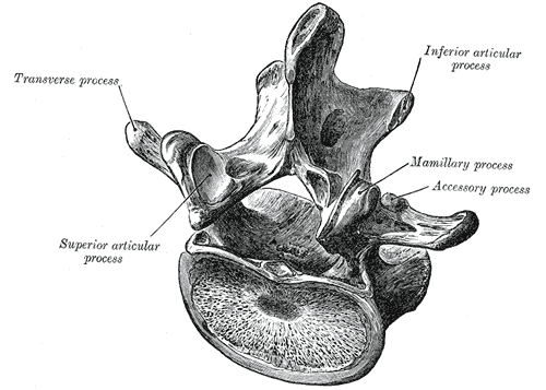
On an oblique X-ray, the lumbar vertebra looks like a Scottish Terrier in profile. Tap each part to reveal the anatomy.
Ear
Eye
Nose
Neck (Pars)
Front Leg
Body
Scoliosis, Cobb Angle & Leg Length
Lateral curvature of the spine. Tap through each stage to see the mechanism happen.
Dextroscoliosis: curve convex to the right. Levoscoliosis: curve convex to the left.
Named for the direction of convexity, not concavity.
>75° = cardiovascular compromise: cor pulmonale from chronic pulmonary hypertension.
Tap to see what happens with a short right leg
The SART Rule (Type I Mechanics)
Sidebend AWAY from the short leg · Rotate TOWARD the short leg
This IS Fryette's Law I (Type I): in a neutral spine, sidebending and rotation occur to opposite sides. The short leg creates a group curve across multiple lumbar segments. Sidebend left, rotate right = opposite directions = Type I.
Diagnosis: L1-L4, N SL RR (neutral, sidebent left, rotated right).
Treatment: Heel lift on the short side. Levels the sacral base, removes the compensatory curve.
Related OMM Concepts
Psoas involvement and L5 sacral torsion mechanics in low back pain.
Muscle Involvement
Psoas & Low Back Pain
Psoas spasm is a major contributor to low back pain. The psoas originates from the lumbar transverse processes and vertebral bodies (T12 to L5), crosses the pelvis, and inserts on the lesser trochanter of the femur.
When in spasm, it pulls the lumbar spine into flexion and the hip into flexion. The patient has difficulty standing upright and walks with a forward lean.
Classic Sign
Hip held in flexion, resists extension
Testing
Positive Thomas testPatient supine, pull one knee to chest. If the opposite thigh lifts off the table, the psoas/hip flexor on that side is tight or in spasm.
Sacral Mechanics
L5 Dysfunction: Forward vs. Backward Torsions
L5 somatic dysfunction is named by the sacral torsion it creates. Understanding the naming convention is the key to diagnosis.
Forward Torsions (Neutral L5)
L on L · R on R
Named axis and rotation go to the SAME side. The sacrum rotates forward on its oblique axis.
Backward Torsions (Non-Neutral L5)
L on R · R on L
Named axis and rotation go to OPPOSITE sides. The sacrum rotates backward on its oblique axis.
Same = Forward = Neutral L5: the letters match (L on L, R on R), the sacrum goes forward, L5 is happy (neutral). Opposite = Backward = Non-Neutral L5: the letters cross (L on R, R on L), the sacrum goes backward, L5 is dysfunctional (non-neutral).
Board-Style Walkthrough
Work through these clinical vignettes. Tap an answer, then tap through the teaching chain.