Three laws that govern how every vertebra moves, and why ERS/FRS notation makes sense once you know them
Medically reviewed by Fatima Ali, DO
Opening Challenge
A 52-year-old woman presents with chronic thoracic pain. Structural examination with the patient standing reveals a smooth group of vertebrae at T4 through T8 that are all sidebent left and rotated right in the neutral position. No single segment shows flexion or extension restriction. Which of the following best describes this finding?
A) Type I dysfunction governed by Fryette's Law I: NSLRR
B) Type II dysfunction governed by Fryette's Law II: ERRSR
C) Type II dysfunction governed by Fryette's Law I: FRLSL
D) Type I dysfunction governed by Fryette's Law III
E) Type II dysfunction at each segment independently
Type I dysfunction, Fryette's Law I: NSLRR. The key findings: neutral position, multiple vertebrae as a group (T4 through T8), sidebent left and rotated RIGHT. That opposite coupling (sidebend and rotation in opposite directions) is the fingerprint of Law I. Law I governs neutral mechanics in groups. Notation: NSLRR (Neutral, Sidebent Left, Rotated Right). Somatic dysfunctions are named for freedom of motion. The treatment addresses the entire group, not a single segment. Neutral + group + opposite coupling = Law I. Non-neutral + single segment + same-side coupling = Law II.
00 · Anatomy Review
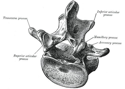
Vertebral Anatomy Review
Tap each card to reveal the anatomy you need before the laws make sense.
⚫Vertebral Body
Structure
Cylindrical weight-bearing anterior part of the vertebra. Largest in the lumbar spine, smallest in the cervical. Stacked bodies form the anterior column that bears axial load.
➡Transverse Processes (TP)
Structure
Lateral projections from each vertebra. In the thoracic spine, they articulate with ribs via costal facets. In the lumbar spine, they are called costal processes.
Board key
A posterior TP on one side means rotation TO that side. The transverse process follows the vertebral body. If the right TP is more posterior, the body has rotated RIGHT. This is the single most tested palpation finding on the clinical medicine.
⚠️Spinous Process (SP)
Structure
Posterior midline projection. Palpable landmark for surface anatomy.
Board trap
SP points OPPOSITE the rotation direction. Right rotation = SP deviates LEFT. Think of the SP as the tail of the vertebra: when the body turns right, the tail swings left. This is a major board trap: students see SP deviated left and write "rotated left." That is the trap. Body rotation is to the RIGHT.
🔄Articular Facets
Structure
Superior and inferior pairs forming zygapophyseal (facet) joints. Their angle determines coupling behavior.
Why this matters
In the thoracic spine, facet angles are more coronal, allowing rotation as the primary motion. In the lumbar spine, facet angles are more sagittal, favoring flexion/extension and limiting rotation. The facet geometry is WHY coupling patterns differ between neutral and non-neutral positions.
🔶Vertebral Foramen
Structure
Central space housing the spinal cord. The cord terminates at L1 (conus medullaris). Below L1, the canal contains only the cauda equina (nerve roots).
Regional differences
Thoracic spine has the smallest foramina relative to cord size, making it vulnerable to compression. Cervical foramina are larger and triangular. Lumbar foramina are large.
🏴Regional Differences
Thoracic
Smallest foramina, rib articulations, kyphotic curve, primary motion is rotation. Coronal facet orientation allows coupled motion in the neutral zone.
Lumbar
Largest vertebral bodies, lordotic curve, primary motion is flexion/extension. Sagittal facet orientation limits rotation. L5 articulates with the sacral promontory at the lumbosacral junction.
Lumbar vertebra, superior view: body (anterior), transverse processes (lateral), spinous process (posterior)
01 · The Framework
Three Laws of Spinal Motion
Two mechanics, one principle about restriction. Master these and every notation clicks into place.
Law I: Group Neutral Mechanics
In the neutral position (neither flexed nor extended), sidebending precedes rotation. They couple to OPPOSITE sides. This affects a GROUP of 3 or more adjacent thoracic/lumbar vertebrae. The asymmetry does NOT change with flexion or extension.
Why is Type 1 always a group? In neutral, no single facet joint is locked or jammed. The facets sit loosely in their articular surfaces, sharing load symmetrically across multiple levels. When the spine sidebends, the coupled rotation is distributed across several segments because no one segment is mechanically captured. The vertebrae tilt together like a row of dominoes leaning the same way. That is why you see a smooth, gentle curve across 3+ segments instead of one abrupt stuck vertebra. The facets are free enough that the mechanical force spreads.
Somatic dysfunctions are named for freedom of motion. Notation: segment, then N (neutral), then sidebend direction, then rotation direction.
Treatment: The entire group is treated together, not segment by segment.
Segment Diagnostic Builder · set the motion, read the diagnosis
superior view (posterior up)
lateral view
Sagittal position
Rotation
Sidebending
Type II · Fryette's Law II
ERRSR
Law II: Single Segment, Non-Neutral
When the spine is in a non-neutral position (flexed or extended), rotation precedes sidebending. They couple to the SAME side. This affects a SINGLE segment.
Why is Type 2 always a single segment? When one vertebra is driven into flexion or extension, its facet joints engage asymmetrically. One side jams (the facet surfaces compress together), and the other side gaps open. This mechanical capture locks that ONE segment into a fixed position. The segments above and below are not captured because the force is concentrated at a single articulation. Think of it like stepping on one link in a chain: only that link bends. The segments around it may compensate with a Type 1 group curve, but the primary lesion is always a single stuck vertebra.
Flexion or extension will either intensify or resolve the asymmetry: if extension resolves it, extension is the direction of ease.
Notation: segment, then position (E or F), then rotation direction, then sidebend direction.
Why facet orientation determines coupling: The angle of the superior articular facets dictates which motions are mechanically linked. Different spinal regions have different facet angles, which is why the same force produces different coupling patterns at different levels.
Region
Facet orientation
Primary motion
Cervical
Backward, Upward, Medial (BUM)
Flexion/extension (sagittal)
Thoracic
Backward, Upward, Lateral (BUL)
Rotation (transverse)
Lumbar
Backward, Medial (BM)
Flexion/extension (coronal limited)
Try it: Use the Segment Diagnostic Builder in the Law I tab. Set position to Flexed or Extended, then set rotation and sidebending to the same side. The readout shows Type II with the full notation. Try setting them to opposite sides: the builder flags it as invalid.
Law III: Restriction in All Planes
Movement of any vertebral segment in one plane alters movement in all other planes. Restriction in one plane = restriction in all planes. Applies to cervical, thoracic, and lumbar.
This explains why TART findings (tissue texture, asymmetry, restriction, tenderness) cluster at the same segment. A segment that cannot extend also cannot fully rotate or sidebend.
F/E
Rotation
Side- bend
Tap any circle to "restrict" that plane. Watch what happens to the others.
The clinical medicine test: neutral = opposite coupling (Law I). Flexed or extended = same coupling (Law II). One question gives you the position, you pick the law.
Feature
Law I (Type 1)
Law II (Type 2)
Position
Neutral
Non-neutral (F or E)
Motion precedence
Sidebending precedes rotation
Rotation precedes sidebending
Coupling
OPPOSITE sides
SAME side
Segments
Group (3+ adjacent)
Single segment
Diagnostic test
Asymmetry UNCHANGED with flex/ext
Asymmetry RESOLVES with flex or ext
Notation
NSRRL
ERRSR or FRLSL
Treatment
Entire group
Single segment
02 · Interactive
Coupling Predictor
Set the spinal position and sidebending direction, then predict which way rotation goes.
Step 1: Set the spinal position
Position
Step 2: Set the sidebending direction
Sidebending
Which direction does the rotation go?
Score:0 / 0
The rule you are building: Neutral = opposite coupling. Sidebending precedes rotation (Law I). Flexed or Extended = same-side coupling. Rotation precedes sidebending (Law II). Run through all 6 combinations until it becomes automatic.
03 · At the Table
The Examination
How to diagnose Type 1 vs Type 2 at the treatment table. Tap each step to reveal.
Examination procedure: from palpation to diagnosis
1
Position patient (seated or prone). Palpate transverse processes of each segment in neutral. What are you looking for?
Tap to reveal
2
You found a posterior TP. Now flex the patient. What are you checking?
Tap to reveal
3
Extend the patient. What are you checking now?
Tap to reveal
Which TP is more posterior = rotation direction. Does asymmetry resolve in flex or ext? If yes = Type 2 (non-neutral). If no = Type 1 (neutral). The position that resolves the asymmetry is the direction of ease.
Asymmetry RESOLVES in flexion or extension: Non-neutral dysfunction (Type 2). The position that resolves it is the direction of ease.
Asymmetry does NOT resolve: Neutral dysfunction (Type 1). No flex/ext component. Group curve.
Diagnosis Machine
Work through 4 randomized scenarios. Palpate, test positions, name the notation.
Scenario 1 of 4
Worked example: T7 NSRRL
Left TP of T7 is more posterior (rotation left). Asymmetry does not change during flex/ext. Ease in right sidebending. Neutral dysfunction. Group curve.
Worked example: T7 ERRSR
Right TP more posterior (rotation right). Asymmetry resolves in extension (direction of ease). Restriction in left sidebending. Type 2 single segment.
04 · Build It
Notation Builder
Two games: build the notation from a description, then decode notation into meaning.
Game A: Build the Notation
...
Position
SidebendS
RotationR
1 of 6
Named for freedom of motion: the notation describes where the segment IS (ease), not where it cannot go (barrier).
Game B: Decode the Notation
05 · Barriers
Barrier Finder
Given a notation, identify ease, restriction, and treatment direction.
Cervical Spine: The Three Exceptions
The cervical spine does NOT follow the thoracolumbar position-dependent switching rule. Each region has its OWN fixed coupling pattern regardless of position. This is a top board trap.
OA (Occiput on Atlas)
OPPOSITE coupling. Always.
Sidebend right = rotation LEFT. Sidebend left = rotation RIGHT. This holds whether the head is neutral, flexed, or extended. The biconvex occipital condyles on the concave atlas facets create this unique geometry.
Think "Type I-like" but do not call it Type I. OA has its own rules.
C2 through C7 (Typical Cervical)
SAME-SIDE coupling. Always.
Sidebend right = rotation RIGHT. Sidebend left = rotation LEFT. Neutral or non-neutral position does not change this. The articular facet orientation locks same-side coupling at every level from C2 to C7.
Think "Type II-like" but do not call it Type II. C2-C7 always couples same-side.
AA (Atlas on Axis, C1-C2)
Rotation only. No sidebend coupling.
The dens (odontoid process) acts as a pivot. C1 rotates around C2 with almost no sidebending component. When a board stem mentions AA or C1-C2, think pure rotation. Do not pair it with sidebending.
The trap: a board stem describes a non-neutral cervical finding and asks what coupling applies. Students default to thoracolumbar rules (non-neutral = same-side). That works for C2-C7 by coincidence but FAILS for OA. OA is opposite in every position. Know the joint, not the rule.
Quick Challenge
Treatment Order
Find Type II primary→Treat it first→Reassess group curves→Treat residual Type I if needed
Walk the chain: why this order matters
1
You find both a Type I group curve (T4-T8) and a Type II single segment (T6 ERRSR). Why not treat the group first?
Tap to reveal
2
What is the relationship between the Type II and the Type I group curve?
Tap to reveal
3
What happens to the group curve after you release the Type II?
Tap to reveal
4
When would you still need to treat the group curve after fixing the primary?
Tap to reveal
The Type II drives the Type I. Fix the driver first. The compensation often self-corrects. If it does not, treat the residual group on reassessment.
06 · Regions
Regional Application
Lumbar and thoracic specifics, landmarks, and the psoas connection.
Lumbar Spine
Largest vertebral bodies, lordotic curve, greatest ROM in flexion/extension. Sagittal facet orientation limits rotation.
Landmarks: Iliac crests sit between L4/L5. Spinal cord terminates at L1 (conus medullaris). Below L1 = cauda equina only.
L5-sacral relationship: L5 sidebends in the same direction as the sacral oblique axis, and rotates opposite to the sacrum on that axis.
Static exam: Prone, palpate TPs, roll fingers cephalad (induces flexion) and caudad (induces extension) to check for flex/ext component at each segment.
Psoas connection: Nonneutral dysfunction at L1/L2, positive Thomas test, pelvic shift contralateral, tender point 1 cm medial to ASIS. These four findings together = psoas dysfunction in clinical practice. Worsens going from sitting to standing.
Thoracic Spine
Smallest foramina, kyphotic curve, greatest ROM in rotation. Coronal facet orientation favors rotation.
Rule of Threes (SP location relative to TP):
Vertebrae
SP position
T1-T3
Same level as TP
T4-T6
Half-segment below TP
T7-T9
One full segment below TP
T10
Same as T7-T9
T11
Same as T4-T6
T12
Same as T1-T3
Costovertebral relationship: Thoracic SD affects ribs and rib SD affects the thoracic spine. Rib heads articulate at the vertebral body and TP.
Sympathetic chain: Ganglia run adjacent to thoracic vertebral bodies. Thoracic SD can affect autonomic tone to visceral organs.
Anatomical Landmarks
These landmarks let you identify vertebral levels by palpation. Tap each card to reveal its level.
Posterior
Vertebral prominence (most prominent SP)
C7. The spinous process of C7 is the most prominent and easily palpated in the posterior midline of the neck.
Posterior
Spine of the scapula
T3. The root of the scapular spine is at the level of the T3 spinous process.
Posterior
Inferior angle of the scapula
T7. With the patient's arms at their sides, the inferior scapular angle sits at the T7 level.
Anterior
Sternal notch (jugular notch)
T2. The suprasternal notch at the top of the manubrium corresponds to the T2 vertebral level.
Anterior
Sternal angle (angle of Louis)
T4. The junction of the manubrium and body of the sternum. Also where rib 2 articulates, making it a key landmark for rib counting.
Anterior
Xiphoid process
T9. The inferior tip of the sternum. Marks the T9 level and the upper boundary of the abdomen.
Posterior
Iliac crests (highest point)
L4-L5 interspace. A horizontal line connecting the iliac crests crosses between L4 and L5. This is the most commonly used landmark for lumbar palpation and LP needle insertion.
Posterior
PSIS (posterior superior iliac spine)
S2. The PSIS sits at the level of the second sacral segment. Often visible as the "dimples of Venus" on the lower back.
07 · Process of Elimination
Elimination Game
Tap each clue to eliminate wrong answers. Last one standing wins.
During examination, a single vertebra at L3 is found in a flexed position. When sidebent left, it also rotates left. Tissue texture changes are localized to L3 only.
Type I dysfunction, Law I
Type II dysfunction, Law II
Neutral group dysfunction
OA-type coupling
Law III violation
🔍Clue 1: Tap to reveal. How many segments are involved?
🔍Clue 2: Tap to reveal. What position is the spine in?
🔍Clue 3: Tap to reveal. What is the coupling direction?
🔍Clue 4: Tap to reveal. Build the notation.
Type II dysfunction, Law II. Single segment (L3) + non-neutral (flexed) + same-side coupling (sidebend and rotate LEFT together) = Law II governs. Notation: FRLSL at L3. Barrier: extension, right rotation, right sidebending.
Pattern recognition: same-side coupling in any non-neutral position = Law II, single segment, ERS or FRS. Opposite coupling in neutral = Law I, group.
08 · Board Walkthrough
clinical Walkthrough
clinical vignettes with full clinical presentations. Decision tree, teaching chain, memory hooks, then quiz.
You palpate a posterior right TP at T8. You flex the patient. The asymmetry stays. You extend. Still stays. What type?
Type 1: asymmetry unchanged in flex/ext = neutral dysfunction
Type 2: single segment = must be non-neutral
Correct. Asymmetry that does not resolve in flexion or extension = neutral dysfunction = Type 1. This is a group pattern (check adjacent levels). The unchanged asymmetry means there is no flex/ext component to name.
Single segment alone does not determine the type. The diagnostic test is whether the asymmetry resolves with flexion or extension. Unchanged = Type 1 (neutral). Resolves = Type 2 (non-neutral). Here the asymmetry stays, so it is Type 1.
You find a primary T6 ERRSR (Type 2) AND a T3-T7 group curve (Type 1). Which do you treat first?
Type 2 primary first, then reassess group
Type 1 group first to reduce the curve
Correct. The Type 2 is the primary lesion driving the compensatory Type 1 group curve. Treat the driver first. The group may self-correct once the primary is released.
Treating the compensation first (the group) is treating the symptom. The group curve will return because the Type 2 driving it is still present. Always treat the primary Type 2 first.
The T6 dysfunction is ERRSR. You choose HVLA (direct technique). What is the thrust direction?
Toward restriction: flexion, left rotation, left sidebending
Toward ease: extension, right rotation, right sidebending
Neutral position, then compression
Correct. ERRSR = ease is extension + right rotation + right sidebend. Barriers are flexion + left rotation + left sidebend. HVLA (direct technique) always thrusts toward the barrier (restriction). Into flexion, left rotation, left sidebending.
That describes indirect technique (going toward ease). HVLA is a direct technique: you engage and thrust through the restrictive barrier, not toward ease.
Neutral plus compression is not the HVLA setup. HVLA requires three-dimensional barrier engagement in all restricted planes before the thrust.
Walk the chain: from palpation to treatment
1
You feel a posterior TP on the right. What does that tell you?
Tap to reveal
2
You flex and extend the patient. The asymmetry resolves in extension. What type is this?
Tap to reveal
3
Rotation right, extension ease. What about sidebending?
Tap to reveal
4
Where is the barrier?
Tap to reveal
5
For HVLA, where do you thrust?
Tap to reveal
Posterior TP = rotation to that side. Resolves in flex/ext = Type 2, that position is ease. Same-side coupling in non-neutral. Barrier is the opposite of ease. HVLA thrusts into the barrier.
Memory Hook 1
Law I vs Law II: how to remember which coupling goes where?
"I for Opposite, II for Same." The Roman numeral I looks like it is standing alone = opposite directions. The Roman numeral II looks like a pair walking together = same direction. Neutral = Law I (opposite). Non-neutral = Law II (same).
Memory Hook 2
How to remember that ERS describes ease, not the barrier?
ERS = where the bone parked, not the getaway route. Crime scene analogy: ERRSR tells you where the vertebra IS (ease). The barrier is the OPPOSITE: the getaway route it cannot take. HVLA chases the getaway (barrier), not the parking spot (ease).
Memory Hook 3
The cervical exceptions: OA vs C2-C7 vs AA?
OA = opposite coupling in any position. C2-C7 = same-side coupling in any position. AA = rotation only, no meaningful sidebend. Do not apply thoracolumbar switching rules to any of these.
Memory Hook 4
Spinous process direction vs rotation direction?
SP is the tail of the vertebra. It swings OPPOSITE the rotation. Rotation RIGHT = SP deviates LEFT. Like a dog turning right, the tail swings left. The transverse process, by contrast, follows the body: prominent on the RIGHT = rotated RIGHT.
Memory Hook 5
The examination test: how to remember Type 1 vs Type 2?
Asymmetry resolves = Type 2 (flex/ext found the ease). Asymmetry unchanged = Type 1 (neutral, no flex/ext component). The one where something changes is Type 2. The one where nothing changes is Type 1.
Lumbar vertebra lateral view
Thoracic vertebra: facet angle drives coupling
Lumbar vertebra superior: sagittal facets
OA joint: opposite coupling
Spinal curvatures
Bone Wizardry is an independent educational resource for visual learning in the medical sciences. It is not affiliated with, endorsed by, or sponsored by any licensing or examination board, contains no real or recalled examination questions, and does not guarantee any educational or examination outcome.
Reviewed by Fatima Ali, DO and Kaitlyn Cocuzzo, MD Sources: Greenman's Principles of Manual Medicine · Atlas of Osteopathic Techniques (Nicholas) · Osteopathic Medicine (Chila) · Gray's Anatomy plates (Wikimedia Commons)