/>
Osteopathic Manipulative Medicine

Fibular Head
Somatic Dysfunction

The seesaw that boards loves to test. Proximal goes one way, distal goes the other.

Opening Challenge
A patient inverts their ankle and now has lateral knee pain. You palpate the fibular head · it's more prominent anteriorly. Quick: which way did the fibular head go?
Scroll to learn
The Seesaw Model

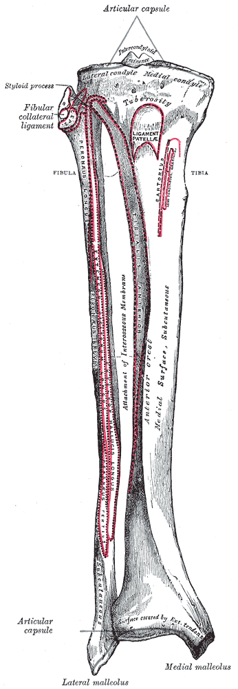
This is the one concept that makes everything else click. The fibula is a seesaw · the interosseous membraneA tough fibrous sheet connecting the tibia and fibula along their entire length. It's the fulcrum of the seesaw · when one end of the fibula moves, the membrane forces the other end to move oppositely. is the fulcrum.

When the proximal fibular head goes anterior, the distal end (lateral malleolus) goes posterior. And vice versa. Always. No exceptions. 🔑Seesaw = See the opposite. Top goes left? Bottom goes right. Always.

Interosseous Membrane PROXIMAL ANT DISTAL POST Fibular Head Lateral Malleolus
Anterior fibular head → posterior lateral malleolus. The seesaw always balances.
Anterior vs Posterior · The Two Dysfunctions
More Common
Anterior Fibular Head
Mechanism: Inversion injury (ankle rolls in)
Proximal: Fibular head anterior + lateral
Distal: Lateral malleolus posterior + medial
Foot posture: Supination (add + inv + PF)
Palpation: More prominent anteriorly at knee
Pain with: Lateral knee pain, peroneal nerve stretch
Less Common
Posterior Fibular Head
Mechanism: Eversion injury (ankle rolls out)
Proximal: Fibular head posterior + medial
Distal: Lateral malleolus anterior + lateral
Foot posture: Pronation (abd + ev + DF)
Palpation: More prominent posteriorly at knee
Pain with: Posterolateral knee pain
Key Fact The foot tells you the ankle, and the ankle tells you the knee. Supination (foot turned in) = anterior fibular head. Pronation (foot turned out) = posterior fibular head. 🔑Supination → Seesaw tips Anterior up top. Or: "Sup? I'm up front." (anterior)
Supination vs Pronation · The Components

Boards loves asking "which motions make up supination?" This is a three-part combo for each. Think of it as a martial arts stance.

Supination = the "inward roll"
Add + Inv + PF
Adduction · foot turns in (toes point medially)
Inversion · sole faces medially (like checking your shoe sole)
Plantarflexion · foot points down (like pressing a gas pedal)
Pronation = the "outward roll"
Abd + Ev + DF
Abduction · foot turns out (toes point laterally)
Eversion · sole faces laterally (showing off your sole)
Dorsiflexion · foot pulls up (like lifting off the gas)
Board Trap Boards gives you the distal findings (lateral malleolus position) and asks about the proximal end. Don't fall for it · seesaw rule. If they say "lateral malleolus is posterior," the fibular head is anterior. Always. They're testing whether you know the seesaw model or just memorized the proximal findings.
How to Diagnose It · Step by Step

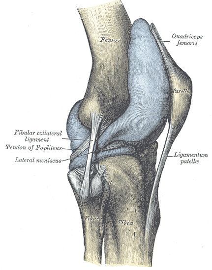
The diagnosis is about palpation comparison. You're checking the proximal tibiofibular jointThe joint where the fibular head articulates with the lateral tibial condyle. It's a synovial plane joint with slight gliding motion. Feel it just below and lateral to the tibial plateau. and comparing both sides.

1
Palpate the Fibular Head Bilaterally
Find the fibular head on both sides. It's just below and lateral to the tibial plateau. Compare prominence · anterior vs posterior. The dysfunctional side will have a more prominent fibular head in one direction.
You palpate both fibular heads. The right one is more prominent anteriorly. What does this tell you?
2
Check the Distal End (Confirm with Seesaw)
Palpate the lateral malleolus. If the fibular head is anterior, the malleolus should be posterior compared to the other side. This is your confirmation · if both ends point the same direction, recheck. The seesaw doesn't lie.
3
Check Foot Posture
Anterior fibular head → foot in supination (turned in, inverted, plantarflexed). Posterior → foot in pronation (turned out, everted, dorsiflexed). The foot is the third confirmation. Three clues, all saying the same thing.
Treatment · Muscle Energy

Both anterior and posterior fibular head dysfunctions use muscle energy techniqueA direct technique where you position the patient at the restrictive barrier, have them push against you (isometric contraction for 3-5 seconds), then take up the new slack. Repeat 3-5 times. The patient does the work · you just hold position and move further into the barrier after each contraction. (MET). It's a direct technique · you push toward the barrier, not away from it.

You're treating an anterior fibular head. Which direction do you push the fibular head for muscle energy?
🧬
Anterior Fibular Head MET
1. Wrap hands around proximal fibula
2. Push fibular head posteriorly
3. Patient dorsiflexes + everts foot against your resistance (activates peroneal muscles which pull fibula posterior)
4. Hold 3-5 sec → relax → take up slack
5. Repeat 3-5 times
🧬
Posterior Fibular Head MET
1. Wrap hands around proximal fibula
2. Push fibular head anteriorly
3. Patient plantarflexes + inverts foot against your resistance (activates muscles that pull fibula anterior)
4. Hold 3-5 sec → relax → take up slack
5. Repeat 3-5 times
Key Fact The patient's foot motion for treatment is the opposite of the dysfunction's foot posture. Anterior fibular head (supinated foot) → treat with dorsiflexion + eversion (pronation components). You're using the muscles to pull it back to normal.
Board Trap They ask "what motion does the patient perform?" and list supination components alongside pronation components. The trick: the patient's motion is always the opposite of the dysfunctional posture. Anterior (supinated) → patient does pronation motions. Posterior (pronated) → patient does supination motions. The patient is undoing the dysfunction with their own muscles.
The Key Concepts · Flip to Learn

Tap each card. Front = the concept. Back = the board-tested detail.

🧭
Anterior Dysfunction
tap to reveal
Anterior Fibular Head
Mechanism: Inversion injury (ankle rolls inward)

Proximal: Fibular head sits anterior + lateral
Distal: Lateral malleolus swings posterior + medial
Foot: Supination (add + inv + PF)

Treat with: Direct MET, push posterior. Patient does dorsiflexion + eversion.
🤵
Posterior Dysfunction
tap to reveal
Posterior Fibular Head
Mechanism: Eversion injury (ankle rolls outward)

Proximal: Fibular head sits posterior + medial
Distal: Lateral malleolus swings anterior + lateral
Foot: Pronation (abd + ev + DF)

Treat with: Direct MET, push anterior. Patient does plantarflexion + inversion.
🔍
MOB / TART Exam
tap to reveal
Palpation Findings
MOB: Motion restriction, Orientation of the fibular head, Bony landmark prominence

TART: Tissue texture change, Asymmetry, Restriction of motion, Tenderness

Key palpation: compare fibular heads bilaterally. The dysfunctional side is more prominent in the direction of displacement. Check the lateral malleolus to confirm seesaw.
🥊
HVLA vs MET
tap to reveal
Treatment Choice
MET (Muscle Energy): Direct technique. Patient isometrically resists in the direction of correction. 3-5 sec contractions, repeat 3-5x. Works via post-isometric relaxation.

HVLA: High velocity low amplitude thrust. Direct technique. Fast thrust toward the restrictive barrier. Contraindicated if hypermobility, fracture, or osteoporosis present.

Boards almost always test MET for fibular head.
Diagnosis + Treatment · Decision Tree
Tap each node to reveal the next branch. Follow the algorithm.
🔍 Patient has lateral knee pain after an ankle injury. Step 1: Palpate fibular head bilaterally.
Fibular head more prominent ANTERIORLY
Dx Anterior Fibular Head Somatic Dysfunction
Confirm with Seesaw Palpate lateral malleolus. Expect posterior position relative to the other side.
Check Foot Posture Foot should be in supination (adducted + inverted + plantarflexed).
Treat: MET Position fibular head at posterior barrier. Patient dorsiflexes + everts foot against resistance. Hold 3-5 sec, repeat 3-5x.
Fibular head more prominent POSTERIORLY
Dx Posterior Fibular Head Somatic Dysfunction
Confirm with Seesaw Palpate lateral malleolus. Expect anterior position relative to the other side.
Check Foot Posture Foot should be in pronation (abducted + everted + dorsiflexed).
Treat: MET Position fibular head at anterior barrier. Patient plantarflexes + inverts foot against resistance. Hold 3-5 sec, repeat 3-5x.
Fibular heads appear SYMMETRIC
No fibular head SD Check for other causes: LCL sprain, proximal tibiofibular joint pathology, common peroneal nerve entrapment, proximal tibia fracture.
Clinical Images

Scroll to see anatomy and clinical context. Tap to enlarge.

Fibula anatomy anterior view
Fibula · Anterior view. Head articulates at proximal tibiofibular joint.
Proximal tibiofibular joint anatomy
Proximal tibiofibular joint. Palpate just below lateral tibial plateau.
Ankle ligament anatomy
Ankle ligament anatomy. Inversion injury stresses lateral ligaments and pulls fibula.
Common peroneal nerve at fibular head
Common peroneal nerve wraps around fibular head. Anterior SD can stretch it.
Interosseous membrane tibia fibula
Interosseous membrane = the seesaw fulcrum. Connects tibia and fibula their full length.
Clinical Vignettes
4 patients with knee or ankle complaints. Don't overthink it · the seesaw does the work for you.
0/4
You know your seesaws.