Von Recklinghausen disease. One faulty copy of NF1 on chromosome 17, one broken tumor-suppressor brake on Ras, and the body grows skin spots, iris freckles, nerve sheath tumors, and the occasional optic nerve glioma. Spot the pattern, never miss it again.
NF1Von RecklinghausenChr 17NeurofibrominCAL1PSO
A 9-year-old boy is brought to clinic because his teacher noticed he keeps closing his right eye while reading. On exam he has eight flat tan macules on his trunk (each larger than a dime), freckling under both arms and in the groin creases, and on slit-lamp multiple small raised pigmented bumps on the iris of both eyes. Visual acuity is 20/200 in the right eye, 20/20 in the left.
What is the most likely diagnosis driving the new vision loss?
Tuberous sclerosis
Sturge-Weber syndrome
Neurofibromatosis type 1 with optic nerve glioma
McCune-Albright syndrome
Neurofibromatosis type 2
Six or more cafe au lait macules + axillary/inguinal freckling + iris hamartomas = NF1 until proven otherwise.
The new monocular vision loss in a child with NF1 is an optic nerve glioma, which is a pilocytic astrocytoma growing along the optic pathway. NF1 patients screen with serial eye exams for exactly this reason.
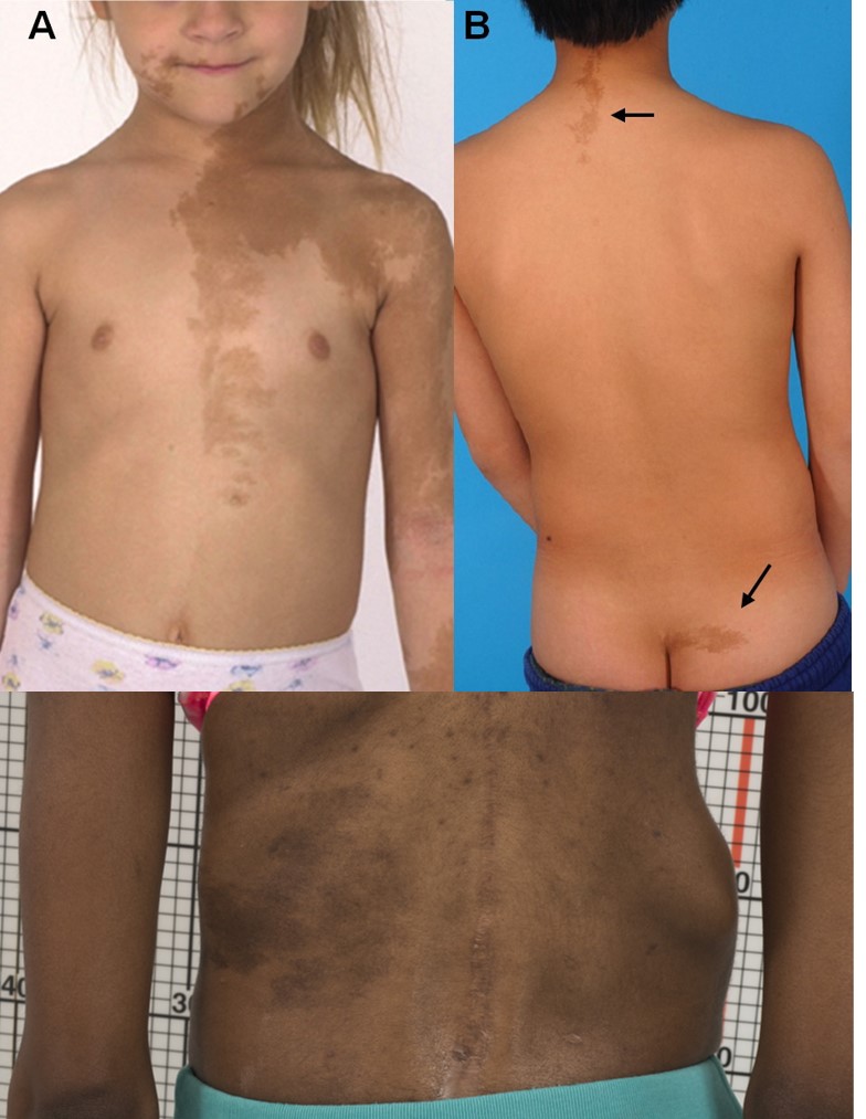
McCune-Albright also gives cafe au lait, but the borders are jagged (coast of Maine) and the spots usually do not cross midline. Tuberous sclerosis gives ash-leaf spots and shagreen patches, not the tan smooth-bordered macules of NF1. NF2 lives on chromosome 22 and announces itself with bilateral vestibular schwannomas (hearing loss), not Lisch nodules and not optic glioma. Sturge-Weber is a port-wine birthmark in the V1 dermatome with leptomeningeal angiomas.
Pattern Anchor
CAL1PSO · The seven-letter fingerprint
Tap each letter. Each one is a finding from the lecture mnemonic and a separate diagnostic clue. Two or more of these together in one patient should make NF1 the first thing you say.
C · A · L · 1 · P · S · O
Tap a letter to reveal the finding
C
Cafe au lait spots
Flat tan macules with smooth borders. Diagnostic threshold: six or more, each larger than 5 mm before puberty or 15 mm after puberty.
A
Axillary & inguinal freckling
Tiny tan spots clustered in the armpits and groin creases. Called the Crowe sign. Sun-protected skin should not freckle. When it does, suspect NF1.
L
Lisch nodules
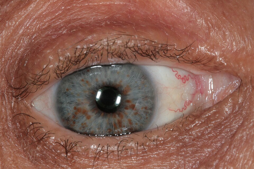
Small raised pigmented hamartomas on the iris. Painless, asymptomatic, visible only on slit-lamp. Appear in the majority of adults with NF1, less common in young children.
1
Chromosome 17 · NF1 gene
NF1 sits at 17q11.2 and encodes neurofibromin, a tumor suppressor that switches off the Ras growth signal. One bad copy is enough to lose the brake (autosomal dominant).
P
Plexiform & cutaneous neurofibromas
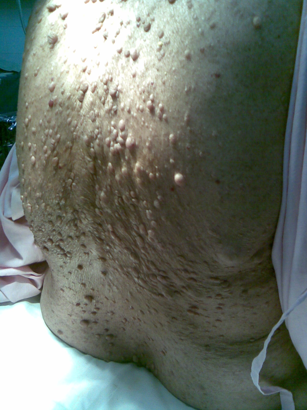
Soft, rubbery tumors of peripheral nerve sheaths. Cutaneous ones bud from the skin in adolescence. Plexiform ones are deep, diffuse, congenital, and carry the malignant transformation risk (MPNST).
S
Skeletal lesions
Sphenoid wing dysplasia (pulsating exophthalmos), scoliosis, and tibial pseudarthrosis (false joint from a congenital long-bone cortical defect). The bones of NF1 break easily and heal wrong.
O
Optic nerve glioma
A pilocytic astrocytoma growing along the optic pathway. Most common CNS tumor in NF1. Lecture hook: NF1 hits the optic pathway; the optic nerve is the headline tumor site. Children with NF1 get serial eye exams to catch it before vision is lost.
Two or more = clinical NF1 by NIH criteria. Six cafe au lait alone in a young child still warrants the workup.
Slit-Lamp Finding
Lisch nodules · freckles on the iris
Photo on the left is real (Wikimedia, slit-lamp view). Schematic on the right is a drawn idealized iris so you can see what the eye is hunting for: raised, pigmented, dome-shaped hamartomas scattered across the iris stroma.
Photo Honesty: real photo + drawn schematic
Photos vary in lighting, angle, and pupil position. The schematic teaches the pattern. No overlays are drawn on top of the photo.
Real photo · slit-lamp
Look for dome-shaped, pigmented bumps scattered across the iris surface. Often easier to find peripherally than near the pupil.
Wikimedia Commons · Dimitrios Malamos · CC BY-SA 4.0
Schematic · idealized iris
Pigmented, dome-shaped hamartomas of melanocytes and fibroblasts. Painless. Sit on the surface of the iris stroma. The bumps you draw are what you scan for on slit-lamp.
Schematic · Bone Wizardry
Why it matters: Lisch nodules do not affect vision. They are pure diagnostic gold; finding even one in a child with cafe au lait spots points to NF1.
Skin Finding
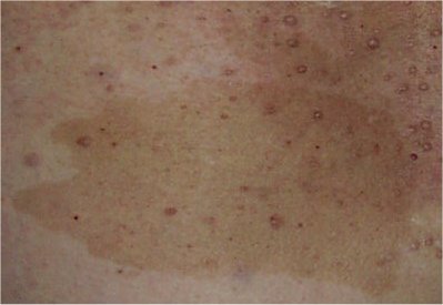
Cafe au lait macules · coast of California
Smooth, flat tan patches with even borders. The shape of the border is the discriminator between NF1 and McCune-Albright: NF1 borders are smooth like the California coast, McCune-Albright borders are jagged like the coast of Maine.
Photo + schematic side by side
Real photo shows actual lesions in their natural variety. The drawn schematic isolates the border quality you are scoring on exam.
Real photo · NF1 patient
A single flat, milk-coffee colored macule with a smooth border. Count six or more for the NF1 criterion.
Wikimedia Commons · CC BY-SA 3.0
Schematic · border anatomy
Same color, different coastline. NF1: smooth, oval, regular. McCune-Albright: jagged, irregular, often respects midline.
Schematic · Bone Wizardry
Criterion: six or more cafe au lait macules, each greater than 5 mm before puberty or greater than 15 mm after, counts as one diagnostic point toward NF1.
Cross-Reference
When the O lights up: optic nerve glioma
An NF1 child with new vision loss, proptosis, or a relative afferent pupillary defect is hiding an optic pathway tumor. Histology is almost always pilocytic astrocytoma. The deep dive on that tumor is one tap away.
NF1 is autosomal dominant with variable expressivity. The gene encodes neurofibromin, a tumor suppressor that turns the Ras growth signal off. Lose one good copy plus a second hit in a cell, and Ras runs without a brake. Tumors follow wherever that cell lineage goes.
Inheritance
Autosomal dominant with variable expressivity. Siblings with the same mutation can look very different. About 50% are de novo mutations, so family history is often blank.
Locus
Chromosome 17q11.2. Lecture mnemonic: NF1 on chromosome 17; NF2 on chromosome 22. Two letters, two numbers, both doubled.
Gene product
Neurofibromin. Acts as a GTPase-activating protein (GAP) for Ras. Translation: neurofibromin tells Ras to stop sending growth signals.
Tumor mechanism
One inherited bad copy plus a somatic hit on the good copy in a single cell: two-hit tumor suppressor loss. Ras stays in the on position. That cell line grows: neurofibroma, glioma, sometimes pheochromocytoma.
Punnett · AD inheritance
Parent (Nf,nf) × Parent (nf,nf)
nf
nf
Nf
Nf nfAffected
Nf nfAffected
nf
nf nfUnaffected
nf nfUnaffected
50% of offspring inherit the mutant allele. One bad copy is enough: the disease shows. Nf = mutant dominant allele, nf = wild-type.
Growth signal→Ras-GTP (ON)
normally:
Ras-GTP→Neurofibromin→Ras-GDP (OFF)
in NF1:
Ras-GTP→No brake→stays ON→tumor growth
Discriminator
NF1 vs NF2 · the two-letter trap
Same word, different disease. NF1 lives on chromosome 17 with skin and iris findings. NF2 lives on chromosome 22 and announces itself with bilateral vestibular schwannomas. The exam expects you to keep them straight.
NF1 is dominant, variable, and surveillance driven
The board trap is treating the skin findings like decoration. They are a tumor suppressor clue. NF1 loss increases Ras signaling, so the patient is not just getting spots. They need optic pathway monitoring, blood pressure checks for renovascular disease or pheochromocytoma, and attention to plexiform neurofibroma growth.
What will you do next time? New pain, rapid enlargement, or neurologic deficit in a neurofibroma means think malignant peripheral nerve sheath tumor until proven otherwise.
Complication Ladder
When NF1 changes tempo, escalate
Stable spots and soft skin tumors are surveillance clues. A changing lesion is different. Pain, rapid growth, weakness, numbness, visual decline, hypertension, or early puberty changes the question from diagnosis to complication triage.
What will you do next time? Vision change points to optic pathway glioma, episodic hypertension points to pheochromocytoma, and painful enlarging nerve-sheath mass points to malignant transformation.
Clinical Images
Cafe au lait macule · NF1 · tap to expand
Cutaneous neurofibromas · tap to expand
Jagged border mimic · compare to NF1 · tap to expand
Test Yourself
clinical vignettes
Five questions, no timer. Each explanation has a clue, a chain, and the one high-yield detail you should walk away with.
Medically reviewed by Kaitlyn Cocuzzo, MD and Fatima Ali, DO · Last updated June 30, 2026 at 1:10 AM ET
Bone Wizardry is an independent educational resource for visual learning in the medical sciences. It is not affiliated with, endorsed by, or sponsored by any licensing or examination board, contains no real or recalled examination questions, and does not guarantee any educational or examination outcome.