6 muscles, 3 nerves, visual field cuts, and the gaze-center lesions that make eye stems finally localize.
THE CASE
Which Muscle Is Out?
Decode the vignette before looking at the answer.
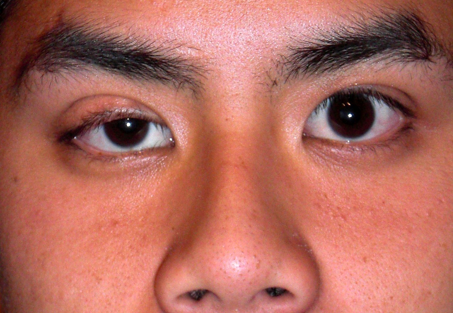
A 32-year-old woman presents with intermittent double vision one week after a bicycling accident. Symptoms worsen when she tries to type on her computer or button her shirts. Exam reveals a right-sided head tilt. Her left eye is deviated upwards, which becomes more prominent when she attempts left eye adduction.
This patient's symptoms are most likely due to impaired innervation to the:
Superior oblique (CN IV). Here is how to decode it:
The contralateral head tilt is the giveaway. She tilts RIGHT because the LEFT superior oblique is weak. The SO normally intortsIntorsion = top of the eye rotating toward the nose. The superior oblique pulls the top of the eye inward. When it is paralyzed, the eye extorts (rolls outward), so the patient tilts their head to compensate. the eye. When it fails, the eye drifts upward (hypertropia), and this gets WORSE on adduction because that is where the SO is supposed to depress the eye.
Why typing and buttoning? Both require looking DOWN and IN, exactly the action the superior oblique performs. That is the clinical pearl: trouble reading or going downstairs = think CN IV.
Lateral Rectus = CN VI (Abducens)🔑LR6 · Lateral Rectus, CN 6. The nerve that abducts. Superior Oblique = CN IV (Trochlear)🔑SO4 · Superior Oblique, CN 4. The pulley muscle. The trochlea is a literal pulley in the orbit. Everything else = CN III (Oculomotor) · SR, IR, MR, IO, levator palpebrae (eyelid), pupil constriction.
CN III does the most. CN IV does one thing. CN VI does one thing. That is the whole map.🔑The obliques are counterintuitive: Superior oblique goes DOWN. Inferior oblique goes UP. They do the OPPOSITE of what their name suggests vertically, because they wrap around the eye at an angle.
THE PALSIES
Six Disorders, One Fingerprint Each
Read the visible fingerprint, then lock the mechanism underneath it.
CN III
👀
CN III Palsy
Oculomotor Nerve
Eye Position"Down and out"
EyelidPtosis
PupilDilated or spared
Causeposterior communicating artery aneurysm or ischemia
CN III innervates SR, IR, MR, IO (eye movement in every direction except lateral and down-in), levator palpebrae (eyelid), and parasympathetic pupilloconstriction fibers (run on the OUTSIDE of the nerve). When CN III is paralyzed, only the lateral rectus (CN VI) and superior oblique (CN IV) are functioning, pulling the eye down and out. The eyelid droops because levator is out.
Pupil Rule
Dilated pupil = compressive: parasympathetics on the outside get compressed first. Posterior communicating artery aneurysm, uncal herniation. Emergency. Get angiography NOW.
Normal pupil = ischemic: vasa nervorum infarcts the inner motor fibers first, spares outer parasympathetics. Diabetes, HTN. Usually resolves in 3 months.
Board Trap
Down-and-out + dilated pupil = posterior communicating artery aneurysm until proven otherwise. Do NOT attribute this to diabetes without imaging. A missed aneurysm is a missed subarachnoid hemorrhage waiting to happen.
CN IV
👇
CN IV Palsy
Trochlear Nerve
DiplopiaVertical
Worst WhenLooking down-and-in
Head TiltContralateral
Cause #1Trauma
🔍 Trouble reading stairs · head tilt AWAY from the lesion
mechanism below
Why This Happens
The Mechanism
The superior oblique depresses the eye when it is adducted (looking toward the nose). When CN IV is paralyzed, the SO cannot depress the adducted eye. The inferior oblique (also elevating during adduction) is now unopposed, so the eye rides up = hypertropia. Worse on adduction because that is exactly when the SO is most needed.
Why Trauma?
CN IV has the longest intracranial course of ANY cranial nerve. It is the ONLY CN that exits from the dorsal brainstem. It is the thinnest cranial nerve. Long, thin, dorsally exposed = gets slammed against the tentorium during closed head injuries.
Board Trap
Head tilt is CONTRALATERAL. Left CN IV palsy = RIGHT head tilt. The patient tilts AWAY from the lesion to compensate for the extorsion. If the stem says trouble reading, typing, buttoning, or going downstairs + head tilt + trauma = CN IV until proven otherwise.
CN VI
👉
CN VI Palsy
Abducens Nerve
DiplopiaHorizontal only
Eye PositionMedial deviation (esotropia)
Can't DoAbduct (look out)
CauseRaised ICP, MS, diabetes
🔍 Most commonly paralyzed CN · false localizing sign in raised ICP
mechanism below
Why This Happens
The Mechanism
Lateral rectus is the ONLY muscle CN VI controls. When it is paralyzed, the medial rectus (CN III) is unopposed and pulls the eye inward. The patient cannot abduct the eye. Horizontal diplopia only, because the vertical muscles (CN III, CN IV) are intact.
False Localizing Sign
CN VI palsy from raised ICP does NOT mean the lesion is at the abducens nucleus (pons). It means the brain is swelling and stretching CN VI along its long course over the petrous bone and clivus. The lesion could be anywhere (cerebellar mass, pseudotumor, hydrocephalus). CN VI is the canary in the coal mine for raised ICP.
Also Think
Wernicke encephalopathy (thiamine deficiency) causes CN VI palsy as part of the classic triad (CN VI palsy, ataxia, encephalopathy). Always think about this in alcoholics or malnourished patients.
MLF LESION
🚬
INO
Internuclear Ophthalmoplegia
LesionMLF (ipsilateral)
Ipsilateral EyeFails to adduct
Contralateral EyeAbducts with nystagmus
ConvergenceINTACT
🔍 Young patient = MS · elderly = brainstem stroke
mechanism below
Why This Happens
The Pathway
For left gaze: Left CN VI nucleus fires (abducts left eye) AND sends interneurons up the right MLF to the right CN III nucleus (adducts right eye). Damage to the right MLF cuts the adduction signal to the right eye on left gaze. Result: left eye abducts fine, right eye CANNOT adduct.
Naming Rule
INO is named for the side that CANNOT adduct. Right INO = right eye can't adduct = right MLF lesion. This is counterintuitive but it is how clinical medicine label it.
Convergence Preserved
Convergence uses a SEPARATE supranuclear pathway that bypasses the MLF. Intact convergence proves CN III itself is working fine. The problem is in the communication, not in the nerve.
1st order: hypothalamus to ciliospinal center of Budge (C8-T2). Causes: Wallenberg syndrome (lateral medullary infarct), cervical cord lesions. 2nd order: ciliospinal center to superior cervical ganglion (rides with brachial plexus, then subclavian/carotid). Causes: Pancoast tumor, cervical rib, neck surgery. 3rd order: superior cervical ganglion to orbit. Causes: carotid artery dissection (painful Horner), cavernous sinus lesion.
Partial vs Complete Ptosis
Horner causes PARTIAL ptosis (sympathetics control Muller muscle, a minor eyelid elevator). CN III palsy causes COMPLETE ptosis (levator palpebrae is totally out). Partial ptosis = think sympathetic. Complete ptosis = think CN III.
Board Alert
Horner + ipsilateral face/neck pain = carotid artery dissection until proven otherwise. This is a stroke emergency. CTA neck immediately.
COMBINED
🙄
One-and-a-Half
INO + CN VI Palsy
Ipsilateral EyeNO horizontal movement at all
Contralateral EyeAbducts only (nystagmus)
LesionIpsilateral paramedian pons
CauseMS, pontine stroke, tumor
🔍 Ipsilateral eye stuck · only the contralateral eye can move (laterally)
mechanism below
Why This Happens
The Anatomy
The ipsilateral CN VI nucleus is destroyed (loses the "one" = ipsilateral gaze palsy) PLUS the ipsilateral MLF is destroyed (loses the "half" = ipsilateral adduction failure, i.e., an INO). Together: the ipsilateral eye cannot move horizontally in any direction. The contralateral eye can ONLY abduct (its lateral rectus via CN VI still works) but it cannot adduct (the MLF that would carry the signal is destroyed on the other side).
How to Count
Normal conjugate gaze = two eyes moving together (2 eyes). The "one" is the ipsilateral gaze palsy (ipsilateral CN VI nucleus destruction). The "half" is the contralateral adduction loss (MLF lesion = INO). Total lost gaze capacity = 1.5 eyes worth of movement.
Unique Feature
Exotropia (wall-eyed) of the contralateral eye may be present at rest because the contralateral medial rectus has no functioning MLF input to maintain alignment. This is called "paralytic pontine exotropia."
REFERENCE
All 6 Muscles at a Glance
Actions, nerves, and what happens when each fails
Muscle
Nerve
Primary Action
When It Fails
Superior Rectus
CN III
Elevation (up)
Can't look up; eye drifts down
Inferior Rectus
CN III
Depression (down)
Can't look down; eye drifts up
Medial Rectus
CN III
Adduction (in)
Can't adduct; eye drifts out
Inferior Oblique
CN III
Elevation in adduction + extorsion
Can't elevate in adduction
Superior Oblique
CN IV
Depression in adduction + intorsion
Hypertropia worse on adduction + contralateral head tilt
Lateral Rectus
CN VI
Abduction (out)
Can't abduct; esotropia (eye turns in)
The obliques are counterintuitive: Superior oblique goes down. Inferior oblique goes up. They do the OPPOSITE of what their name suggests in the vertical plane because they wrap around the eye at an angle, pulling it from a posterior-medial direction.
CN III Palsy
CN IV Palsy
CN VI Palsy
CN III · Oculomotor Palsy
The big one. Does the most, breaks the loudest.
What It Controls
SR, IR, MR, IO, levator palpebrae, pupil constriction (parasympathetics on OUTSIDE of nerve)
Classic Presentation
Ptosis (droopy eyelid) + eye "down and out" (lateral rectus and superior oblique unopposed)
"Down and out" with dilated pupil = posterior communicating artery aneurysm until proven otherwise. Get angiography NOW. Diabetic CN III spares the pupil because ischemia affects the inner vasa nervorum first, parasympathetics run on the outside of the nerve.
CN IV · Trochlear Palsy
The most commonly injured nerve by trauma. Only does one muscle.
What It Controls
Superior oblique only. One nerve, one muscle.
Classic Presentation
Vertical diplopia + trouble looking down-and-in. Contralateral head tilt to compensate.
Why trauma? CN IV trifecta of vulnerability: longest intracranial course, ONLY CN that exits from the dorsal brainstem, thinnest cranial nerve. It slams against the tentorium during head injuries.
Board Trap
Head tilt is CONTRALATERAL. Left CN IV palsy = RIGHT head tilt. The patient tilts AWAY from the lesion to compensate for the extorsion. Trouble reading/typing/buttoning + head tilt + trauma = CN IV until proven otherwise.
CN VI · Abducens Palsy
The most commonly PARALYZED cranial nerve. Long course = vulnerable to ICP.
What It Controls
Lateral rectus only. One nerve, one muscle.
Classic Presentation
Horizontal diplopia only + can't abduct the eye. Eye turns IN (esotropia). Diplopia worst looking toward the affected side.
Key Feature
Medial deviation at rest (medial rectus unopposed). Horizontal diplopia ONLY, no vertical component.
CN VI palsy is a "false localizing sign" in raised ICP. It does not mean the lesion is at CN VI. It means the brain is swelling and stretching the nerve along the clivus. Do not be tricked into localizing to the pons.
PATHWAY LAB
Field Cuts and Gaze Centers
Anchor the anatomy, then force the localizer.
Orbit mapLR6 and SO4 are the exceptions. Everything else is CN III.Field-cut mapNasal retina crosses at the chiasm. Temporal retina stays ipsilateral.Ptosis clueComplete ptosis points to CN III. Partial ptosis with miosis points to Horner.
Visual Field Localizer
Pick the lesion. The output names the field loss and the board clue.
Start with crossing. If the lesion is before the chiasm, one eye loses vision. If it is after the chiasm, both eyes lose the same side of the world.
Conjugate Gaze Localizer
Pick the broken command center. The output separates nerve, nucleus, tract, and cortex.
Start with one eye or both eyes. A peripheral nerve palsy breaks one eye. A gaze center lesion breaks conjugate movement of both eyes.
ALGORITHM
Patient Has Diplopia · Where Is It?
First separate monocular optics from binocular misalignment, then localize the nerve or pathway.
1
What happens when either eye is covered?
Double vision persists with one eye covered
Double vision disappears when either eye is covered
2
Binocular diplopia. What direction are the two images separated?
Horizontal only (side-by-side images)
Vertical or oblique component (images stacked or tilted)
3
Horizontal diplopia. Can the patient abduct (look laterally) the affected eye?
No → Eye stays medially deviated (esotropia)
No → Eye can't adduct (look inward), deviates outward
3
Vertical diplopia. Is there ptosis (droopy eyelid)?
Yes → Complete ptosis present
No ptosis → vertical diplopia only
3
Can't adduct but convergence is intact. Where is the lesion?
Yes → Convergence is intact, adduction only fails on gaze
No → Convergence also fails
3
Ptosis + vertical diplopia. What is the pupil doing?
Dilated pupil (mydriasis)
Normal (pupil-sparing)
3
No ptosis, vertical diplopia. Does it worsen on downward-inward gaze?
Yes → Worse looking down-and-in, patient has head tilt
No → Different pattern
CHALLENGE
Which Muscle Moves the Eye?
Click the direction. Name the PRIMARY muscle. Get it wrong, it shakes.
Click a direction to see which muscle moves the eye there
↖ Up-Left
↑ Up
↗ Up-Right
← Left
👁
→ Right
↙ Down-Left
↓ Down
↘ Down-Right
TEST YOURSELF
Clinical Vignettes
Twenty-five patients with eye movement, visual field, and gaze-localizer problems. Figure out what is broken.
Medically reviewed by Kaitlyn Cocuzzo, MD and Fatima Ali, DO · Last updated July 5, 2026 at 8:04 PM ET
Bone Wizardry is an independent educational resource for visual learning in the medical sciences. It is not affiliated with, endorsed by, or sponsored by any licensing or examination board, contains no real or recalled examination questions, and does not guarantee any educational or examination outcome.