Colon Polyps & Malignant Risk

Villous adenoma, Lynch, FAP, Peutz-Jeghers, Fearon-Vogelstein, surveillance intervals

The Adenoma to Carcinoma Sequence

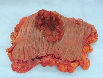
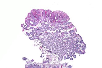
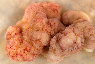
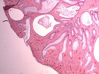
Watch the same crypt drift toward cancer. Bigger, more villous, more dysplastic means more risk. Size and villous architecture are the two things you actually act on.

Normalflat mucosa
Tubularsmall, stalked, low risk
Tubulovillousmixed architecture
Villouslarge, sessile, highest risk
Carcinomainvades the basement membrane
Low malignant potentialHigh malignant potential
From the Attending

Three polyps walk into a colonoscopy. One is a tubular adenoma, one is tubulovillous, one is villous. Which one do you watch? The answer you pick tells me everything about whether you understand the actual biology here. Not the name. The biology.

The Trap
Most students pick tubular adenoma as the risky one because it is the most common. More common does not mean more dangerous. Villous architecture rewires the risk. Know the difference before you touch the walkthrough bank.

Polyp Risk Lineup

Tap a card to see the full profile.

Zero Risk
Hyperplastic Polyp
Small, pale, sigmoid/rectum. Sawtooth architecture. No dysplasia. Board answer: no cancer risk.
Hyperplastic polyp: saw-tooth glands, no dysplasia, no malignant potential. Found in sigmoid and rectum. Exception: large hyperplastic polyps in right colon may actually be sessile serrated adenomas misclassified. Small sigmoid hyperplastics = safe. Follow-up: resume 10-year standard screening.
Low Risk <5%
Tubular Adenoma
80% of all adenomas. Tubular glands, pedunculated. Low risk unless ≥1cm or HGD.
Tubular adenoma: most common adenoma. Round tubular glands. Often pedunculated (on a stalk). Risk under 5%. Becomes "advanced" if ≥1cm, ≥25% villous component, or high-grade dysplasia present. 1-2 small tubular adenomas: 5-7 year surveillance. 3+ adenomas: 3 years.
Moderate 7-10%
Tubulovillous Adenoma
Mixed architecture: 25-75% villous. Intermediate risk. Often larger and sessile.
Tubulovillous: mixed architecture, 25-75% villous component. Risk roughly 7-10%. Often found in rectosigmoid. Classified as advanced adenoma. Surveillance: 3 years after resection. Board pearl: if the question says "tubulovillous", the answer is probably 3-year surveillance.
Very High 10-40%
Villous Adenoma
Frond-forming, sessile, large. Secretory diarrhea + hypokalemia. Highest adenoma risk.
Villous adenoma: the board villain. Finger-like fronds, sessile (no stalk), often in rectum. CRC risk 10-40%. Secretes sodium and potassium → massive watery diarrhea + hypokalemia (McKittrick-Wheelock if severe). Continues during fasting (secretory pattern). Surgical resection if not amenable to endoscopic removal.
Right-Sided Risk
Sessile Serrated Adenoma
Flat, right colon, serrated glands. Methylation pathway → sporadic MSI-H CRC.
Sessile serrated adenoma (SSA): right colon, flat/sessile, serrated architecture with crypt dilation. BRAF mutation → CpG island methylation → MLH1 silencing → MSI-H → sporadic right-sided CRC. Looks like hyperplastic on colonoscopy but is NOT. Board trap: sporadic MSI-H CRC without family history = SSA pathway, not Lynch germline.
No Adenoma Risk
Inflammatory Pseudopolyp
IBD-related. Granulation tissue regenerating after ulceration. No malignant potential by themselves.
Inflammatory pseudopolyp: not a true adenoma. Regenerative mucosa and granulation tissue forming during IBD flares. No inherent malignant potential. Note: UC itself carries CRC risk (surveillance colonoscopy every 1-2 years after 8 years of disease), but these polyps specifically are NOT precancerous.

The Transformation Cascade

Fearon-Vogelstein adenoma-carcinoma sequence. Tap step by step.

🧬
Normal Colonic Epithelium
Baseline: APC and p53 intact, KRAS wild-type
Orderly crypt architecture. Stem cells at base, differentiation toward the lumen. Nothing has broken yet.
⚠️
Early Adenoma
Loss of APC (chr 5q) → unchecked Wnt signaling
APC normally destroys beta-catenin. Lose APC, beta-catenin floods the nucleus, drives proliferation. This is the initiating hit. FAP patients inherit one broken APC copy at birth.
📈
Intermediate Adenoma
KRAS mutation (codon 12) → constitutive RAS activation
KRAS locks RAS in the "on" position. Growth signal never turns off. Polyp enlarges. This is why KRAS-mutated cancers don't respond to anti-EGFR therapy. RAS is already stuck downstream.
🟠
Late Adenoma (Villous Features)
Loss of SMAD4/DCC (chr 18q) → TGF-beta signaling failure
SMAD4 and DCC normally mediate TGF-beta growth inhibition. Loss removes the brake. Polyp develops villous architecture. Now large, frond-forming, high risk.
💀
Carcinoma
Loss of TP53 (chr 17p) → no apoptosis checkpoint
p53 is the last line of defense. Without it, cells with severe DNA damage survive and proliferate. Invasive carcinoma forms. The sequence: APC → KRAS → SMAD4/DCC → p53. That is the entire board answer in one line.

Hereditary Syndromes

Lynch Syndrome (HNPCC) MLH1 / MSH2 / MSH6 / PMS2
Polyp typeAdenomatous (few, but high malignant risk)
How it worksMismatch repair deficiency → microsatellite instability (MSI-H)
CRC locationRight colon (ascending, proximal)
Amsterdam II3 relatives, 2 generations, 1 diagnosed <50, no FAP
Extra-colonicEndometrial (most common), ovarian, gastric, urinary tract
ScreeningColonoscopy every 1-2 years from age 20-25
Trap
Sporadic MSI-H right-sided CRC (from sessile serrated adenoma, BRAF mutation, MLH1 methylation) looks like Lynch but is NOT. Lynch = germline MMR mutation. Sporadic = somatic MLH1 methylation. Check BRAF V600E: if mutated, it is sporadic, not Lynch.
Familial Adenomatous Polyposis APC (chr 5q21, tumor suppressor)
Polyp burdenHundreds to thousands of tubular adenomas carpeting the colon
CRC risk100% by age 40 if untreated
Screening startAnnual flexible sigmoidoscopy from age 10-12
TreatmentProphylactic colectomy in teens/early 20s
Gardner variantFAP + osteomas + desmoid tumors + CHRPE + epidermoid cysts
Turcot variantFAP + medulloblastoma (posterior fossa, Homer-Wright rosettes)
Peutz-Jeghers Syndrome STK11 / LKB1
Polyp typeHamartomatous (branching smooth-muscle core), small bowel predominant
Classic findingMelanotic macules on lips, buccal mucosa, and digits
ComplicationIntussusception (small bowel polyps acting as lead point)
Cancer riskGI, pancreatic, breast, ovarian, testicular (Sertoli cell)
Board memoryLips + pigment + small-bowel obstruction = STK11
Cowden Syndrome PTEN (phosphatase, chr 10q23)
Polyp typeHamartomatous polyps (GI tract)
Classic findingsMacrocephaly, trichilemmomas (hair follicle tumors on face)
Cancer riskBreast (most common), thyroid (follicular type), endometrial
vs Peutz-JeghersNo melanotic macules; macrocephaly is the Cowden giveaway
Turcot Syndrome APC (FAP type) or MMR gene (Lynch type)
FAP variantAPC mutation + medulloblastoma (posterior fossa, Homer-Wright rosettes)
Lynch variantMMR mutation + glioblastoma multiforme (hemispheres, pseudopalisading necrosis)
Board ruleFAP = medulloblastoma. Lynch = glioblastoma. Keep them separate.
From the Attending

Turcot is a forced-pairing question. They give you the brain tumor and ask you to name the gene. If the brain tumor is posterior fossa with rosettes: medulloblastoma = APC = FAP. If it is a hemisphere tumor with necrosis: glioblastoma = MMR = Lynch. Every time.

Surveillance Interval Calculator

What did colonoscopy find?

Clinical Findings

Colorectal adenocarcinoma
Adenocarcinoma from adenoma
Tubular adenoma histology
Tubular adenoma (histo)
Villous adenoma histology
Villous adenoma (histo)
Juvenile polyp histology
Juvenile polyp (histo)
Clinical image

Memory Hooks

Tap to reveal.

Villous adenoma = runs you dry
Think "Villous = Voluminous diarrhea." Secretes liters per day, takes your potassium with it. The only polyp that kills you before it turns into cancer.
FAP adenoma carpet
Hundreds of polyps by age 20. If you see "hundreds of polyps" in a stem, write APC before you read the rest. CRC by 40 if untreated = colectomy in the 20s. Gardner adds jaw lumps and desmoids. Turcot adds medulloblastoma.
Lynch goes right
Lynch syndrome hits the right colon (ascending). The question gives you family history of CRC plus endometrial cancer plus MSI-high pathology. Amsterdam II: 3 relatives, 2 generations, 1 under 50, no FAP.
Lips + pigment = STK11
Peutz-Jeghers = melanotic freckles on lips and buccal mucosa. Hamartomatous polyps in small bowel causing intussusception. STK11/LKB1 mutation. Multiorgan cancer risk: GI, pancreas, breast, ovary, testis.
Advanced adenoma = 3 years
Advanced adenoma criteria: ≥1cm OR ≥25% villous component OR high-grade dysplasia. Any one of these = 3-year surveillance. If you see "1.2cm tubular adenoma" stop there. That is advanced. That is 3 years. Done.
Sigmoidoscopy finds polyp? Go look at everything
Flexible sigmoidoscopy only sees the left colon. Find a polyp = full colonoscopy. The rest of the colon is unseen. This is a classic boards "next step" question answer.

Board-Style Walkthrough

Question 1 of 25
Loading...