A cirrhotic liver is a scarred, stiff filter. Blood backs up behind it, and the pressure spills out as one complication after another: varices, ascites, infection, confusion, kidney failure. The exam never asks "what is cirrhosis." It drops you a decompensated patient and asks for the next move. So we learn the cascade, and the one action that defuses each branch.
Medically reviewed by Fatima Ali, DO & Kaitlyn Cocuzzo, MD✦elite
Before you scroll
A 54-year-old man with alcohol-associated cirrhosis is admitted with two days of worsening abdominal distension and a low-grade fever. He is not confused. Temperature is 38.1 C, blood pressure 104/64 mmHg. The abdomen is distended with shifting dullness. A diagnostic paracentesis is performed: the ascitic-fluid neutrophil (PMN) count is 480 cells/mm3. Which of the following is the most appropriate next step?
What number makes the diagnosis?
An ascitic PMN count of 250/mm3 or higher in a cirrhotic equals spontaneous bacterial peritonitis. His count is 480, so SBP is confirmed. He does not need a second tap to "be sure."
What is the treatment bundle?
Empiric IV cefotaxime (a third-generation cephalosporin), started now, before culture results. AND IV albumin (1.5 g/kg day 1, 1 g/kg day 3), because albumin in SBP cuts the rate of hepatorenal syndrome and death.
Why not the other moves?
Ciprofloxacin prophylaxis is for AFTER the episode is treated, not instead of treating it. Lactulose treats encephalopathy, which he does not have. Delaying antibiotics to "confirm" can kill him. PMN 250 or more equals SBP equals cefotaxime plus albumin, today.
Scroll ↓ first, what cirrhosis actually is
Start With The Scar
What Cirrhosis Is, And What Causes It
Cirrhosis is the end stage of chronic liver injury: diffuse fibrosis plus regenerative nodules that wreck the architecture. Two things follow. The liver stops working (failure), and blood cannot get through it (portal hypertension). Tap a cause to see its board clue.
From the Attending
Stellate cells are the culprits. Chronic injury wakes them up, they lay down collagen, and the soft liver turns into a brick wall of fibrosis and nodules. Now two problems run in parallel. The cells that survive cannot do enough work, so you lose synthetic function: low albumin, high INR, high bilirubin. And blood cannot squeeze through the scar, so pressure climbs upstream. Hold those two ideas. Every single complication on this page comes from one or the other. Failure or pressure. That distinction drives everything.
Compensated vs decompensated
Compensated = scarred but quiet. Decompensated = the first time it spills over.
A patient can carry compensated cirrhosis for years with normal-looking labs. Decompensation is defined by the first appearance of ascites, variceal bleeding, hepatic encephalopathy, or jaundice. That transition is the prognosis cliff: median survival drops from over a decade to about two years. The clinical medicine love to test the moment a stable patient tips over.
Four events define decompensation: ascites, variceal bleed, encephalopathy, jaundice. The first one is the cliff edge.
Alcohol
The most common cause in the West. AST:ALT ratio greater than 2:1 ("a toast to alcohol," AST high). Heavy chronic use.
MASLD / MASH
Metabolic-dysfunction (fatty) liver disease. Obesity, type 2 diabetes, metabolic syndrome. Now a leading cause; ALT often greater than AST early.
Chronic HCV / HBV
Chronic viral hepatitis. HCV is a top driver of cirrhosis and hepatocellular carcinoma; HBV can cause HCC even without cirrhosis.
Hemochromatosis
Iron overload. "Bronze diabetes," high ferritin, high transferrin saturation, HFE gene. Iron poisons the liver, pancreas, heart, skin.
Wilson disease
Copper overload, under age 40. Kayser-Fleischer rings, low ceruloplasmin, neuropsychiatric signs. Liver plus brain.
Alpha-1 antitrypsin
Misfolded protein traps in hepatocytes (PAS-positive globules) and leaves the lungs unprotected: liver cirrhosis plus panacinar emphysema.
PBC / PSC
Primary biliary cholangitis: middle-aged woman, itching, anti-mitochondrial antibody. Primary sclerosing cholangitis: man with ulcerative colitis, "beads on a string," p-ANCA.
Autoimmune hepatitis
Young or middle-aged woman, high IgG, anti-smooth-muscle and ANA antibodies. Responds to steroids.
The stigmata: tap the body
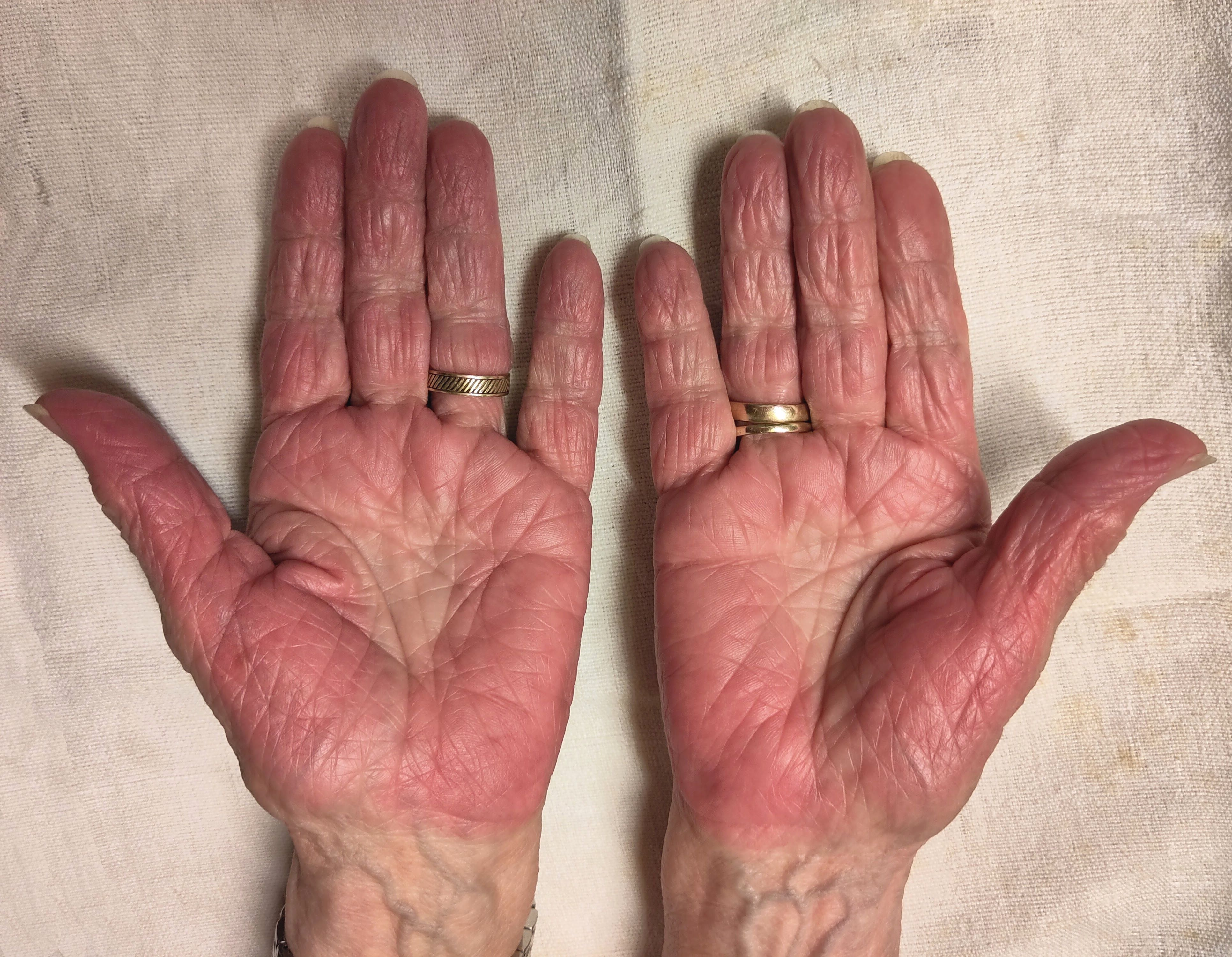
Many physical signs come from hyperestrogenism (the scarred liver cannot clear estrogen). Tap each marker to learn what it is and why it happens. These are the exam's free points: a stem describes the body and expects you to think "liver."
Tap a glowing marker
Each gold dot is a classic finding. Tap to learn what it is and the mechanism behind it.
0 of 9 markers explored
Tap to reveal the estrogen connection
Spider angiomata, palmar erythema, gynecomastia, and testicular atrophy all trace back to one thing: the failing liver cannot clear estrogen, so estrogen runs high. High estrogen widens skin vessels (spiders, red palms) and feminizes the body (breast tissue, shrunken testes). One mechanism, four signs.
The Signature Game · Pressure Becomes Complications
The Portal Pressure Cascade
This is the whole topic in one engine. Pump the portal pressure up. As it climbs past the threshold, the complications light up one by one. Tap each lit branch to get the single management move that defuses it. Light all five to finish.
Portal pressure (HVPG, mmHg)
5
Normal portal pressure is about 5 mmHg. Clinically significant portal hypertension begins at 10. Varices and bleeding need higher still. Keep pumping.
Pressure is still normal.
Raise the portal pressure to start lighting up the cascade. Each branch unlocks at the pressure where that complication actually appears.
The one chain that ties it together
Scar → blood cannot pass → portal pressure rises → the body opens collateral veins (varices) and leaks fluid (ascites). Splanchnic vessels dilate, which drops the effective blood volume, so the kidneys clamp down (hepatorenal) and aldosterone rises (more ascites). Meanwhile the bypassed blood skips the liver's detox, so ammonia reaches the brain (encephalopathy), and stagnant ascites gets infected (SBP). One rising number, five downstream disasters.
Portal pressure is the faucet. Open it and the same five leaks always appear: varices, ascites, SBP, encephalopathy, hepatorenal.
The Fluid · First Question Is Always SAAG
Ascites and the SAAG
When you tap a belly full of fluid, the first lab that matters is the SAAG: serum albumin minus ascitic albumin. It splits every cause of ascites into two camps. Sort each cause into high gradient (portal hypertension) or low gradient (everything else).
The number that splits the room
SAAGSerum-Ascites Albumin Gradient. The serum albumin minus the ascitic-fluid albumin. It estimates portal pressure: a big gradient means a big back-pressure pushing protein-poor fluid out. ≥ 1.1 = portal hypertension. SAAG < 1.1 = NOT portal hypertension.
High gradient means the back-pressure is so high it forces protein-poor fluid across the vessel wall, leaving a big albumin difference between blood and belly. That is cirrhosis (most common), plus heart failure and Budd-Chiari. A low gradient means the leak is not pressure-driven: the peritoneum itself is the problem, as in cancer (peritoneal carcinomatosis), TB peritonitis, or pancreatitis.
High gradient, high pressure. 1.1 is the wall: at or above it, blame the portal vein.
SAAG ≥ 1.1
Portal hypertension: the pressure pushed the fluid out
SAAG < 1.1
Not portal hypertension: the peritoneum is the problem
Drag (or tap a cause, then a bucket) to sort all 6. The high-gradient list is the portal-pressure family; everything else is low gradient.
How you actually treat cirrhotic ascites
Step 1: sodium restriction (under 2 g/day). The fluid is a salt-and-water problem, so you turn off the tap. Step 2: diuretics, the classic pair is spironolactone plus furosemide in a 100:40 ratio. Spironolactone leads because cirrhosis drives high aldosterone. Step 3 (refractory): large-volume paracentesisDraining the ascitic fluid with a needle. When you remove a large volume (over 5 liters), give IV albumin to prevent post-paracentesis circulatory dysfunction (the circulation collapses into the suddenly empty space). with IV albumin, or a TIPS.
Salt down, spironolactone plus furosemide, then big taps with albumin. Spiro leads because aldosterone is high.
The Infection · One Number Decides
The SBP PMN Counter
Spontaneous bacterial peritonitis is ascites that got infected with no obvious source. There is no debate and no waiting: it lives or dies on the neutrophil count in the tap. Slide the count and watch the threshold trip.
Drag: ascitic-fluid PMN (neutrophil) count
80cells/mm3
Below threshold
A PMN count under 250 is not SBP. Keep an eye out, but no empiric antibiotics yet. Push the count up to cross the line.
The whole SBP playbook
Diagnose: ascitic PMN ≥ 250/mm3 (often a single organism on culture, classically E. coli or other gut flora). Always tap BEFORE antibiotics. Treat: empiric IV cefotaxime (third-generation cephalosporin) PLUS IV albumin (1.5 g/kg day 1, 1 g/kg day 3). The albumin is the high-yield twist: it prevents hepatorenal syndrome and lowers mortality. Prevent (after an episode): long-term oral norfloxacin or ciprofloxacin.
250 is the magic number. Tap first, then cefotaxime plus albumin. Albumin saves the kidneys.
From the Attending
Here is the trap they set. The cirrhotic with ascites looks fine, maybe a low-grade fever, maybe just a little more confused. Do not wait for a rigid abdomen, because in SBP it is often soft and quiet. Any cirrhotic with ascites who deteriorates in ANY way gets a diagnostic tap. Count the neutrophils. If it is 250 or more, you do not need cultures back to start cefotaxime. And do not forget the albumin. Miss the albumin and the kidneys follow the liver down. Find the source. Tap the belly. Every time.
The Bleed · Order Is Everything
Build the Variceal Bleed Bundle
A cirrhotic vomiting bright red blood is a portal-hypertension emergency. The exam tests the order and the "don't forget" steps. Drag the four actions into the sequence you actually run during an acute variceal bleed.
1
Drop the very first step here
2
Drop step 2 here
3
Drop step 3 here
4
Drop step 4 here
Start octreotide (lowers splanchnic / portal pressure)Resuscitate: 2 large-bore IVs, transfuse to Hgb ~7Give prophylactic IV ceftriaxoneEndoscopic band ligation
Drag (or tap a step, then a slot) to build the order. Fill all four to lock it in.
Why this exact order
Resuscitate first, because a dead patient cannot be scoped. Two large IVs and blood, targeting a hemoglobin around 7 (over-transfusing raises portal pressure and worsens bleeding). Octreotide next: it constricts splanchnic flow and drops portal pressure, slowing the bleed before endoscopy. Ceftriaxone is the step everyone forgets. Cirrhotics who bleed get infected, and infection worsens bleeding and outcomes, so prophylactic antibiotics are standard in every variceal bleed. Band ligation is the definitive endoscopic fix. If bleeding is refractory: balloon tamponade as a bridge, then TIPSTransjugular Intrahepatic Portosystemic Shunt: a stent connecting the portal vein to a hepatic vein, dumping high-pressure portal blood into the systemic circulation. It relieves pressure but lets ammonia bypass the liver, so it can precipitate encephalopathy..
Resuscitate, octreotide, ceftriaxone, band. The forgotten one is the antibiotic, and it is not optional.
Prophylaxis (before and after)
Primary prophylaxis (varices found, never bled): nonselective beta-blocker (nadolol, propranolol, or carvedilol) OR band ligation. The beta-blocker lowers portal pressure. Secondary prophylaxis (already bled once): nonselective beta-blocker PLUS serial band ligation. Both, because a re-bleed is deadly.
Nonselective beta-blocker or banding to prevent. After a bleed: both.
The Last Two Branches · Confusion And Kidney Failure
Encephalopathy and Hepatorenal Syndrome
Two complications of the bypassed, failing liver: ammonia reaches the brain, and splanchnic dilation starves the kidneys. Tap a card to open its file; opening one dims the other so you study one at a time.
Caput medusae (dilated periumbilical veins)Ascites (abdominal distension)Spider angioma (hyperestrogenism)Palmar erythema (red palms)Esophageal varices and band ligation (illustration)Scleral icterus (jaundice)
🧠
Hepatic Encephalopathy
ammonia to the brain, find the trigger
▼
Why it happens
Bypassed and failing liver cannot clear ammonia, which crosses into the brain. Reversible. Bloodwork shows elevated ammonia (but you treat clinically).
Signs
Asterixis (a flapping tremor), confusion, day-night reversal, progressing to coma.
Treat
Lactulose titrated to 2 to 3 soft stools/day (traps ammonia as ammonium in the gut for excretion), plus rifaximin (kills ammonia-making gut bacteria).
Critical step
FIND THE PRECIPITANT: GI bleed, infection / SBP, constipation, dehydration, hypokalemia, excess protein, or a recent TIPS. Treating only the ammonia and missing the trigger fails the question.
Hook
Lactulose to the bowel movements, rifaximin to the bugs, and hunt the trigger. Hypokalemia is the sneaky one.
🥬
Hepatorenal Syndrome
a diagnosis of exclusion, kidneys are fine
▼
Why it happens
Splanchnic vasodilation drops the effective arterial volume, so the kidneys are hypoperfused and clamp down. The kidney tissue itself is normal.
The tell
Acute kidney injury in advanced cirrhosis with a very low urine sodium, bland urine sediment, and NO improvement after stopping diuretics and giving albumin volume.
Diagnosis
Of exclusion: rule out prerenal azotemia (responds to fluids), ATN (muddy-brown casts, high urine sodium), and obstruction first.
Treat
Albumin plus a vasoconstrictor (midodrine + octreotide, or terlipressin). Definitive cure is liver transplant.
Hook
Kidneys look normal, urine sodium is near zero, and fluids do not fix it. Squeeze the splanchnic bed; transplant the liver.
Two more things the clinical medicine bolt on
HCC surveillance: every cirrhotic gets ultrasound every 6 months (with or without AFP) to catch hepatocellular carcinoma early. MELD-Na score (bilirubin, INR, creatinine, sodium) prioritizes patients for liver transplant. Higher = sicker = higher on the list. Coagulopathy: the liver makes clotting factors, so cirrhotics have a high INR and low platelets (thrombocytopenia from splenic sequestration). This matters for the osteopathic section coming up.
Ultrasound every 6 months for cancer. MELD-Na ranks the list. High INR plus low platelets = bleed risk.
Build the Plan
Walk a Decompensated Admission
One cirrhotic patient, one decision at a time. Guess each branch before it opens. This is the reasoning the exam wants when it asks for the next step in a decompensated patient.
From the Attending
When a cirrhotic crashes, run the cascade in your head. New fluid? Tap it and count the neutrophils. New confusion? Lactulose, and hunt the trigger, do not just chase the ammonia. Rising creatinine? Stop the diuretics, give albumin, and ask if it is hepatorenal. Bleeding? Resuscitate, octreotide, antibiotics, scope. Every emergency on this page is one branch of the same tree. Name the branch and the treatment is already written. Know your clues.
The Osteopathic Lens · And A Hard Stop
OMT in Liver Disease
The viscera have autonomic and lymphatic supply you can address with OMT as an adjunct. But cirrhosis carries a specific, testable contraindication. Learn both.
The autonomics of the upper GI / hepatobiliary tract
Sympathetics: the liver, gallbladder, stomach, and proximal gut are supplied by the greater splanchnic nerve, from T5 to T9 (right side emphasized for the liver/biliary tree). A Chapman point or viscerosomatic reflex can show up paraspinally in that range. Parasympathetics: the vagus nerve supplies the gut down to the splenic flexure, driving motility and secretion.
Liver and biliary sympathetics live at T5 to T9 (greater splanchnic). The vagus runs parasympathetics to the mid-gut.
What OMT can add (as an adjunct)
Gentle techniques can support GI motility and ease lymphatic congestion alongside medical management: rib raising and paraspinal soft tissue to normalize sympathetic tone, mesenteric / ganglion release for motility, and lymphatic pump techniques to help mobilize congested fluid. None of this replaces diuretics, antibiotics, lactulose, or transplant. It is adjunctive, not curative.
Rib raising for sympathetics, mesenteric release for motility, lymphatic pump for congestion. Adjunct, never the cure.
The contraindication they test
HVLA is contraindicated in the coagulopathy and thrombocytopenia of cirrhosis.
Cirrhotics have a high INR (the liver cannot make clotting factors) and low platelets (the big spleen traps them). A high-velocity, low-amplitude thrust into a patient who cannot clot risks bleeding, hematoma, and injury. The same caution applies to any forceful or high-force technique. Reach instead for gentle, indirect, and lymphatic approaches: counterstrain, myofascial release, balanced ligamentous tension, gentle lymphatic pump.
No HVLA when they cannot clot. High INR plus low platelets equals gentle, indirect techniques only.
From the Attending
This is a single-best-answer trap waiting to happen. The stem gives you a cirrhotic with back stiffness and an INR of 1.9 and platelets of 60,000, then offers HVLA as a choice. Do not take it. A patient who cannot clot does not get a thrust. Pick the gentle, indirect technique. Coagulopathy plus thrombocytopenia equals no HVLA. Don't overthink it.
Don't Miss The Branch
Five Quick Calls
Five rapid questions pulled from a bigger pool, reshuffled every visit. Cross out (right-click / long-press) and highlight (double-click / double-tap) as you read.
clinical Practice
Walk the Cases
Full clinical vignettes, one at a time, in a shuffled order. Progress saves to your account on this device. Cross out (right-click / long-press) and highlight (double-click / double-tap) as you go.
From the Attending
These vignettes are written the way the real exam writes them: a known cirrhotic with one new finding that points to one branch of the cascade. Cover the choices, find the new clue, name the complication, and only then read the options. Read every explanation, not just the one you missed. That is how the pattern locks in.
Tip: kill the wrong choices first, then read the explanation chain for every option.
First Aid for the clinical medicine Step 1. Gastrointestinal pathology and pharmacology: cirrhosis, portal hypertension, ascites and SAAG, spontaneous bacterial peritonitis, hepatic encephalopathy, hepatorenal syndrome, octreotide, beta-blockers, TIPS.
Harrison's Principles of Internal Medicine. Cirrhosis and its complications; management of portal hypertension and variceal hemorrhage.
American Association for the Study of Liver Diseases (AASLD). Practice guidance on ascites, spontaneous bacterial peritonitis, and hepatic encephalopathy in cirrhosis.
Greenman, Principles of Manual Medicine; Chila (ed.), Foundations of Osteopathic Medicine. Visceral autonomics, lymphatic technique, and contraindications to HVLA.
Reviewed by Fatima Ali DO and Kaitlyn Cocuzzo MD. Vignettes are original clinical teaching cases; demographics, values, and answer order are written for practice. Always confirm management against the current guideline at the point of care.
Bone Wizardry is an independent educational resource for visual learning in the medical sciences. It is not affiliated with, endorsed by, or sponsored by any licensing or examination board, contains no real or recalled examination questions, and does not guarantee any educational or examination outcome.
That was the free half
You see the cascade. Now make every branch automatic.
The stigmata, the pressure cascade, and the SAAG sorter are yours free. Everything that turns "I get it" into a 10-second answer on exam day lives in Elite:
The SBP PMN counter and the full treatment bundle
The variceal bleed sequence and prophylaxis rules
Encephalopathy and hepatorenal syndrome, side by side
The decompensated-admission decision tree
The osteopathic layer plus board vignettes that save progress