General Surgery · GI
A blocked dead-end tube, a pain that walks from the navel to the right lower corner, and a clock counting down to rupture. Learn the migration, the signs, the imaging algorithm, and the one time you drain instead of cut.
The Setup
Everything about appendicitis flows from one event: the only way out gets plugged. Walk the chain once and the whole disease falls into place.
Start with the plug. The appendix is a narrow dead-end tube hanging off the cecum. Block its only opening and mucus keeps pouring in with nowhere to go → pressure climbs inside the tube → the tube's own veins get squeezed shut → the wall starves of blood (ischemia) → gut bacteria invade the dying wall (transmural inflammation) → if nobody steps in, the wall dies through and bursts. That cascade usually runs over about 48 to 72 hours, which is why timing matters.
Now the pain. Early on, the swollen tube only sends a vague visceral signal the brain localizes to the midline near the navel (the T10 dermatome). Hours later the inflammation burns through to the parietal peritoneum, the sensitive lining of the belly wall sitting right over the appendix. Those somatic nerves give a crisp, one-finger pain at McBurney pointOne third of the way along a line from the front hip bone (ASIS) to the umbilicus. The classic surface marker for the appendix.. That walk from vague-belly-button to sharp-right-lower-corner is the signature.
What does the plug? It depends on age. Flip each card.

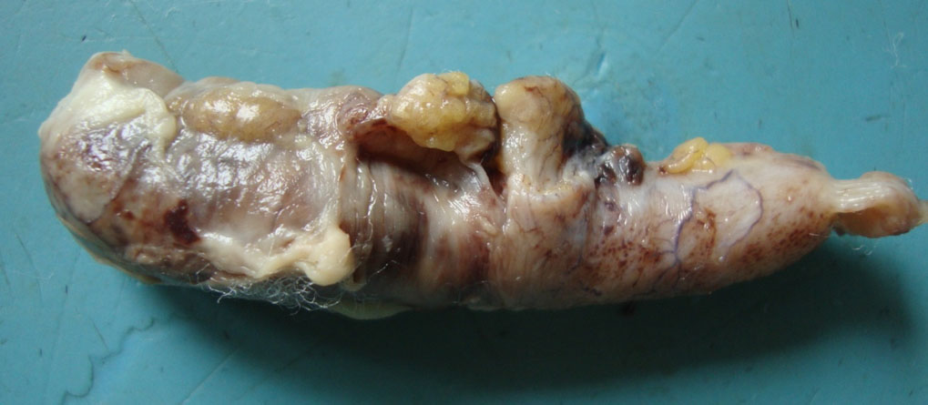


At the Bedside
Appendicitis has a script. The history follows an order, and the exam has named maneuvers that tell you where the appendix is hiding.
Order matters in the history. Classic appendicitis goes: appetite dies first (anorexia is nearly universal), then pain, then maybe a single bout of vomiting after the pain. Compare that to gastroenteritis, where vomiting and diarrhea lead and the pain is diffuse and crampy. A patient who is genuinely hungry should make you doubt appendicitis (the so-called "hamburger sign"). Fever is usually low grade; a high spiking fever points to perforation or abscess.
The exam names the location. The appendix can point in different directions, and each maneuver lights up a different hiding spot. Tap each sign.
Psoas sign means right lower quadrant pain when the examiner extends the right hip. Extension stretches the psoas. If the appendix is retrocecal and inflamed, it irritates the muscle as it moves.
Translation: right-sided psoas sign is not a psoas treatment cue. It is an appendicitis clue when it travels with fever, anorexia, guarding, or leukocytosis.
Confirming It
The diagnosis is mostly clinical, but the imaging choice changes with the patient in front of you. Work the algorithm, then learn the look-alikes.
Labs. Expect a mild rise in white cells (about 10,000 to 18,000) with a left shift, and a bumped CRP. A white count well above 18,000 hints at perforation. The single most important early lab in a woman who could be pregnant is a beta-hCG, because a positive test rewrites the entire workup.
The imaging algorithm. Three forks decide the test. Try each step before you reveal it.
The mimics. Right lower quadrant pain has a crowded differential. Two classics live on the next two cards; the table catches the rest.
| Mimic | What separates it |
|---|---|
| Ectopic pregnancy | Positive beta-hCG with an empty uterus on transvaginal ultrasound. Can rupture and bleed. Check the pregnancy test first. |
| Ovarian torsion | Sudden severe unilateral pain, adnexal mass, absent flow on Doppler. A surgical emergency for the ovary. |
| Ruptured ovarian cyst | Sudden pain often at mid-cycle (Mittelschmerz), free fluid, beta-hCG negative. |
| Crohn ileitis | Chronic diarrhea, weight loss, prior episodes; terminal ileum inflamed on imaging. |
| Nephrolithiasis | Colicky flank-to-groin pain, the patient writhes, hematuria, stone on CT. |
| Cecal diverticulitis | Older patient, focal cecal wall thickening; can look identical on CT. |
The Plan
Most appendicitis goes to the operating room. The board points live in the exceptions: the walled-off abscess, the normal appendix, and the fever on day six.
Uncomplicated appendicitis. The cure is to take the appendix out, usually laparoscopic appendectomy with antibiotics around the operation. Antibiotics alone is a real option in selected adults without a fecalith, but roughly 4 in 10 come back needing surgery within a few years, so for clinical practice the answer for uncomplicated disease is appendectomy.
Free perforation with diffuse peritonitis. A sick patient with a rigid belly and free air goes the other way fast: resuscitate, start antibiotics, and take them for a prompt appendectomy and washout.
The walled-off abscess or phlegmon. When someone shows up late (more than 5 days) with a firm right lower quadrant mass and CT shows a contained collection with a thick rind, the smart move flips. Operating into glued, angry tissue risks tearing the cecum and turning a small operation into a bowel resection. So you cool it down: antibiotics plus image-guided percutaneous drainage of a sizable abscess, then an interval appendectomy weeks later once the inflammation settles.
The normal appendix at surgery. You opened up and the appendix is clean. Most surgeons still remove it (so a future right lower quadrant pain is not blamed on an organ that is already gone), then hunt for the real cause: run the last 2 feet of ileum for a Meckel diverticulum, inspect for Crohn terminal ileitis, and check the ovary and tube in women. If you find an inflamed Meckel, resect it. If you find Crohn with an inflamed cecal base, classically leave the appendix to avoid a stump leak.
Prove It
Eight original clinical vignettes, 5 dealt per round, answer choices shuffled, never repeating within a round. Tap a wrong answer first to see why it almost works, then read the glowing clues.
