The first major branch of the abdominal aorta at T12. Three branches, six board traps, and the watershed zones that kill colons.
Quick Challenge
A patient with a posterior duodenal ulcer presents with massive upper GI bleeding. Which artery is most likely eroded?
The gastroduodenal artery (GDA) runs directly behind the first part of the duodenum. A posterior duodenal ulcer erodes through the duodenal wall into the GDA, causing massive hemorrhage. The GDA is a branch of the common hepatic artery, which comes from the celiac trunk.
↓ scroll to explore
Celiac trunk and its three branches
Branches of celiac axis in detail
Superior mesenteric artery territory
Portal venous system
Abdominal aorta: all major branches
The Three Branches
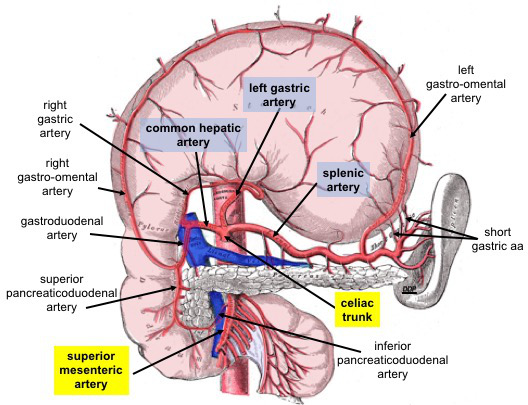
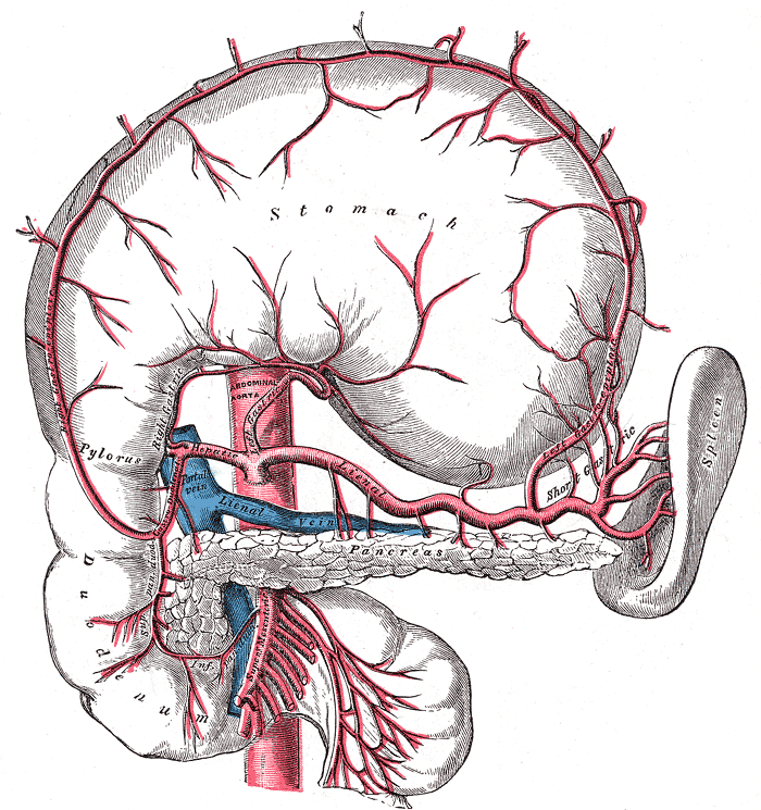
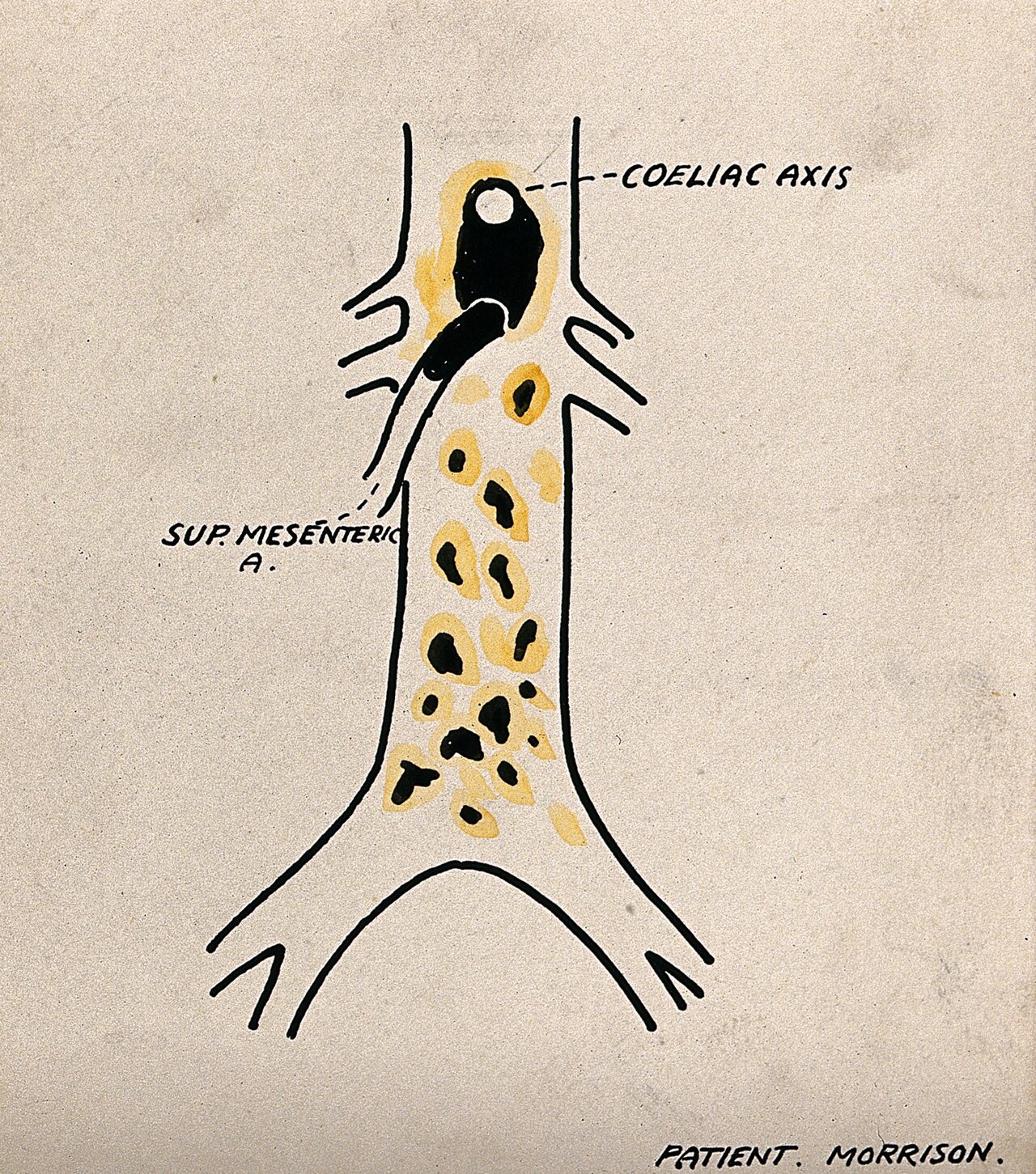
The celiac trunk exits the aorta at T12, immediately below the aortic hiatus. It trifurcates into left gastric, splenic, and common hepatic.
Smallest Branch
Left Gastric Artery
Runs along the lesser curvature of the stomach. Gives off esophageal branches to the distal esophagus before turning along the stomach.
Territory
Lesser curvature of stomach + distal esophagus
Board Pearl
The left gastric vein (coronary vein) drains into the portal vein. In portal hypertension, blood backs up into esophageal veins via this route, causing esophageal varices.
Left Gastric = the varices artery. Coronary vein backs up into esophageal veins when portal pressure rises. Left gastric connects the celiac to esophageal varices.
Largest Branch
Splenic Artery
Runs along the superior border of the pancreas (tortuous path). Gives off short gastric arteries and the left gastroepiploic artery before reaching the splenic hilum.
Key Branches
Short gastrics (fundus) + left gastroepiploic (greater curvature) + pancreatic branches + splenic hilum
Board Pearl
Short gastric arteries supply the gastric fundus. After splenectomy, fundal ischemia is possible if short gastrics are ligated.
Splenic artery rides the pancreas like a snake on a log. Top border, tortuous path. It supplies the spleen, fundus (short gastrics), and left side of the greater curvature (left gastroepiploic).
Most Tested Branch
Common Hepatic Artery
Splits into the gastroduodenal artery (GDA) and the proper hepatic artery. The proper hepatic then gives off the right gastric and divides into right and left hepatic arteries. The cystic artery branches from the right hepatic.
GDA Branches
Right gastroepiploic (greater curvature) + superior pancreaticoduodenal (head of pancreas + duodenum)
Board Trap
Which artery supplies the gallbladder? Cystic artery, from right hepatic, from proper hepatic, from common hepatic, from celiac trunk. Five links in the chain.
Common hepatic is the board question factory. GDA hides behind duodenum (bleeds in posterior ulcers). Cystic artery runs through the triangle of Calot (cystic duct + common hepatic duct + liver edge).
Mesenteric Territories
Eliminate the wrong arteries. Which vessel supplies each gut segment?
Elimination Round 1
The jejunum, ileum, ascending colon, and proximal 2/3 of the transverse colon are supplied by which artery?
Celiac Trunk
SMA
IMA
Internal Iliac
Elimination Round 2
The distal 1/3 of the transverse colon, descending colon, sigmoid colon, and upper rectum are supplied by which artery?
SMA
Celiac Trunk
IMA
Pudendal
Elimination Round 3
The splenic flexure is vulnerable to ischemia because it sits at the border of which two arterial territories?
Celiac + SMA
SMA + IMA
IMA + Internal Iliac
Celiac + IMA
Watershed Zones
Areas between two arterial territories that are vulnerable to ischemia during hypotension.
SMA / IMA Border
Splenic Flexure (Griffiths' Point)
Where the SMA territory (middle colic, via marginal artery) meets the IMA territory (left colic). This is the most common site of ischemic colitis in hypotensive patients.
Clinical Scenario
Elderly patient post-aortic surgery develops bloody diarrhea and left-sided abdominal pain. The splenic flexure is the first segment to lose perfusion.
Splenic flexure = the dead zone between two arterial kingdoms. When blood pressure drops, the border territory starves first. Think of it as no-man's-land.
IMA / Internal Iliac Border
Rectosigmoid Junction (Sudeck's Point)
Where the superior rectal artery (terminal IMA) meets the middle and inferior rectal arteries (internal iliac). Second most common watershed zone.
Clinical Significance
Ligation of the IMA during sigmoid colectomy or aortic surgery risks ischemia here if collateral flow is poor.
Sudeck's point: where the IMA hands off to the internal iliac. If the IMA gets ligated during surgery, the rectosigmoid junction is the segment that may not survive.
Portal Hypertension
Portosystemic Anastomoses
When portal pressure rises, blood detours through connections between the portal and systemic venous systems. The four classic sites:
1. Esophageal
Left gastric vein (portal) connects to esophageal veins (systemic via azygos). Result: esophageal varices.
2. Rectal
Superior rectal vein (portal via IMV) connects to middle/inferior rectal veins (systemic via internal iliac). Result: hemorrhoids (internal).
3. Paraumbilical
Paraumbilical veins (portal via round ligament) connect to superficial epigastric veins (systemic). Result: caput medusae.
4. Retroperitoneal
Colic/splenic veins (portal) connect to renal/lumbar veins (systemic). Occur at retroperitoneal bare areas.
Portal hypertension creates four escape routes: esophageal varices, hemorrhoids, caput medusae, retroperitoneal shunts. The left gastric vein is the key link for the deadliest one (varices).
Board-Style Walkthrough
25 clinical vignettes. Answers shuffle each round. Right-click or long-press to cross out options. Double-tap to highlight.