
Fryette's Laws
The two rules that govern all thoracic somatic dysfunction diagnosis. Get these wrong, every notation falls apart.
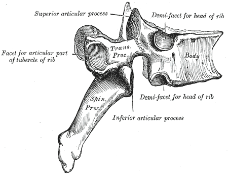

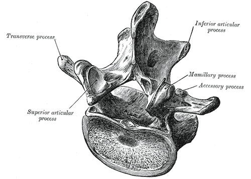

A = Asymmetry (one transverse process sits more posterior than its pair).
R = Restriction of motion (the segment will not move into one direction).
T = Tenderness (the patient's report of pain on palpation; the most subjective).
Any ONE of the four can be present, but the diagnosis of somatic dysfunction needs at least one objective finding. Tenderness alone is not enough.
Inhalation dysfunction: the rib stays elevated, will not drop on exhalation. The most caudad rib of a group holds the key.
Exhalation dysfunction: the rib stays depressed, will not rise on inhalation. The most cephalad rib holds the key.
Rib angles also sit directly over the paravertebral sympathetic chain, which is why rib-angle pressure is used for sympathetic modulation.
OPPOSITE directions
Written: N SR RL
SAME side
FRSL = Flexed, Rotated Left, Sidebent Left
Rotation vs sidebending: Opposite directions
Number of vertebrae: Multiple (group)
Cause: Postural compensation
Onset: Chronic, gradual
Example: T4-T8 as a group, neutral, sidebent right, rotated left. Written: T4-T8 N SR RL
Rotation vs sidebending: Same side
Number of vertebrae: Single
Cause: Traumatic or acute injury
Onset: Often acute
Example: T6 stuck in extension, rotated right, sidebent right. Written: T6 ERRSR
Notation System
How to read and write thoracic SD diagnoses. Every clinical medicine OMM question is either asking you to decode a notation or pick the right one.
Step 1: Flex the spine. Does the TP become symmetric or asymmetric?
Step 2: Extend the spine. Same question.
Step 3: Where the asymmetry is worse = position of dysfunction (F or E).
Result: Rotation is always toward the side where the TP is more posterior (more prominent).
ERRSR: Stuck in extension, rotated right, sidebent right.
Barrier is in: flexion, left rotation, left sidebending.
Thrust: into flexion + left rotation + left sidebend.
FRLSL: Stuck in flexion, rotated left, sidebent left.
Thrust: into extension + right rotation + right sidebend.
Thoracic HVLA Techniques
Three core techniques. Each targets a different region of T1-T12. Know which technique goes where and why.
Physician: Passes arm under the patient's axilla. Places the opposite hand at the target transverse process, palm up under the patient's back.
Action: Rolls patient toward physician to load the contact hand against the TP. Leans body weight through the patient's crossed arms to deliver the thrust on exhalation.
Best for: Mid-thoracic T4-T10. Good for most Type II ERS/FRS dysfunctions in this range.
Physician: Stands behind patient. Reaches through the patient's arms from behind. Thenar eminences contact the transverse processes at the dysfunctional level.
Action: Patient exhales. Physician delivers an anterior thrust through the thenar contacts on exhalation.
Best for: Upper thoracic T1-T4. The neck-interlaced position locks out the cervical spine and focuses force at the target level.
Physician: Applies thumb or pisiform contact to the transverse process of the dysfunctional vertebra. Body positioned lateral to the patient.
Direction: Thrust directed anteriorly and slightly cephalad at approximately 45 degrees.
Best for: Lower thoracic T6-T12. Especially useful when supine positioning is contraindicated.
This is NOT: ligament tearing, bone cracking, or a sign of damage. It is the same mechanism as knuckle cracking.
Post-procedure soreness: Normal. Similar to post-exercise soreness. Resolves within 24-48 hours.
Viscerosomatic Reflexes
Thoracic SD is not always musculoskeletal. The board wants you to link spinal levels to organs and reason backward from spine to viscera.
| Spinal Level | Associated Organs | Clinical Clue |
|---|---|---|
| T1-T4 | Heart (T1-T5), Upper extremity (C8-T1 for hand/forearm), Lung apex | Chest pain, palpitations, arm numbness |
| T2-T7 | Lungs, Upper GI, Esophagus | Dyspnea, dysphagia, regurgitation |
| T5-T9 | Heart (T1-T5), Stomach, Esophagus | Epigastric pain, heartburn, cardiac referred pain |
| T6-T9 | Gallbladder (right-sided), Liver, Stomach | Right upper quadrant pain, jaundice, nausea after fatty meals |
| T10-T11 | Small bowel, Appendix (T10 most classic), Kidney | Periumbilical pain (early appendicitis), cramping, flank pain |
| T11-L2 | Large bowel, Kidney, Adrenal, Ureter | Flank pain, hematuria, colicky abdominal pain |
| T10-L1 | Uterus, Ovary, Testis | Pelvic pain, dysmenorrhea, testicular torsion referred pain |
Rule: Treat the spine AND work up the cardiac cause in parallel. Do not dismiss chest pain as musculoskeletal because there is a spinal finding.
Somatic findings at T10 in a patient with fever and periumbilical pain: think appendicitis. The SD at T10 is the somatic expression of the visceral injury. Treat the appendix.
Walk a segment to its diagnosis
Answer before you reveal. Each step locks once you commit.
Memory hooks
Tap each card to bring it into focus.
Neutral Opposites, Non-neutral Sames
Restriction names what it cannot do
Posterior process points at the rotation
Green light to fatty meals: T6 to T9 on the right
Case Challenge
One case, three reveals. Tap each question to uncover the reasoning. Cover the answer and think first.
Type II (Non-group) somatic dysfunction: T6 ERSR
The single vertebra is stuck in an extended position (restricted when moved into more extension = it is already AS FAR into extension as it will go, and motion testing confirms this). Rotated right and sidebent right = same side = Type II mechanics (Law II: F or E yields same-side coupling).
Written: T6 ERSR = Extended, Rotated Right, Sidebent Right.
T6 is within the T5-T9 (stomach, esophagus) and T6-T9 (gallbladder, liver) viscerosomatic zones.
Combined with his epigastric discomfort and indigestion, this warrants evaluation for peptic ulcer disease, GERD, or gallbladder pathology (biliary colic, cholecystitis) before attributing his symptoms to musculoskeletal causes.
The spinal SD at T6 is likely a secondary somatic manifestation of an upper GI or hepatobiliary process.
T6 ERSR: stuck in extension, rotated right, sidebent right.
Engage the barrier: the opposite of the position of dysfunction.
Flexion (opposite of extension) + left rotation (opposite of right) + left sidebending (opposite of right).
Technique choice: T6 is mid-thoracic, so the Dog Technique (crossed arm, supine) is most appropriate. Position the physician's hand under the T6 transverse process. Flex the patient's thoracic spine slightly, introduce left rotation and left sidebending, then thrust on exhalation through the flexion-left rotation-left sidebending barrier.
Clinical note: Given the potential visceral involvement, OMM is adjunctive. Address the underlying GI or hepatobiliary cause first.
Board Drill
Four clinical questions. No multiple-select. No copied content. Work through each before revealing the explanation.
Board Walkthrough
Original clinical vignettes, one at a time, shuffled and never repeated until the bank exhausts. Right-click or long-press an option to cross it out. Double-click or double-tap to highlight.
