Five dysfunctions of the sphenobasilar synchondrosis: two physiologic, three traumatic. Know the axes, the naming rules, and which ones can happen normally.
Opening Challenge
A 27-year-old recreational soccer player has frontal headaches and neck pain that began after a ball struck the left side of her head. Vault-hold palpation feels like a skewed parallelogram: the examiner's index fingers drift right, while the little fingers drift left. Which SBS strain pattern is present?
A) Right lateral strain
B) Inferior vertical strain
C) Left lateral strain
D) Left torsion
Left lateral strain. Lateral strain makes the head feel like a parallelogram because the sphenoid and occiput shear sideways around parallel vertical axes. In the vault-hold shorthand, the little fingers point toward the named side and the index fingers move opposite. Little fingers left plus index fingers right means the sphenoid base is named left. The trap is naming it for the index-finger drift.
↓
01 · The Foundation
Primary Respiratory Mechanism
Toggle between flexion and extension to see what every structure does in each phase.
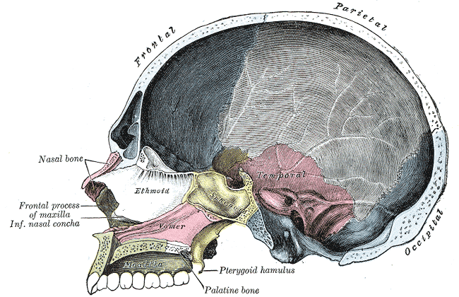
Sagittal section: the SBS is the sphenoid-occipital junction that rises in cranial flexion and falls in cranial extension.
⬆️
SBS Rises
The sphenobasilar synchondrosis moves superiorly. Picture pushing up on a rubber ball from below: it squishes wider and shorter. That is cranial flexion.
🔄
Paired Bones Externally Rotate
Temporal and parietal bones rotate outward, spreading the vault. When the ball squishes wider, the sides flare out. Same thing.
↔️
AP Diameter Shortens
As it widens transversely, the front-to-back distance shrinks. The ball gets fatter but shorter. Wider + shorter = flexion.
△
Sacrum Counternutates
The sacral base tips posteriorly (counternutation = sacral flexion). The sacrum rocks back as the cranium widens. They are coupled through the dural tube.
⬇️
SBS Falls
The SBS descends inferiorly. Now let go of the ball: it springs back to tall and narrow. The skull narrows transversely and lengthens AP. Opposite of flexion.
🔃
Paired Bones Internally Rotate
Temporal and parietal bones rotate inward. The sides pull back in as the vault narrows. Everything tightens and lengthens.
↔️
AP Diameter Lengthens
As the skull narrows, the AP distance increases. Narrower + longer = extension. The skull stretches out front-to-back.
▽
Sacrum Nutates
Sacral base tips anteriorly (nutation = sacral extension). S in Spine: the spine straightens as the sacrum nutates. Extension phase tightens and lengthens everything.
Cranial Rhythmic Impulse
6 · 12 cycles/min
CRI is the palpable rhythm of the PRM. Slower than respiration, faster than heart rate variability.
Board shortcut: Flexion = SBS up, skull wide, paired bones OUT, sacrum counternutation. Extension = everything reverses. The midline bones (sphenoid, occiput, ethmoid, vomer) flex and extend. The paired bones (temporal, parietal) externally and internally rotate.
Sphenoid bone: the greater wings are the index-finger contact in the cranial vault hold.
Memory hook: "Flexion Flares." In flexion, everything flares outward. Paired bones externally rotate. Skull widens. Sacrum base goes posterior. Extension is the opposite: everything narrows back in.
01b · See It Move
Craniosacral Mechanism
Toggle between extension and flexion. Watch the SBS, spinal cord, and sacrum move together.
Extension: SBS descends. Skull narrows transversely, lengthens AP. Sacrum nutates (base anterior). S in Spine: the spine straightens.
03 · The Five Dysfunctions
SBS Dysfunction Types
Tap each tab. Know the naming rule, the axis, and whether it is physiologic or traumatic.
Model Lab
SBS Hand-Motion Model
Pick a strain pattern and watch the same vault-hold contacts move. Index contacts ride the sphenoid greater wings. Little-finger contacts ride the occiput. The board trick is deciding which contact actually names the pattern.
Left lateral strain
Index contacts drift right while little-finger contacts drift left. That side-to-side parallelogram is the lateral strain pattern.
Index: right
Little fingers: left
Board name: little fingers point to the named side. Left lateral strain.
Torsion
The sphenoid and occiput rotate around the anteroposterior (AP) axis in OPPOSITE directions. One greater wing goes up, the other goes down.
Naming: Named for the HIGH greater wing of the sphenoid. If the left wing is superior, it is a left torsion.
Example: Left torsion = left greater wing superior, right greater wing inferior. The sphenoid and occiput are twisting around the AP axis like wringing a towel.
Classification:Physiologic. Can occur during normal cranial motion. No trauma required.
PhysiologicAP AxisOpposite rotationNamed for HIGH wing
Memory: "Torsion Twists, named for the Top." Torsion = twist around AP axis. Named for the TOP (high) greater wing.
Sidebending-Rotation (SB-R)
The sphenoid and occiput sidebend in the SAME direction but rotate around vertical axes in OPPOSITE directions. This creates a convexity on one side of the SBS.
Naming: Named for the convexity (the sidebend direction). If the SBS is convex to the left, it is a left SB-R.
Example: Left SB-R = SBS convex to the left. Both bones sidebend left (same direction), but they rotate around their vertical axes in opposite directions.
Classification:Physiologic. Can occur during normal cranial motion.
PhysiologicVertical axis rotationSame sidebend, opposite rotationNamed for convexity
Memory: "SB-R: Same sideBend, named for the Round side." The convex (round) side names the dysfunction. Both bones lean the same way.
Vertical Strain
This is the part the boards love. Vertical strains move on TWO parallel transverse axes, and the sphenoid and occiput rock the same rotary direction around them. Their tops tip the same way, not the mirror-image way of normal flexion.
Because flexion and extension are named in mirror image for the two bones, that one motion reads as one bone flexed and the other extended. That is why the notation is SFOE or SEFO and never both-flexed. The net result is the sphenoid base riding superior or inferior relative to the occiput.
Naming: Named for the sphenoid, and the notation spells out what BOTH bones are doing in two letters each.
Superior vertical strain (SFOE):Sphenoid Flexed, Occiput Extended. The sphenoid base rides superior. Classic cause: an anterior central blow to the frontal bone driving the sphenoid base up.
Inferior vertical strain (SEFO):Sphenoid Extended, Occiput Flexed. The sphenoid base rides inferior. Trauma can come from anterior or posterior to the SBS.
Classification:Non-physiologic. Always a trauma finding.
Non-physiologicTwo parallel transverse axesSame direction (not opposite)Named for sphenoid
Memory: "Sphenoid up is Superior, written SFOE." Sphenoid Flexed, Occiput Extended = SFOE = superior. Flip both letters for the mirror: SEFO = inferior. Sphenoid letter is always first, and the sphenoid always names the strain.
Lateral Strain
The sphenoid and occiput translate laterally in opposite directions around two parallel vertical axes. This is a shear: one bone slides left while the other slides right.
Naming: Named for the side the sphenoid (basisphenoid) translates toward relative to the basiocciput. If the sphenoid shifts left, it is a left lateral strain. Read the SPHENOID direction, never the occiput.
Mechanism: Most commonly from birth trauma, also a lateral blow to the head. The sphenoid and occiput slide past each other like tectonic plates.
Classic sign in children: a "parallelogram head" with facial asymmetry. The skew of the basisphenoid against the basiocciput skews the whole face.
Symptoms: may cause headache and visual disturbances.
Classification:Non-physiologic. Requires trauma.
Non-physiologicLateral shearOpposite lateral shiftsNamed for sphenoid
Memory: "Lateral strain makes a paralleLogram, and the sphenoid Leads the name." Named for whichever side the sphenoid slides toward. Birth trauma plus a parallelogram-shaped head in a kid is the classic board pairing.
Vault Hold Decoder: which side names the lateral strain?
Left lateral strain. Index fingers ride the greater wings of the sphenoid. Little fingers ride the occiput. In lateral strain, those contacts shear in opposite horizontal directions and the head feels like a parallelogram. For the common board hand-position rule: little fingers point toward the named side, index fingers move opposite.
Compression
The SBS is jammed together. No flexion or extension can occur at the synchondrosis. The cranial mechanism is essentially locked.
Naming: Just "compression." No left or right, no superior or inferior. The joint is stuck.
Mechanism: Most commonly from birth trauma (difficult delivery, forceps). Can also result from severe head trauma at any age.
Clinical: The CRI will be diminished or absent on palpation. The cranium feels rigid, with no inherent motion at the SBS.
Classification:Non-physiologic. Always trauma.
Non-physiologicNo axisJammed / lockedBirth trauma
Memory: "Compression = Crushed shut." No motion. No direction. Just a jammed SBS. Think of a newborn skull squeezed through a difficult delivery.
Master rule: Physiologic = Torsion and SB-R (can happen during normal cranial motion). Non-physiologic = Vertical Strain, Lateral Strain, Compression (require trauma). If the board question says "birth trauma" or "difficult delivery," think compression first.
04 · Classify
Physiologic vs Non-Physiologic
Tap each dysfunction to sort it into the correct bucket. Get all five right.
Classify each SBS dysfunction as physiologic (can occur during normal cranial motion) or non-physiologic (requires trauma). Tap a card, then tap the bucket it belongs in.
Torsion
Superior Vertical Strain
Sidebending-Rotation
Lateral Strain
Compression
Physiologic
Non-Physiologic
The split: Only two are physiologic: torsion and sidebending-rotation. They involve the normal axes of cranial motion (AP axis and vertical axis). The other three require an external force to distort the SBS into a position it was never designed to reach.
05 · Cranial Bones
Paired vs Unpaired
Tap each card to reveal what happens to the bone during cranial flexion.
🧠Sphenoid
Classification
Unpaired / midline
In Flexion
Undergoes flexion. The greater wings move laterally and inferiorly. The SBS rises.
Board note
Midline bones flex and extend. They do NOT externally/internally rotate. The sphenoid is the key bone for naming torsion, vertical strain, and lateral strain.
🧠Occiput
Classification
Unpaired / midline
In Flexion
Undergoes flexion. The occiput moves with the sphenoid at the SBS. The squamous portion moves posteriorly.
Board note
Occiput and sphenoid together define the SBS. Their relative motion determines the dysfunction type.
👂Temporal (paired)
Classification
Paired
In Flexion
Undergoes external rotation. The mastoid tips move medially and the petrous portions widen the skull laterally.
Board note
Paired bones externally rotate in flexion, internally rotate in extension. Always. This is the opposite of what midline bones do (which flex/extend).
🔀Parietal (paired)
Classification
Paired
In Flexion
Undergoes external rotation. The parietals flare laterally, widening the cranial vault.
Board note
Along with the temporals, the parietals demonstrate the "Flexion Flares" pattern: paired bones rotate outward during flexion.
👁️Ethmoid
Classification
Unpaired / midline
In Flexion
Undergoes flexion with the other midline bones. Descends slightly with sphenoid flexion.
Board note
Rarely tested directly, but know it is midline and follows the flexion/extension pattern, not external/internal rotation.
▲Vomer
Classification
Unpaired / midline
In Flexion
Undergoes flexion. Moves with the sphenoid as the SBS rises.
Board note
Like the ethmoid, rarely a standalone question. Its value is knowing it belongs in the midline group.
Midline: flex/extend↔Paired: ext/int rotate
The clean split: Unpaired midline bones (sphenoid, occiput, ethmoid, vomer) undergo flexion and extension. Paired bones (temporal, parietal) undergo external and internal rotation. In flexion, paired bones externally rotate. In extension, they internally rotate. Never cross these categories on a board question.
06 · Surface Anatomy
Cranial Landmarks
Where the sutures meet. Tap each card for the bones that converge, the old fontanelle, and the board trap underneath.
Lateral skull: pterion sits anterior at the temple; asterion sits posterior behind the ear.
⚠️Pterion
Bones that meet (the "H")
Junction of the greater wing of the sphenoid, the temporal, the parietal, and the frontal bones. The sutures form an H shape. Located just behind and above the zygomatic arch (roughly at the temple).
Why it is dangerous
The middle meningeal artery runs deep to the pterion. A fracture here can tear the artery and produce an epidural hematoma, the classic lucid-interval bleed. The pterion is the thinnest part of the lateral skull.
Board trap
Lateral head trauma plus "talk and die" lucid interval points to pterion fracture and middle meningeal artery, NOT a bridging vein (that is the subdural at a different site).
Red box marks the pterion on a real skull: the H where four bones meet at the temple.
👂Asterion
Bones that meet
Junction of the occipital, temporal, and parietal bones. It is where the occipitomastoid, parietomastoid, and lambdoidal sutures come together.
Location
Sits just behind the ear. A surgical landmark for the posterolateral skull and the transverse-sigmoid sinus junction underneath.
Board trap
Do not confuse with pterion. Pterion is anterior (temple, four bones, H, middle meningeal artery). Asterion is posterior (behind the ear, three bones, three sutures).
Red box marks the asterion behind the ear: occipital, parietal, and temporal bones.
△Bregma
Bones / sutures that meet
Junction of the sagittal suture, the coronal suture, and the metopic (frontal) suture. Sits at the top of the vault in the midline.
Was a fontanelle
This was the anterior fontanelle, the largest one. It closes by roughly 18 to 24 months. A bulging anterior fontanelle in an infant signals increased intracranial pressure.
Board trap
Anterior fontanelle needs THREE sutures: sagittal plus coronal plus metopic. Leaving out the metopic suture is the classic half-right miss.
Bregma boxed at the top of the vault (coronal meets sagittal); pterion, asterion, and lambda shown for context.
▽Lambda
Bones / sutures that meet
Junction of the sagittal suture and the lambdoid suture, at the back of the vault in the midline. Named for its resemblance to the Greek letter lambda.
Was a fontanelle
This was the posterior fontanelle. It closes early, by about 2 to 3 months, much sooner than the anterior fontanelle.
Board trap
Sagittal plus lambdoid is the POSTERIOR fontanelle at lambda. Sagittal plus coronal plus metopic is the ANTERIOR fontanelle at bregma. Mixing the two is the most common suture error.
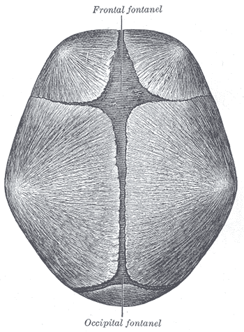
Neonatal vault: bregma is the anterior fontanelle; lambda is the posterior fontanelle.
The one that kills: the pterion overlies the middle meningeal artery. A blow to the temple that fractures it can produce an epidural hematoma with a lucid interval. Of all four landmarks, this is the high-yield emergency.
Squamous vs petrous, no contest: Squamous = squama = "scale," the same root as squamous epithelium (flat cells). So the squamous temporal is the flat, scale-thin lateral plate, the thinnest part of the skull, which is exactly why a blow at the pterion tears the middle meningeal artery. Petrous = petra = "rock," the hardest bone in the body: the dense pyramid that hides and protects the inner ear, with the tentorium cerebelli anchored along its ridge. Flat scale on the outside, rock on the inside.
Front vs back, fast: Pterion = front (temple), four bones (sphenoid, temporal, parietal, frontal), H shape, middle meningeal artery. Asterion = back (behind the ear), three bones (occipital, temporal, parietal), three sutures. Bregma = top-front midline, old anterior fontanelle. Lambda = top-back midline, old posterior fontanelle.
05 · Hands-On
The Vault Hold
The gold standard hand position for cranial diagnosis. Know exactly where every finger goes.
Index (2) greater wing of the SPHENOID
Middle (3) squamous TEMPORAL, approximating the zygomatic process
Ring (4) petrous TEMPORAL, near the mastoid process
Little (5) squamous OCCIPUT, medial to the occipitomastoid suture
Thumbs (T) crossed over the sagittal suture, resting on the frontal bone
Exam-precise placement, digit by digit: walk it from the index finger back. 2nd (index) sits on the greater wing of the sphenoid. 3rd (middle) sits on the squamous portion of the temporal bone, just in front, approximating the zygomatic process. 4th (ring) drops to the petrous portion of the temporal bone near the mastoid process. 5th (little) lands on the squamous portion of the occiput, kept medial to the occipitomastoid suture. The thumbs cross over the sagittal suture and rest lightly on the frontal bone, off the head, acting as a fulcrum, not a contact.
What to feel: During flexion, the greater wings rise and flare (the index fingers lift and spread). The vault widens under your fingers. During extension, the wings descend and narrow. Torsion: one greater wing reads higher than the other under the index fingers. SB-R: one side of the vault bulges into your hand.
07 · Board Practice
clinical Walkthrough
28 clinical vignettes with clue highlights, per-choice breakdowns, and tap-through teaching chains.
Medically reviewed by Fatima Ali, DO and Kaitlyn Cocuzzo, MD · Last updated July 5, 2026 at 8:04 PM ET
Bone Wizardry is an independent educational resource for visual learning in the medical sciences. It is not affiliated with, endorsed by, or sponsored by any licensing or examination board, contains no real or recalled examination questions, and does not guarantee any educational or examination outcome.