The only structure that passes above piriformis. The artery of the hip abductors. And the favorite trap on any answer sheet where the real answer is the medial circumflex femoral.
Quick Challenge
A 7-year-old boy has 8 weeks of insidious left thigh pain and a limp, with no trauma. He has restricted abduction and internal rotation, the leg is held in external rotation, and the hip is forced into external rotation when you flex it. A pelvic radiograph shows widening of the left hip joint. Which artery, if compromised, best explains the underlying problem?
This is Legg-Calve-Perthes disease, idiopathic avascular necrosis of the femoral head in a child. The femoral head lives on the medial circumflex femoral artery, whose retinacular branches wrap the posterior femoral neck. The superior gluteal artery is the trap. The boy cannot abduct, so the brain jumps to the abductor artery. But the restriction is a painful, swollen joint, not starved abductor muscle. The superior gluteal feeds the abductors. It does not feed the head.
↓ scroll to explore
Meet The Artery
Tap each tab. Where it starts, how it leaves the pelvis, and what it feeds. Everything you need to own this vessel on its own.
The gluteal arteries: the superior runs above piriformis, the inferior below it.
Where It Starts
Largest Branch Of The Posterior Division
The internal iliac artery splits into an anterior and a posterior divisionThe posterior division of the internal iliac gives the iliolumbar, lateral sacral, and superior gluteal arteries. The superior gluteal is its terminal continuation, so it is the biggest branch.. The superior gluteal artery is the largest branch of the posterior division and is essentially its terminal continuation.
Family Tree
Internal iliac → posterior division → superior gluteal (the big one). Sister branches of that division: iliolumbar and lateral sacral.
Board Pearl
If a stem asks for the artery that is the terminal continuation of the posterior division of the internal iliac, it is the superior gluteal. Same root, the internal iliac, that gives the inferior gluteal off the anterior division.
SuPERIOR gluteal comes off the POSTERIOR division. Two big P sounds that travel together. The inferior gluteal drops off the anterior division instead.
The internal iliac branches: the superior gluteal is the big terminal one off the posterior division.
How It Exits
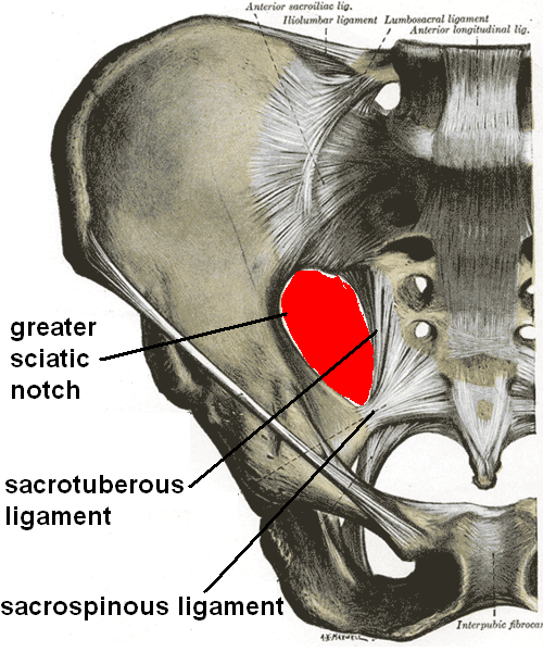
The One Structure Above Piriformis
It leaves the pelvis through the greater sciatic foramenThe greater sciatic notch of the hip bone is converted into a foramen by the sacrospinous ligament. Piriformis runs through it and splits it into a small space above and a larger space below., passing above the piriformis. It is the ONLY structure that exits above piriformis. Just outside the pelvis it splits into a superficial branch and a deep branch.
Superficial vs Deep
Superficial branch: into the overlying gluteus maximus. Deep branch: runs between gluteus medius and minimus, the heart of the abductor supply.
Board Trap
Everything else in the gluteal region (inferior gluteal, sciatic nerve, pudendal vessels and nerve, posterior femoral cutaneous nerve) exits BELOW piriformis. Above piriformis is a one-tenant building.
Piriformis: the muscle that splits the foramen into the door above and the door below.
What It Feeds
The Hip Abductors
Its deep branch supplies gluteus medius, gluteus minimus, and tensor fasciae latae, the muscles that abduct the hip and stabilize the pelvis when you stand on one leg. The superficial branch feeds the superficial part of gluteus maximus.
Territory
Gluteus medius and minimus and tensor fasciae latae (the abductors) plus superficial gluteus maximus. Not the femoral head.
If It Is Injured
Think function loss in the muscles it feeds: abductor weakness and a Trendelenburg pattern, not avascular necrosis of the femoral head. Different territory, different story.
Medius, minimus, tensor fasciae latae: the abductor crew. The superior gluteal is their landlord. Cut the landlord and the abductors weaken, the hip does not rot.
Gluteus medius and minimus: the abductors the superior gluteal artery feeds.
Above Or Below The Piriformis
Piriformis splits the greater sciatic foramen into two doors. Tap each door to see who walks through. This one image separates the superior gluteal from its look-alike, the inferior gluteal.
The greater sciatic foramen: piriformis runs through it, making one space above and one below.
Piriformis is the gatekeeper. One door above, one door below.
Above piriformis: one tenant. The superior gluteal artery (with its vein and the superior gluteal nerve) is the ONLY thing that exits here. The superior gluteal nerve is why injury here also threatens abduction: it powers gluteus medius, minimus, and tensor fasciae latae.
Below piriformis: the crowd. Everything else exits here: the inferior gluteal artery and nerve, the sciatic nerve, the pudendal nerve and internal pudendal vessels, the posterior femoral cutaneous nerve, and the nerves to obturator internus and quadratus femoris. The inferior gluteal artery runs WITH the sciatic nerve and feeds gluteus maximus.
Superior is above, inferior is below. Boring, and exactly why it works. The superior gluteal is the lonely tenant on the top floor; everyone else crowds the ground floor with the sciatic nerve.
The Abduction Red Herring
Here is the exact trap that makes smart students pick the superior gluteal. Walk into it on purpose, then watch it fall apart.
Back to the boy. The stem says restricted abduction. Your gut fires: abduction means the abductor muscles, the abductors are fed by the superior gluteal artery, so the answer must be superior gluteal. Before you commit, tap what is ACTUALLY limiting his abduction.
The restriction is mechanical. The femoral head is undergoing avascular necrosis, the joint is irritable and swollen, and a sore, distended hip simply will not move through its range. That is why he guards abduction and internal rotation and holds the leg in external rotation. It is a painful joint, not a starved muscle.
If the superior gluteal artery were the problem, you would see abductor weakness and a Trendelenburg gait over time, not 8 weeks of insidious pain with joint-space widening on film. Superior gluteal injury weakens the abductors. It does not rot the head.
Kill shot: it feeds the abductors, not the head. Restricted abduction here is a sore joint, not a starved one.
Abduction restricted does not equal abductor artery. A child who cannot abduct a painful hip is protecting a joint. A patient whose superior gluteal is cut cannot HOLD the pelvis level: that is Trendelenburg, a different sentence entirely.
Find The Real Artery
Two quick decisions to lock the right vessel, then knock out all four distractors on the answer sheet one at a time.
Legg-Calve-Perthes: the dead, flattened femoral head that the right artery would have fed.
Decision 1. The thing that is actually dying in this boy is the femoral head. So the artery we care about is the one that feeds what?
Right idea to anchor on the dying structure. The femoral head is the necrotic part, so we want its blood supply, not the muscle supply. Abductor supply is the superior gluteal, and that is the trap. Next decision unlocks below.
Decision 2. Which artery wraps the posterior femoral neck and sends retinacular branches into the head?
Medial circumflex femoral artery. It curves around the back of the femoral neck, and its retinacular branches are the dominant supply to the head in a child past infancy. Compromise it and the head loses its blood: avascular necrosis. That is the answer the stem is built around.
Knock Out The Distractors
Five arteries are on the answer sheet. Four are wrong. Tap each one to hear why it dies, and find the single artery that feeds the femoral head.
Superior gluteal
Inferior gluteal
Deep femoral (profunda)
Artery of ligamentum teres
Medial circumflex femoral
Medial Circumflex = Marrow Circulation of the head. It hugs the back of the neck. The superior gluteal hugs the abductors. Two different jobs, two different vessels.
Clinical Walkthrough
Original board-style vignettes. Five appear per round, shuffled, never repeating until the bank is exhausted. Right-click or long-press to cross out options. Double-tap to highlight.
Bone Wizardry is an independent educational resource for visual learning in the medical sciences. It is not affiliated with, endorsed by, or sponsored by any licensing or examination board, contains no real or recalled examination questions, and does not guarantee any educational or examination outcome.