Intro
Subjects MnemonicsCluecards Practice Questions Tutoring Physicians Prescryption
ANATOMY · THE FOVEAL TRAP

Artery of the Ligamentum Teres

The tiny vessel that runs inside the ligament of the head of the femur. It promises to feed the head and delivers a dimple. Here is why it is the seductive wrong answer in a limping child.

Quick Challenge

A 7-year-old boy has 8 weeks of insidious left thigh pain and a limp, no trauma. He has restricted abduction and internal rotation, the leg is held in external rotation, and the hip forces into external rotation on flexion. A pelvic radiograph shows widening of the left hip joint. This is early avascular necrosis of the femoral head. Loss of which vessel best explains it?

This is the classic pediatric hip picture, and the trap is built for you: the patient is a child, so your gut reaches for the "child artery," the artery of the ligamentum teres. Do not take the bait. Even in a 7-year-old, the head runs on the retinacular branches of the medial circumflex femoral artery. The foveal artery feeds only a small dimple around the fovea and is already fading at this age. Whole-head necrosis is a medial circumflex femoral problem, every time.
↓ scroll to explore

The Vessel Itself

A complete tour of this little artery: where it comes from, where it goes, what it feeds, and the catch that turns it into a board trap. Tap the tabs.

Coronal section of the hip joint showing the ligament of the head of the femur running from the fovea to the acetabular fossa
The ligament of the head spans the joint from the fovea to the acetabular fossa, and the foveal artery hitches a ride inside it.
Where it starts
A Branch of the Obturator

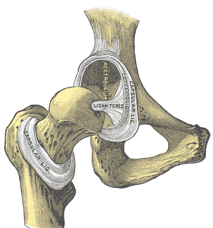
The artery of the ligament of the head of the femur is a small branch of the obturator artery, specifically its posterior divisionThe obturator artery splits into anterior and posterior divisions around the obturator foramen. The posterior division sends the acetabular branch into the hip joint., which sends an acetabular branch into the joint. In some people it arises instead from the medial circumflex femoral artery.

Parent vessel
Obturator artery (posterior division, acetabular branch). Variant origin: medial circumflex femoral artery.
The obturator is a pelvic artery, not a thigh artery. That alone tells you this vessel reaches the head through the inside of the joint, not by wrapping the neck like the real workhorse does.
Where it travels
Through the Ligamentum Teres

It runs inside the ligament of the head of the femur (the ligamentum teres), traveling from the acetabular fossa to the head, and enters the bone at the fovea capitis, the small central pit on the head.

Route
Acetabular fossa → inside the ligamentum teres → enters the femoral head at the fovea capitisThe fovea capitis is the little dimple on the medial head where the ligamentum teres anchors. It is the only spot the foveal artery can reach..
The ligament is a rope across the joint, and the artery hitches a ride on it to one tiny landing spot: the fovea. One rope, one dock. That is the whole journey.
What it feeds
Only a Dimple

It perfuses a small region of the femoral head around the fovea, nothing more. It is most relevant in young children, and even then it is a minor contributor. With age it regresses and is often insignificant in adults.

Territory
A small zone of the head immediately around the fovea capitis. Not the neck, not the dome.
Lifespan
Minor even in childhood. Shrinks and often closes off with age, so in adults it usually contributes little or nothing.
Fovea means a small favor. The foveal artery does a small favor (a dimple of perfusion) when you are young, then quietly retires. Never promote it to the main supply.
The board trap
Not Dominant, Even in a Kid

Here is the catch that the boards love: people assume a child must run on the "child artery." Wrong. Even in a 7-year-old, the head is fed mostly by the retinacular branches of the medial circumflex femoral artery. Whole-head necrosis (the Perthes pattern) reflects loss of that medial circumflex femoral supply, not the foveal artery.

It feeds a dimple, not the dome. A vessel that supplies a pinhead zone cannot kill the whole head. If the entire head dies, look at the artery wrapping the neck.

Dimple vs Dome

One picture settles the whole argument. Cut each artery and watch what dies. The femoral head is pink where it has blood and gray where it does not.

Upper end of the femur showing the head, neck, and the fovea capitis
The fovea capitis is the small pit on the head where the foveal artery enters. Everything around it is dome.
CUT AND COMPARE
The foveal artery reaches one little spot. The medial circumflex femoral wraps the neck and fans over the whole head. Pick one to cut.
DOME DIMPLE MCFA
Route
Obturator artery to the ligamentum teres to the fovea capitis.
Pattern
Feeds a dimple, never the dome, and regresses with age.
Pearl
Whole-head necrosis is medial circumflex femoral territory, never the foveal artery.
Radiograph of Legg-Calve-Perthes disease of the left hip
Perthes: whole-head necrosis
Pelvic radiograph of Legg-Calve-Perthes disease
Perthes pelvis: collapse of the head

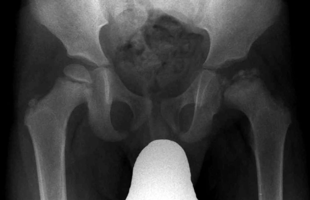
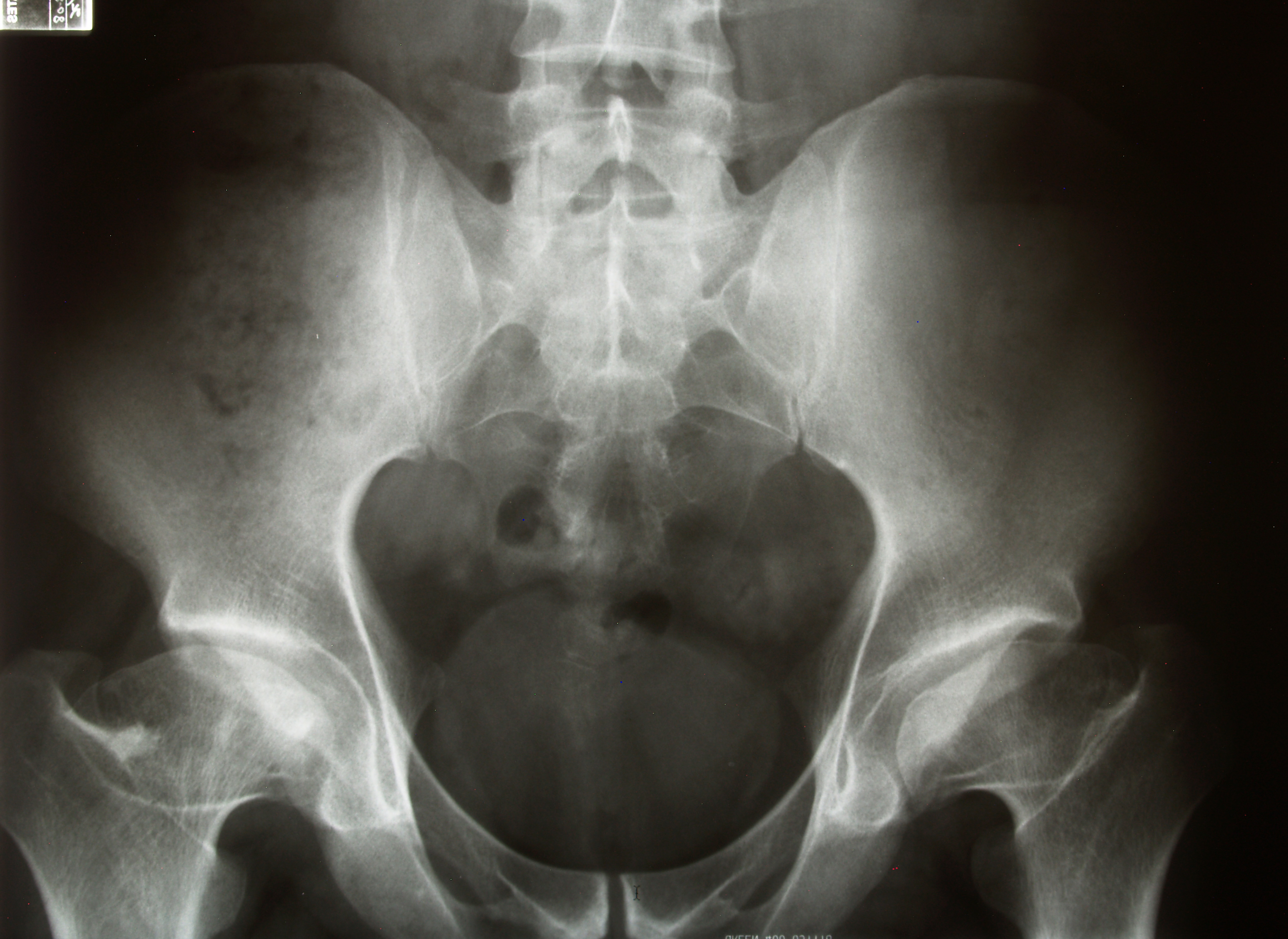
Whole-head necrosis on film: this is the medial circumflex femoral pattern, never a foveal artery problem.

The Five-Artery Lineup

This is the board set. One is the answer. Four are seductive wrong picks. Tap each one for its territory and its quick kill.

Anterior view of the hip joint capsule and ligaments sheathing the femoral neck
The capsule and ligaments sheath the femoral neck, the road the medial circumflex femoral retinacular branches climb to feed the head.
The trap on this page
Artery of the Ligamentum Teres

Reaches only the fovea, contributes little even in childhood, and regresses with age. It is tempting because the patient is a child, but it cannot explain death of the whole head.

It feeds a dimple, not the dome. Pinhead territory cannot cause whole-head necrosis.
The correct answer
Medial Circumflex Femoral Artery

Wraps around the posterior femoral neck and sends up retinacular branches that are the main blood supply to the head and neck, in adults and in children. Lose this and the whole head dies. That is Perthes, a displaced neck fracture, or a posterior hip dislocation.

Wraps the posterior neck, retinacular branches feed the head. Whole-head necrosis points here, every time.
Too far upstream
Deep Femoral Artery

The deep femoral arteryAlso called the profunda femoris. It is the large branch of the femoral artery that supplies the thigh muscles and gives rise to both circumflex femoral arteries. (profunda femoris) is the parent that gives off the circumflex femoral arteries, and it supplies the thigh muscles. It is the source, not the vessel that actually reaches the head.

It is the parent company, not the worker on site. The branch that touches the head is the medial circumflex femoral, not its parent.
Wrong neighborhood
Inferior Gluteal Artery

Supplies the buttock and runs alongside the sciatic nerve as its companion artery. It is a posterior pelvic vessel, not a femoral head vessel.

It is a buttock and sciatic-nerve artery. It never reaches the femoral head.
Powers the wrong thing
Superior Gluteal Artery

Supplies the hip abductors (gluteus medius and minimus, tensor fasciae latae). Injury gives a TrendelenburgA Trendelenburg sign is pelvic drop on the unsupported side because the hip abductors on the stance leg are weak. It is an abductor problem, not a femoral head problem. pattern, not avascular necrosis of the head.

It powers the abductors, not the head. Think Trendelenburg, not necrosis.

Beat the Trap

Commit to an answer before you reveal it. Guess each step, then check yourself.

Step 1. Quick gut check: does the foveal artery feed the whole femoral head, or just the fovea?
Right. The foveal artery only reaches a dimple around the fovea, and that contribution fades with age. A vessel this small cannot necrose the entire head.
Step 2. So when the whole head dies in a child, which artery's loss explains it?
Exactly. Even in childhood the retinacular branches of the medial circumflex femoral are the dominant supply. Whole-head necrosis equals medial circumflex femoral, not the foveal artery.
Step 3. The medial circumflex femoral wraps which surface of the femoral neck?
Correct. It wraps the posterior neck, which is exactly why a displaced femoral neck fracture or a posterior hip dislocation can sever it and cause avascular necrosis.

Sticky Hooks

Tap each card to bring it into focus. These are the lines that keep the foveal artery in its lane.

The Tease
Ligamentum teres is the tease. It runs all the way to the head and then only feeds a dimple. All promise, no delivery.
tap
Small Favor
Fovea means a small favor. The foveal artery does a small favor when you are young, then regresses. Never the main supply.
tap
Most Crucial
Read MCFA as Most Crucial Femoral-head Artery. It wraps the back of the neck, and its retinacular branches are the real supply.
tap
Kids Too
It is a child, so it must be the kid artery? No. Kids run on the MCFA too. The foveal artery is a backup that is already fading.
tap

Test Yourself

Five vignettes pulled from a larger bank, shuffled each visit. No timer. Get them wrong here so you never miss them on the day that counts.

Clinical Walkthrough

Board-style vignettes. Answers shuffle each round. Right-click or long-press to cross out an option. Double-tap to highlight one.

VIGNETTE 1
Educational anatomy review · Bone Wizardry · Last updated June 2026
Bone Wizardry is an independent educational resource for visual learning in the medical sciences. It is not affiliated with, endorsed by, or sponsored by any licensing or examination board, contains no real or recalled examination questions, and does not guarantee any educational or examination outcome.