Tap a glowing zone to read the lesion.
TSC drops a hamartoma in every system · that is the whole shape of the disease.
Brain
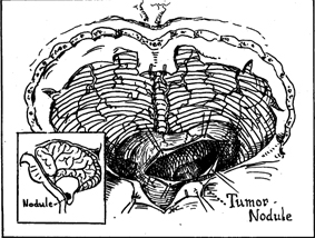
Glial hamartomas, cortical tubers, SEGA
Subependymal nodules and cortical tubers cause seizures (often infantile spasms) and intellectual disability. SEGA (subependymal giant-cell astrocytoma) sits at the foramen of Monro and can obstruct CSF.
Pearl: SEGA at the foramen of Monro is the TSC brain tumor everyone gets quizzed on. New-onset hydrocephalus in a TSC patient = SEGA blocking the foramen.
Heart
Rhabdomyoma
Benign muscle tumor often picked up on prenatal ultrasound. Frequently regresses on its own by age 4. Can cause arrhythmias or outflow obstruction in infancy.
Pearl: Cardiac rhabdomyoma in a fetus or infant → check for TSC. It is the only phakomatosis with a cardiac tumor.
Kidney
Angiomyolipoma (AML)
Fat + vessels + smooth muscle. Often bilateral. Big AMLs bleed catastrophically (Wunderlich hemorrhage). Renal cysts may also be present.
Pearl: Painless flank mass or hematuria in a young TSC patient → image the kidneys. AML > 4 cm = consider embolization or mTOR inhibitor.
Skin
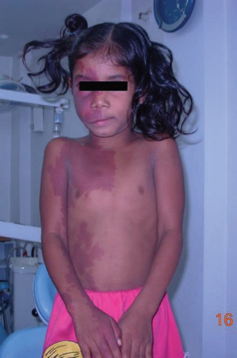
Ash-leaf spots · shagreen · angiofibromas
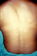
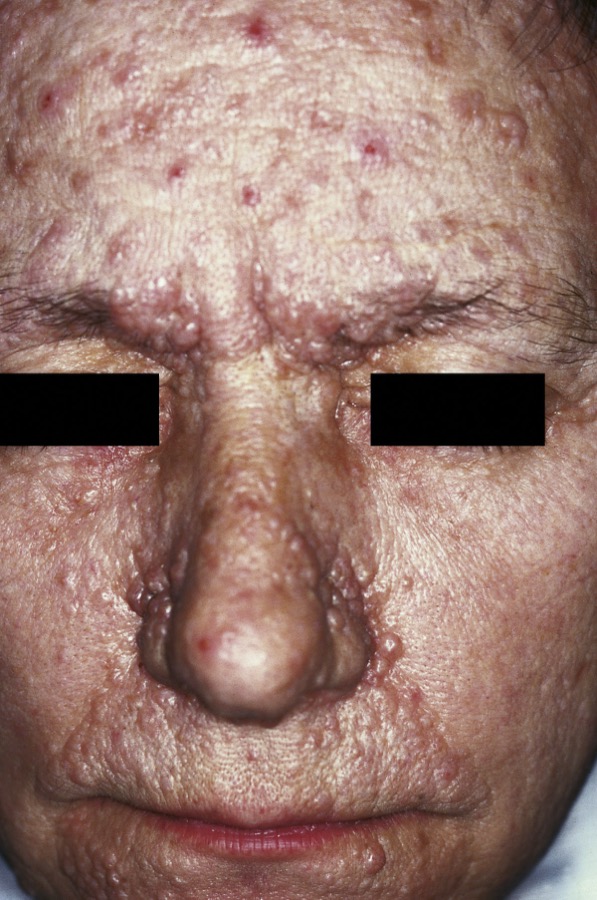
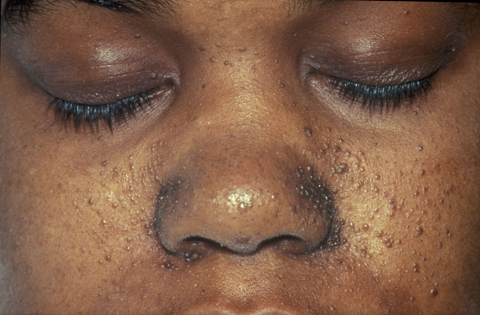
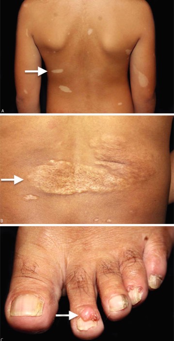
Ash-leaf spots: hypopigmented macules, brightest under Wood lamp. Shagreen patch: leathery raised plaque, usually lumbar. Facial angiofibromas (adenoma sebaceum): red papules across nose + cheeks, post-puberty.
Pearl: A baby with infantile spasms? Get a Wood lamp. Ash-leaf spots are often the first visible sign of TSC.