One little vessel feeds the entire femoral head in older kids and adults. Cut it and the bone dies. This is the answer to Legg-Calve-Perthes, hip dislocation, SCFE, and the dreaded femoral neck fracture.
Quick Challenge
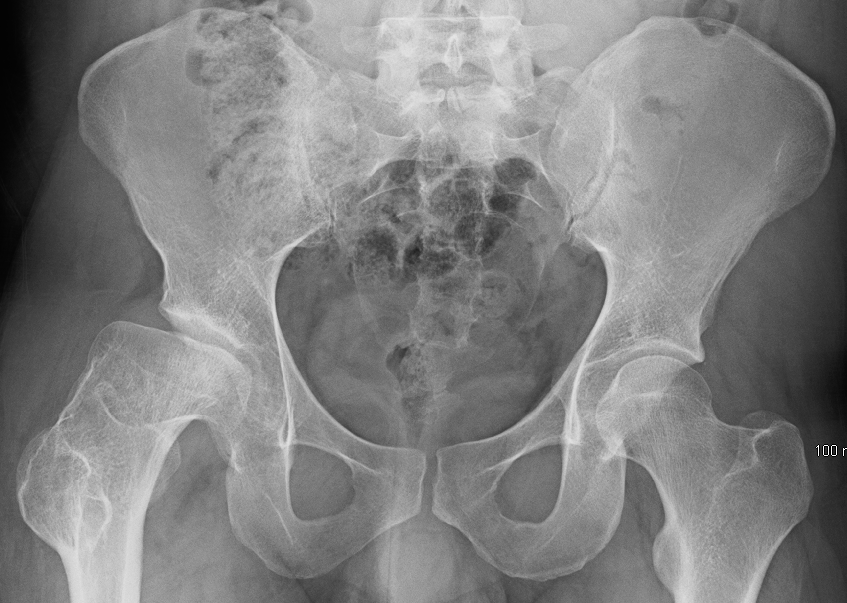
A 7-year-old boy has 8 weeks of insidious left thigh pain and a limp, no trauma. Exam: lumbar group curve (sidebent left, rotated right), normal knee, left leg held in slight external rotation, restricted abduction and internal rotation of the thigh, and forced external rotation when the hip is flexed. Pelvic film: widening of the left hip joint. Which vessel is most likely affected?
This is Legg-Calve-Perthes: idiopathic avascular necrosis of the capital femoral epiphysis in a young child. The blood feeding that epiphysis arrives through the medial circumflex femoral artery (MCFA) and its retinacular branches climbing the femoral neck. Lose that supply and the head dies. The lumbar group curve is a compensatory red herring, not the lesion. Scroll down and watch the blood travel.
↓ scroll to trace the blood
The Lifeline
Watch the Head Die
The femoral head has no backup once the growth plate seals off the metaphyseal vessels. Press play and follow Leo's blood up the neck, then watch what happens when the MCFA is cut.
Chapter One · The Limp
LEO
AGE 7
"Eight weeks of a limp and a sore left thigh. No fall. He just stopped running at recess."
Chapter Two · The Supply
The MCFA climbs the posterior femoral neck and sends retinacular arteries up under the synovium to feed the head.
Chapter Three · The Dark
No retinacular flow, no oxygen, no bone repair. The capital epiphysis becomes avascular necrosis: the radiographic widening and later flattening you saw on Leo's film.
PatternLose the MCFA → avascular necrosis of the capital epiphysis
PearlIn older kids and adults the MCFA owns the head. The foveal artery is a tiny backup that fades.
Perthes hip: the blood-starved head has flattened and the joint space looks too wide. This is Leo's film.A real femoral head after its blood supply died. The dome has collapsed.
The Lineup
Five Vessels, One Answer
Every distractor in a femoral-head question is a real artery near the hip. Tap each one and learn the single sentence that kills it. The MCFA is the only one that owns the head.👑Lateral = Limb (anterior thigh). Medial = Main supply to the head.
Two circumflex arteries circle the femoral neck. The medial one is the branch that climbs onto the head.
The Correct Answer
Medial Circumflex Femoral Artery
Origin
Usually a branch of the deep femoral artery (profunda femoris); sometimes directly off the common femoral.
Course
Passes posteriorly between iliopsoas and pectineus, then wraps the posterior/medial femoral neck.
Branches
Its deep branch gives the posterosuperior and posteroinferior retinacular arteriesThese run up the femoral neck under reflections of synovial membrane (the retinacula of Weitbrecht), so they hug the bone and tear with neck fractures and dislocations. that climb the neck under the synovial retinacula.
Supplies
The main blood supply to the femoral head and neck in older children and adults. A key limb of the cruciate anastomosisA cross-shaped arterial network behind the hip linking MCFA, lateral circumflex femoral, inferior gluteal, and first perforating artery. It is collateral for the thigh, not enough to save the head..
Why it wins: it is the one vessel whose branches physically climb onto the femoral head. Cut it and the head has no other reliable supply.👑Medial Circumflex = Main Circulation to the head. The lateral one is the imposter.Femoral head AVN question = MCFA. Every time.
Distractor 1
Deep Femoral Artery
What it is
The profunda femoris: the large branch of the femoral artery that supplies the thigh muscles and gives off the circumflex vessels.
Why it tempts
It is the parent of the MCFA, so it feels "close enough." It is too proximal: it never reaches the head itself.
You know how naming the river when they asked for one small creek gets you a wrong answer? The deep femoral is the river. The MCFA is the tributary that actually reaches the head. The deep femoral is the parent, not the vessel that perfuses the head.
Distractor 2
Artery of the Ligamentum Teres
What it is
The foveal artery (off the obturator), running in the ligamentum teres to a tiny patch of head around the fovea.
Why it tempts
It does enter the head, and in very young children it contributes. By later childhood and adulthood it regresses to almost nothing.
Think of it like a garden hose pointed at one corner of a football field. It waters that corner, not the whole field. The foveal artery feeds a postage stamp near the fovea, not the head. It fades with age.
Distractor 3
Inferior Gluteal Artery
What it is
Branch of the internal iliac that exits below piriformis to supply gluteus maximus and travel with the sciatic nerve.
Why it tempts
It is a minor contributor to the cruciate anastomosis, so it sits "in the neighborhood" of the hip.
It is the wrong neighborhood entirely: that is the buttock, not the head. Its only hip role is a minor anastomotic backup. Inferior gluteal feeds glute max and the sciatic companion, not the femoral head.
Distractor 4
Superior Gluteal Artery
What it is
Branch of the internal iliac exiting above piriformis to supply the abductors: gluteus medius, minimus, and tensor fasciae latae.
Why it tempts
Leo had restricted abduction, so the abductor artery looks attractive. That restriction is mechanical pain, not ischemia.
The exam is baiting you: it pairs an abductor finding with the abductor artery. But the abductors are sore from a sick joint, not starved of blood. Superior gluteal feeds the abductors, not the head. Restricted abduction here is pain, not infarct.
Trace the Risk
Guess Before You Reveal
Same artery, four classic ways to wreck it. Commit to an answer before each branch opens.
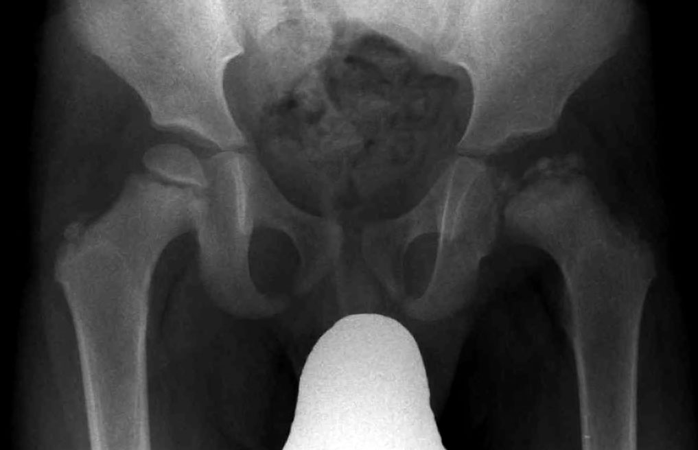
Another Perthes hip on plain film: the classic 7-year-old picture from Step 1.
Step 1: In a 7-year-old, which vessel carries most of the blood to the femoral head?
By this age the foveal (ligamentum teres) artery has mostly regressed. The MCFA and its retinacular branches do the real work. That is exactly why an idiopathic supply failure here produces Perthes.
Step 2: A 70-year-old falls and sustains a displaced subcapital (intracapsular) femoral neck fracture. Why is AVN the feared complication?
The retinacular arteries hug the neck under the synovium, so a high (subcapital) fracture shears them off and the head loses its MCFA supply. This is why displaced intracapsular fractures in the elderly get a replacement rather than fixation.
Step 3: A young man in a head-on collision has a flexed, adducted, internally rotated, shortened limb. Which injury threatens the MCFA?
Flexed, adducted, internally rotated, and shortened is the classic dashboard posterior hip dislocation. The MCFA wraps the posterior neck, so a posterior dislocation kinks or tears it. Reduce it fast (within hours) to save the head.
Memory Vault
Hooks That Stick
Tap a card to unblur the hook. These are the lines that get you to MCFA in three seconds on test day.
Medial vs Lateral
Medial Circumflex = Main Circulation to the head. The lateral circumflex mostly feeds the front of the thigh and trochanter.
tap to reveal
Anterior vs Posterior approach
Surgeons fear the posterior approach because the MCFA runs posteriorly. "Posterior puts the supply on the line."
tap to reveal
The foveal trap
Ligamentum teres artery is a kid's spare tire that deflates. By adulthood it is nearly useless, so it is never the head's main supply.
tap to reveal
The four ways to kill it
Perthes, SCFE, Fracture (neck), Dislocation (posterior). Four roads to AVN, one artery at the end of each.
tap to reveal
Board Trap
Do not pick the lateral circumflex femoral artery. It is the larger vessel, so it feels like the "main" one, but it mostly supplies the anterior thigh and the trochanteric region. The head belongs to the medial circumflex.👑Bigger is not better here. The smaller medial circumflex is the one that climbs onto the head. If a stem mentions femoral head ischemia and offers both, the medial one wins.
Board Walkthrough
One Vignette at a Time
Six original board-style cases. Pick an answer, then open the wrong choices to see exactly why each one dies. Shuffle for a fresh order.
Under the microscope: dead trabecular bone with empty spaces where living cells should be. This is the endpoint every case below is racing to prevent.
1 OF 6
Rapid Quiz
Don't Kill the Hip
Five quick patients walked into your clinic. Get them to the right artery. Miss one and the explanation hands you the cheat code so you never miss it again.
Bone Wizardry is an independent educational resource for visual learning in the medical sciences. It is not affiliated with, endorsed by, or sponsored by any licensing or examination board, contains no real or recalled examination questions, and does not guarantee any educational or examination outcome.