Every cranial nerve has exactly one exit point. Know the bone, name the nerve.
Board Vignette: A 24-year-old unrestrained driver strikes the windshield during a head-on collision. He is confused, has severe facial swelling, multiple broken teeth, and a large forehead hematoma. CT of the head shows a fracture through the crista galli of the ethmoid bone. Three days later he cannot identify the smell of coffee. Which structure's damage BEST explains this finding?
A. Middle meningeal artery
B. Olfactory nerve (CN I)
C. Oculomotor nerve (CN III)
D. Trigeminal nerve (CN V)
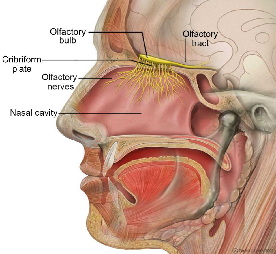
The crista galli sits on top of the cribriform plate of the ethmoid bone. The olfactory nerve fibers (CN I) thread up through tiny holes in that plate to reach the olfactory bulbs above. When the crista galli fractures, it drags the cribriform plate with it, shearing those fibers. No fibers, no smell. The middle meningeal artery lives in the sphenoid (foramen spinosum), CN III exits the sphenoid too (superior orbital fissure), and CN V has three exits, all in the sphenoid. Ethmoid = CN I. Full stop.
The Olfactory Bedside Test
Pick a substance. Pick a nostril (occlude the other). Run the test. Some choices are traps. The cribriform plate is the punchline.
Pick A Substance
Tap a nostril to occlude it · test the OTHER side.
From the Attending
CN I tests olfaction only. Ammonia and alcohol pads stimulate CN V trigeminal pain fibers, not olfactory. If a patient with anosmia says they smell ammonia, they still have intact CN V · CN I is the dead one. Cribriform plate fracture = bilateral anosmia + CSF rhinorrhea.
Ethmoid vs. Sphenoid
Two bones. Most of the board traps live in one of them.
ETHMOID
SPHENOID
TEMPORAL & OCCIPITAL
Ethmoid Bone
Anterior cranial fossa floor · the "sieve bone"
CN exits hereCN I (Olfactory)
Key foramenCribriform plate (hundreds of tiny holes)
LandmarkCrista galli separates L & R olfactory bulbs · falx cerebri attachment
Fracture resultAnosmia + possible CSF rhinorrhea
Board trapSmells like a CN V question because the face hurts → it's always CN I when the ethmoid breaks
Sphenoid Bone
Middle cranial fossa · "the great bone" · most CN exits
CN exits hereCN II, III, IV, V1, V2, V3, VI
Optic canalCN II (Optic) → vision loss, APD
Superior orbital fissureCN III, IV, V1, VI → orbital apex syndrome
Three bones own the skull base. Ethmoid = anterior fossa = CN I only.Sphenoid = middle fossa = II through VI (all the orbital traffic + V branches).Temporal + occipital = posterior fossa = VII through XII (sound + lower CN). When a stem says "head trauma with anosmia and CSF rhinorrhea," you don't think CN V because the face hurts · you think cribriform plate of the ethmoid sheared CN I and the meninges. The bone tells you the nerve.
Skull Base Map
Tap any foramen. One exit. One nerve. One bone.
INTERIOR VIEW · SKULL BASE FROM ABOVE · TAP A FORAMEN
FORAMEN
Anterior (Ethmoid)
Middle (Sphenoid)
Posterior (Temporal/Occipital)
From the Attending
The crista galli is a bony fin sticking up from the ethmoid · the falx cerebri hooks onto it like a tent rope. That's why a high-impact frontal blow can shear the falx, the cribriform plate, and CN I all at once. Stem says "frontal head trauma with bilateral periorbital ecchymosis (raccoon eyes) and loss of smell" · that's anterior cranial fossa fracture through the cribriform plate. CSF leaking through dural tears = rhinorrhea. The crista galli is the landmark, not the lesion.
Fracture Finder
Pick the fracture site. Commit before the answer drops.
1
The patient has a basilar skull fracture. CT shows the break is in the floor of the ANTERIOR cranial fossa. New complaint: no smell. Which bone is cracked?
Ethmoid bone (anterior fossa floor)
Sphenoid bone (middle fossa floor)
Temporal bone (posterior fossa)
2
Correct. The ethmoid's cribriform plate is the floor of the anterior fossa. Now: the patient also notices clear fluid dripping from his nostril. What's leaking?
CSF (beta-2 transferrin positive)
Arterial blood from the middle meningeal artery
3
Separate case: unrestrained passenger, eye deviates "down and out," ptosis, pupil dilated and fixed. The fracture is in the SPHENOID. Which exit point is involved?
Superior orbital fissure (CN III exits here)
Optic canal (CN II exits here)
Foramen ovale (CN V3 exits here)
From the Attending
Anosmia workup is a two-question funnel. (1) Conductive or sensorineural? Nasal blockage (URI, polyps, deviated septum) blocks odorants from reaching the bulb · the nerve still works. (2) If the airway is clear, is it neural? Trauma to the cribriform plate, COVID/post-viral, neurodegenerative disease (Parkinson, Alzheimer), or a frontal mass compressing the bulb. clinical medicine rarely test "rhinitis" · they test the trauma + neurodegenerative angles. Anosmia + frontal lobe signs = think olfactory groove meningioma until ruled out.
Wrong Answer Autopsy
Every distractor has a board trap. Tap to flip and find it.
Middle Meningeal Artery
Why it feels right: It's in the skull. Trauma can rupture it. Very famous board answer.
The Trap: Facial trauma + skull fracture. "MMA injury?" Feels correct. 4.2% of board-takers chose this.
flip for the board trap →
Board Trap
The problem
The middle meningeal artery exits the skull through the foramen spinosum, which is in the sphenoid bone, not the ethmoid. An ethmoid fracture cannot directly damage it.
The rule
MMA + epidural hematoma = temporal bone (pterion), not ethmoid. If the question says "ethmoid" or "crista galli," the answer is NEVER the MMA.
Oculomotor Nerve (CN III)
Why it feels right: Big nerve. Common board answer. Comes up in trauma all the time.
The Trap: "Skull fracture + cranial nerve" → brain goes to "CN III palsy." 5% chose this.
flip for the board trap →
Board Trap
The problem
CN III exits through the superior orbital fissure in the sphenoid. A CN III palsy presents with down-and-out gaze, ptosis, and a blown pupil. This patient has none of that.
The rule
Ethmoid fracture + anosmia = CN I. CN III palsy requires a sphenoid fracture at the superior orbital fissure, a posterior communicating artery aneurysm, or uncal herniation.
Trigeminal Nerve (CN V)
Why it feels right: Face hurts. CN V is the sensory nerve of the face. Makes anatomical sense.
The Trap: Facial pain + facial trauma → "Must be the facial sensation nerve." 12.7% chose this.
flip for the board trap →
Board Trap
The problem
CN V has THREE exits, all in the sphenoid: V1 exits the superior orbital fissure, V2 exits the foramen rotundum, V3 exits the foramen ovale. None of them are in the ethmoid bone.
The rule
Facial pain is the distractor. The clue is anosmia + ethmoid. Pain after trauma is expected. Loss of smell is the specific finding that points to CN I.
Memory Hooks
Tap to unblur. Test yourself first.
🧀
Ethmoid = The Sieve
Ethmoid comes from the Greek word for "sieve." The cribriform plate has hundreds of holes like a kitchen strainer. CN I fibers thread through like spaghetti through a colander. Break the sieve, lose the smell.
tap to reveal
🕷️
Sphenoid = The Spider Bone
The sphenoid is shaped like a bat or spider with wings. It has the most CN exits of any bone: CN II through VI all exit through the sphenoid. If you're not sure which bone, guess sphenoid and you'll be right more often than not.
tap to reveal
🎸
Jugular = J9-10-11
The jugular foramen contains CN 9, 10, and 11 (plus the jugular vein). Think "JVP" but for cranial nerves: Jugular holds 9, 10, 11 (Vagus Plus). A jugular foramen lesion = hoarse voice (X) + dysphagia (IX) + dropped shoulder (XI).
tap to reveal
🏎️
Temporal = 7 & 8
The temporal bone houses the inner ear AND the facial nerve. "TeMporal = 7 & 8" because 7 is the facial nerve (moves the face) and 8 is vestibulocochlear (hears). A temporal bone fracture gives you Bell's palsy + deafness. Both in one shot.
tap to reveal
👅
XII in the Occipital
CN XII (Hypoglossal) exits through the hypoglossal canal in the occipital bone. XII moves the tongue. Occipital is the back of the skull. Easy to forget because it's far from the others. Remember: "XII lives alone in the back."
tap to reveal
💨
CSF Rhinorrhea Clue
Clear fluid dripping from the nose after head trauma = cribriform plate fracture until proven otherwise. Test it: beta-2 transferrin is only in CSF (not nasal mucus). A positive test confirms the dura is torn and CSF is leaking into the nasal cavity through the ethmoid.
tap to reveal
Clinical Anatomy
Tap to expand. Know what the real structures look like.
ETHMOID BONE · Anterior View · tap to expand
CN I ANATOMY · Olfactory Nerve Pathway · tap to expand
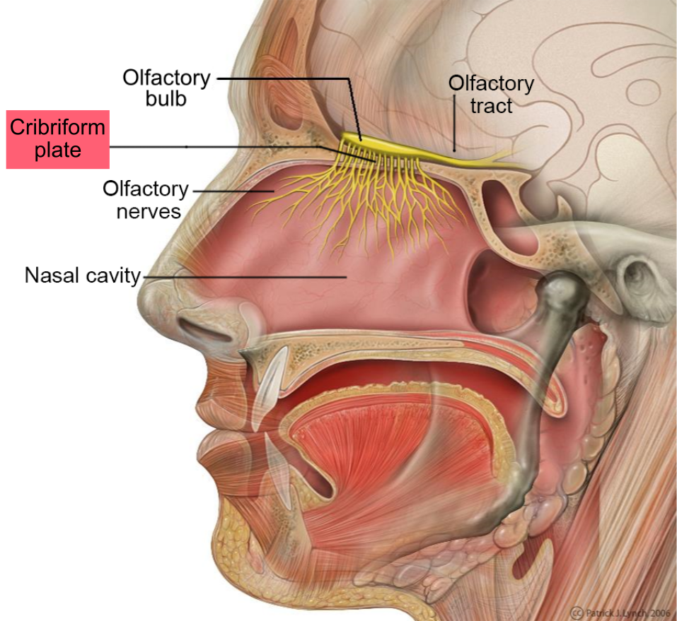
CRIBRIFORM PLATE · Highlighted Anatomy · tap to expand
Prove It
25 original board vignettes. One at a time. Tap each teaching beat to reveal the chain.
From the Attending
CN I stems disguise themselves. Watch for: head trauma + anosmia + CSF rhinorrhea (cribriform plate fracture), frontal lobe signs + anosmia in a middle-aged adult (olfactory groove meningioma), elderly + progressive anosmia + tremor or memory loss (Parkinson / Alzheimer prodrome), post-URI sudden anosmia (post-viral neuronal). The bone you're being asked about is almost always the ethmoid · the cribriform plate is the only window CN I has out of the skull. Bone → nerve → clinical. Every time.
Bone Wizardry is an independent educational resource for visual learning in the medical sciences. It is not affiliated with, endorsed by, or sponsored by any licensing or examination board, contains no real or recalled examination questions, and does not guarantee any educational or examination outcome.