CARDIOLOGY
Pericardium
vs Pleura
Both cause sharp chest pain. One gets better when you lean forward. One leaves a trail on the ECG. Know the tells.
The Setup
Read the Room First
A patient just walked in. One click. No hints. Make the call before you see the chain.
You are right that pericarditis causes sharp chest pain. And you are right that both pericardial and pleural pain get worse with inspiration. That is exactly why this question exists on boards.
Think of it like two car alarms going off in the same parking lot: same sound, different cars. Pericarditis has its own calling card: the pain gets significantly better when you lean forward (the inflamed pericardium pulls away from the surface below it), and the ECG shows diffuse saddle-shaped ST elevation with PR depression. This patient has neither. She has an O2 sat of 88%, cyanosis, and dullness at the left base. That is not an angry pericardium. That is a pulmonary embolism that has infarcted a wedge of lung, and the overlying parietal pleura is now irritated.
The Discriminator
Pleura vs Pericardium
Same sharp pain. Completely different tells. Tap each structure to load its profile.
| Feature | Pleuritic Pain |
|---|---|
| Pain quality | Sharp, stabbing, well-localized over the involved pleural surface |
| Worse with | Inspiration (deep breath), coughing, movement. Patient splints (takes shallow breaths to minimize pain). |
| Position effect | No significant positional relief. Lying on the affected side may reduce breathing depth and slightly help, but no dramatic change with posture. |
| Friction rub | Pleural friction rub over the affected area. Heard as a leathery creaking sound synchronous with breathing. KEY Rub disappears when patient holds breath (lungs stop moving = surfaces stop rubbing). |
| ECG | Normal, or reflects the underlying cause: S1Q3T3 pattern or sinus tachycardia in PE; no specific pleural ECG pattern |
| Classic causes | PE with pulmonary infarction (OCP + hypoxia), pneumonia / parapneumonic pleuritis (fever + cough + consolidation), pneumothorax (sudden + hyperresonant), malignant pleural effusion, viral pleuritis |
| Innervation | Intercostal nerves (T2-T12) for the costal and anterior parietal pleura. Phrenic nerve (C3-C5) for the central diaphragmatic pleura. REFERRED PAIN Phrenic irritation refers to the shoulder tip (ipsilateral). |
| Feature | Pericardial Pain |
|---|---|
| Pain quality | Sharp or pressure-like; may radiate to the left shoulder or trapezoid ridge (phrenic nerve territory); often constant with a positional overlay |
| Worse with | Supine position + inspiration. Lying flat brings the pericardium into contact with the inflamed surface. Both worsen it. |
| Position effect | BETTER LEANING FORWARD (elbows on knees). This is the single most important tell. Leaning forward lifts the pericardium away from the posterior mediastinal structures and the inflamed parietal pericardium. THE TELL |
| Friction rub | Pericardial friction rub: scratchy, high-pitched, triphasic (atrial systole + ventricular systole + early diastole). Best heard at the left sternal border, patient leaning forward, at end-expiration. KEY Rub persists when patient holds breath (heart keeps moving). |
| ECG | Diffuse concave-upward (saddle-shaped) ST elevation in all leads EXCEPT aVR (which shows reciprocal ST depression). PR depression in most leads (pathognomonic). This pattern respects NO coronary territory. |
| Classic causes | Viral/idiopathic (most common; post-URI by ~2 weeks), Dressler syndrome (autoimmune, 2-8 weeks post-MI or cardiac surgery), uremic pericarditis (dialysis patients, BUN >60), SLE/RASystemic lupus erythematosus and rheumatoid arthritis both cause pericarditis via immune complex deposition in the pericardium. SLE pericarditis can recur and is part of the serositis seen in the disease. |
| ECG trick | Pericarditis ST vs STEMI ST: Pericarditis = diffuse + concave (like a smile). STEMI = territorial + convex (like a frown). Plus: pericarditis has PR depression. STEMI does not. |
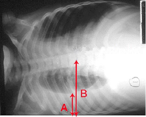
Clinical Images


The Map
Where Does the Signal Go?
Tap each mode. Watch the nerve paths light up. The shoulder tip connection is the one boards love to test.
Interactive Anatomy
Pain Locator
Somatic pleural pain vs positional pericardial pain
The Tell
Three Clues. One Diagnosis.
A 26-year-old man with sharp anterior chest pain sits down in your exam room. Work through the signs to confirm the diagnosis.
Board Walkthrough
Eight Patients. Same Sharp Pain.
Original vignettes. One at a time. The answer chain is on the other side of your choice.
Pericardium or pleura? Lean-forward or no? ECG or no ECG? Let's find out what stuck.
Good instinct: pericarditis also causes sharp chest pain. But think of the two types of chest inflammation as two different warning lights on a dashboard. Pericarditis lights up the "position" light: pain dramatically better leaning forward, and the ECG shows diffuse saddle ST + PR depression. This patient has neither. She has O2 sat 88%, cyanosis risk (OCP), and left-base dullness. That is a PE that has infarcted lung tissue, irritating the overlying parietal pleura. OCP + hypoxia + no positional relief = PE with pleural irritation. Not pericarditis.
OCP = hypercoagulable state. Hypercoagulable state = DVT/PE risk. PE with distal pulmonary infarction irritates the overlying parietal pleura (somatically innervated by intercostal nerves). Result: sharp, well-localized, respiratory-driven pleuritic pain. The dullness at the left base reflects the infarct-related effusion or consolidation. Parietal pleura = somatic pain = sharp + respiratory + no positional relief.
Esophageal pain (spasm, rupture) is midline, often triggered by swallowing or eating, and comes with dysphagia or odynophagia. It does not cause hypoxia. It does not cause dullness at the lung base. The entire hemodynamic and respiratory picture here points away from the esophagus. Esophagus: midline, swallowing-related, no hypoxia. This is none of those.
Myocardial ischemia in a 24-year-old woman is exceptionally rare without major risk factors. And myocardial pain does not track strictly with respiration the way this patient's does. The O2 sat of 88% would be unusual in ACS unless there is massive MI with cardiogenic shock. The picture here is overwhelmingly pulmonary vascular, not myocardial. Myocardial ischemia: crushing, central, radiates to arm or jaw, not respiratory. This is not that.
Classic presentation: young male, post-viral, leaning forward relieves pain, fever, diffuse concave ST elevation + PR depression, normal O2 sat. Most pericarditis is viral (Coxsackievirus B being the most common) or idiopathic. The leaning-forward sign + ECG pattern is the board-ready way to distinguish this from every other cause of chest pain. Post-viral + leaning-forward relief + diffuse saddle ST + PR depression = acute pericarditis.
PE is a great diagnosis to entertain for chest pain + fever in a young person. But PE causes pleuritic pain from pleural irritation, and that pain does NOT improve with leaning forward. Also, PE with hypoxia has O2 sat below normal, not 99%. And the ECG in PE shows sinus tachycardia or the S1Q3T3 pattern, not diffuse concave ST elevation across essentially every lead with PR depression. Leaning-forward relief + PR depression = pericarditis. PE does not do this.
Spontaneous pneumothorax in a young man would cause sudden pleuritic pain with decreased breath sounds and hyperresonance on the affected side. The pain would be constant and respiratory-driven but would NOT dramatically improve by leaning forward. Pneumothorax does not cause the ECG findings described here. And fever is not typical. Spontaneous PTX: hyperresonant + no breath sounds. This patient's O2 is 99% and the ECG tells the whole story.
ST elevation on ECG + chest pain = STEMI reflex. But STEMI has territorial ST elevation: LAD occlusion would hit V1-V4 (anterior), not leads I, II, aVF, V2-V6 simultaneously. More importantly, STEMI has NO PR depression, and STEMI pain does NOT improve with leaning forward. Also, STEMI in a 22-year-old without risk factors post-viral URI requires extraordinary evidence. Diffuse ST across multiple territories + PR depression + leaning-forward relief = pericarditis, not STEMI. Every time.
When pneumonia consolidates the lung, the infection often spreads to involve the visceral pleura. The adjacent parietal pleura (which IS somatically innervated) then becomes inflamed and produces sharp, respiratory-driven pain. This is parapneumonic pleuritis: pleuritis as a direct consequence of pneumonia. The pain is perfectly localized to the affected side because the intercostal nerves carry the signal precisely. Pneumonia + pleuritis = parietal pleural irritation. The consolidation on CXR confirms the source.
Pericarditis from direct extension of pneumonia is possible but rare. More importantly, pericarditis would NOT produce well-localized RIGHT-SIDED chest pain that tracks exactly with breathing. Pericarditis produces anterior chest pain that is better leaning forward and leaves a specific ECG signature (diffuse ST + PR depression). This patient's ECG shows sinus tachycardia only. No PR depression, no diffuse ST. Parapneumonic pleuritis is the common mechanism. Pericardial extension is rare and would show pericarditis ECG findings.
Esophageal spasm causes substernal, midline, squeezing chest pain often related to swallowing. It is not lateralized to one side. And a fever, productive cough, and right lower lobe consolidation are not part of esophageal disease. This is a classic pneumonia case with parapneumonic pleuritis. Esophageal spasm = midline + swallowing-triggered. This woman has right-sided pain with a consolidation. Not esophageal.
Musculoskeletal chest pain from coughing is a real thing, but it does not explain fever, consolidation, and productive cough. More importantly, musculoskeletal pain would be most tender to palpation, and palpation of the chest wall would reproduce or worsen the pain. Parapneumonic pleuritis does not reproduce on palpation the same way. The CXR consolidation seals this. Consolidation + fever + productive cough = pneumonia with pleuritis. Musculoskeletal strain does not explain a consolidation.
The vagus nerve is a major visceral sensory highway for the thorax, but its referred pain patterns go to the epigastrium and jaw area, not the shoulder. Vagal stimulation explains why a heart attack can feel like indigestion. But the shoulder tip is phrenic territory, not vagal. Vagus nerve refers to the epigastrium and jaw. Shoulder tip = phrenic nerve. Different nerve, different dermatome.
The intercostal nerves (T1-T11) innervate the costal and peripheral diaphragmatic pleura. They produce well-localized lateral chest pain that tracks exactly with the affected rib level. T4 refers to the nipple line, not the shoulder. The shoulder tip is C3-C5 territory, well above T4. Intercostal nerves = localized chest wall pain. Shoulder tip = C3-C5 = phrenic. Not T4.
C3, C4, C5 keep the diaphragm alive. The phrenic nerve also innervates the central diaphragmatic pleura. When the diaphragmatic pleura is inflamed (as it is here, given the left diaphragmatic density and effusion), the phrenic nerve carries that irritation up to the C3-C5 spinal segments. The brain interprets it as coming from the dermatome at those levels: the shoulder tip. The shoulder is not actually injured. It is referred pain from the diaphragm. Phrenic nerve (C3-C5) = central diaphragmatic pleura = referred pain to the ipsilateral shoulder tip.
The brachial plexus (C5-T1) supplies motor and sensory function to the arm. Brachial plexus problems cause arm weakness, numbness in specific dermatomes, and Horner syndrome if T1 is involved. Brachial plexus injuries do not cause referred shoulder pain from intrathoracic pathology. The connection to the diaphragmatic pleura goes through the phrenic nerve, not the brachial plexus. Brachial plexus = arm/hand function. Diaphragmatic pleura refers through the phrenic, not brachial plexus.
Anterior ST elevation in V1-V4 only = LAD territory = STEMI. STEMI does not cause leaning-forward relief. And STEMI ST elevation is convex upward (like a frown), not concave (like a smile). The presentation here screams pericarditis, not STEMI. If this were LAD occlusion, she would not feel better by leaning forward. STEMI ST elevation is territorial and convex. Pericarditis ST elevation is diffuse and concave. Know the shape.
Leaning-forward relief is pathognomonic for pericardial irritation. Given that, the expected ECG is the pericarditis pattern: diffuse concave-upward (saddle-shaped) ST elevation in essentially all leads except aVR, with PR depression in the same leads where ST is elevated (aVR mirrors these changes with ST depression and PR elevation). Post-viral timing of 10 days fits perfectly. Leaning-forward relief = pericarditis = diffuse saddle ST + PR depression. These two travel together.
S1Q3T3 is the classic (though not very sensitive) ECG finding in pulmonary embolism: right heart strain from sudden increase in pulmonary resistance. PE causes pleuritic pain from pleural irritation. PE pain does NOT improve with leaning forward. And PE would cause hypoxia, not O2 sat of 98%. S1Q3T3 = PE. Leaning-forward relief = pericarditis. These are different diseases with different ECGs.
A normal ECG is possible in very early pericarditis (Stage I has not yet developed, or changes are subtle). But given the full clinical picture (leaning-forward relief, fever, post-viral, normal O2), pericarditis is the working diagnosis and the expected ECG changes are the diffuse ST + PR depression pattern. A normal ECG in this context would not change the diagnosis. Normal ECG is possible but Stage I pericarditis ECG changes are expected and boards will test the expected finding.
Right upper sternal border is where you auscultate the aortic valve. A pericardial rub would not be particularly audible here, and keeping the patient supine is the worst position for a pericarditis patient (that is when their pain is worst). The pericardium is most accessible to the stethoscope at the left sternal border, and leaning forward brings it closer to the anterior chest wall. Pericardial rub: left sternal border, leaning forward, end-expiration. Not the aortic area.
The apex supine position is the sweet spot for mitral valve sounds (and for S3/S4 gallops). A pericardial rub can sometimes be heard at the apex but is far louder at the left sternal border. Keeping the patient supine actively suppresses the rub in pericarditis because that position increases pericardial contact. And "mid-systole" is single-component thinking: pericardial rubs are triphasic (atrial systole + ventricular systole + early diastole). Pericardial rub = triphasic at left sternal border, not single-component at apex.
Three simultaneous conditions amplify the pericardial rub: (1) Leaning forward brings the pericardium closer to the anterior chest wall. (2) Left sternal border is directly over the pericardium. (3) End-expiration means the lungs are mostly deflated, removing the air buffer between the pericardium and the stethoscope. Use the diaphragm piece (not the bell) and press firmly. This is a high-pitched, scratchy, multi-component rub. Left sternal border + leaning forward + end-expiration = maximum pericardial rub. This is the exam technique.
Left lateral decubitus + apex + inspiration is the classic trick for enhancing the mitral stenosis rumble. Rolling the patient onto their left side brings the mitral valve closer to the chest wall and the bell of the stethoscope. Do not mix up your cardiac auscultation positions: left lateral decubitus = mitral stenosis. Leaning forward at end-expiration at the left sternal border = pericardial rub. Left lateral decubitus = mitral stenosis rumble. Forward lean + LSB + end-expiration = pericardial rub. Know both.
Stent thrombosis is a legitimate fear after PCI. But think about what it would look like: crushing substernal chest pain, ST elevation in the anterior territory only (LAD distribution), cardiogenic shock, troponin surge matching the infarct territory, and absolutely no friction rub. A friction rub means two surfaces are rubbing together. Stent thrombosis does not produce a friction rub. Stent re-occlusion = STEMI-like picture + no friction rub. Dressler = friction rub + fever + 2-8 weeks post-MI. These are different pictures.
Dressler syndrome (post-cardiac injury syndrome) is an autoimmune pericarditis that develops 2-8 weeks after myocardial injury (MI, cardiac surgery, or cardiac trauma). The necrotic myocardium releases intracellular antigens that stimulate an immune response against the pericardium and pleura. Result: fever, pleuritic chest pain, pericardial friction rub, elevated inflammatory markers (ESR), and the pericarditis ECG pattern. Treat with NSAIDs plus colchicine. Fever + friction rub + elevated ESR + 2-8 weeks post-MI = Dressler syndrome. The immune system attacking the dead zone.
Bacterial purulent pericarditis is a rare, life-threatening infection, usually from direct extension (osteomyelitis, esophageal perforation) or hematogenous seeding. It would have much higher fever, the patient would look much sicker (septic), and the timing of 4 weeks post-MI does not fit a hospital-acquired infection. The post-MI 4-week timing with inflammatory markers is the Dressler syndrome fingerprint. Bacterial pericarditis = septic, high fever, rapid deterioration. Dressler = subacute, classic post-MI timing, responds to NSAIDs.
PE is worth considering in any post-MI patient because immobility + hypercoagulability from illness are both present. But PE causes PLEURAL pain (from infarction), which would not produce a pericardial friction rub. PE would also cause hypoxia, and its ECG shows sinus tachycardia or S1Q3T3, not diffuse ST elevation with PR depression. The pericardial rub in this case is specific to pericardial inflammation. Pericardial friction rub = pericardial inflammation. PE cannot produce a pericardial friction rub.
Great instinct: scratchy, creaking chest sound after a viral illness = pericarditis is the first thought. But here is the key: a pericardial friction rub PERSISTS when the patient holds their breath, because the HEART keeps moving. Pericardium rubs against itself with every heartbeat, not every breath. When this patient holds his breath and the sound disappears, that tells you the sound is generated by breathing movement alone. That means the two pleural surfaces are the source. Rub persists with breath-hold = pericardial. Rub disappears with breath-hold = pleural. This rub disappeared. Pleural.
Tietze syndrome (costochondritis with swelling) causes anterior chest wall tenderness and pain, reproducible by pressing on the costochondral junctions. It does not produce an auscultable creaking or scratching sound with breathing. And fever + viral URI + a sound that disappears with breath-holding is textbook pleuritis, not costochondritis. Costochondritis = tender to palpation, no audible rub. Pleuritis = audible rub that disappears with breath-hold.
Aortic dissection produces a tearing, ripping pain radiating to the back, with hemodynamic instability and (sometimes) an aortic regurgitation murmur at the right sternal border. It does not produce a creaking sound that tracks with breathing and disappears when breath is held. The post-viral fever also makes dissection extremely unlikely. Aortic dissection: tearing + back radiation + not respiratory. This is not aortic dissection.
The two-second test: ask the patient to hold their breath. Rub disappears = PLEURAL (the visceral and parietal pleural surfaces only rub when breathing moves them apart and together). Rub persists = PERICARDIAL (the heart never stops, so the pericardial surfaces keep rubbing even without breathing). Here: breath-hold = rub gone = pleural friction rub from pleuritis. Post-viral + fever + left lateral location = viral pleuritis. Breath-hold: rub gone = pleura. Rub stays = pericardium. Two-second bedside test, done.