OMM · Manipulation

HVLA Principles

High-velocity, low-amplitude thrust. The most tested OMM technique and the one with the most absolute contraindications.

Opening Challenge
A 78-year-old woman with rheumatoid arthritis presents with neck stiffness and a C2 somatic dysfunction. The attending offers to perform HVLA to the cervical spine. You object. Why are you correct?
Correct. Rheumatoid arthritis is an ABSOLUTE contraindication to cervical HVLA because RA can erode the transverse ligament and the odontoid process of C2, creating atlantoaxial instability. An HVLA thrust here could cause cervical cord injury or death. This applies specifically to the upper cervical spine. Other RA-affected joints also represent relative contraindications.
Not quite. The correct answer is B. RA is an ABSOLUTE contraindication to cervical HVLA because the disease erodes both the odontoid process and the transverse ligament at C1-C2. Gentleness is irrelevant once structural instability is present. Age alone is not the issue, and chiropractors face the same contraindication.
Section 1 of 7

The Mechanism

What HVLA actually is, how the pop happens, and how it differs from other OMM techniques

HVLA is a direct technique. You take up all the slack in the restricted direction until you reach the restrictive barrier, then apply a short, high-velocity thrust through that barrier with a low amplitude of movement. The key: you go into the restriction, not away from it.

The Core Formula
Position at the restrictive barrier (all slack removed) → Apply a high-velocity, low-amplitude thrust through the barrier → Gas cavitation occurs → Joint gaps, mechanoreceptors fire, paraspinal muscles reflexively relax.

The audible and palpable "pop" is nitrogen cavitationRapid drop in intra-articular pressure creates a gas bubble in the synovial fluid that collapses, producing the audible pop. Same physics as cracking your knuckles.. The joint spaces briefly gape, stretch the periarticular capsule and ligaments, and stimulate mechanoreceptors that reflexively inhibit paraspinal muscle hypertonicity. The pop is satisfying but NOT required for therapeutic benefit.

L
HVLA: Lumbar
Lateral recumbent position
Patient lies lateral recumbent. The physician rotates the upper body and lower body in opposite directions to localize force at the restricted lumbar segment. When the barrier is engaged, a quick rotational thrust pops the zygapophyseal joint. The classic "lumbar roll." Most common HVLA taught in first year.
T
HVLA: Thoracic
Supine dog or prone thrust
Two main approaches: Supine "dog technique" (arms crossed on chest, physician uses patient's body as lever to gap the segment from behind) or prone with physician contact on the transverse process. Upper thoracics can also use the "Kirksville Crunch" in seated position. The transverse process contact localizes the force.
C
HVLA: Cervical
Supine or seated · Maximum caution
Supine or seated. The cervical spine has the most absolute contraindications of any region due to cord proximity, vertebrobasilar arteries, and ligamentous instability risk. Always screen for vertebrobasilar insufficiency before cervical HVLA. Upper cervical HVLA (C0-C2) carries the greatest risk and has the longest list of absolute contraindications.
Direct vs. Indirect: Memory Anchor
HVLA = DIRECT (go INTO the restriction). Counterstrain and myofascial release = INDIRECT (move AWAY from restriction). MET = mixed (patient force, then physician takes up new slack). in clinical practice: "physician thrusts through barrier" = HVLA.
Labeled cervical vertebra superior view
Cervical vertebra · facets
Atlas and axis articulation showing the dens
C1-C2 · dens · transverse lig.
Lumbar vertebrae anterior view
Lumbar segments
Regions of the vertebral column
Column regions
Lateral view of the vertebral column
Spine · lateral

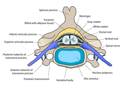
Watch where the thrust actually travels. Neutral sits in the middle of the easy range. In a healthy joint, motion stops at the physiologic barrier (the active end range). The hard stop past it is the anatomic barrier (bone on bone: cross it and you tear tissue). In dysfunction, a restrictive barrier forms early, short of the physiologic limit. HVLA takes up slack to the restrictive barrier, then thrusts a few millimeters through it toward the physiologic barrier. That gap is where cavitation happens.

Neutral Restrictive (comes early in dysfunction) Physiologic (active end range) Anatomic (damage past here) HVLA thrust cavitation: nitrogen pop
Start: The joint sits at neutral. Tap "Take up slack" to engage the restrictive barrier, the first firm resistance felt on the way into the restriction.
Lever Length: Long vs Short
Long lever = force applied far from the segment (using an arm, the head, or the pelvis as the handle); easier to generate force but less precise. Short lever = force applied directly at the segment (a finger or thumb on the transverse process); more localized and specific. The thrust still travels the same few millimeters through the restrictive barrier either way.
Section 2 of 7

Absolute Contraindications

The highest-yield section for clinical practice. Know all nine. Cold.

Definition: Absolute Contraindication
Absolute = HVLA is NEVER performed at that site regardless of skill, experience, or gentleness. These are conditions where ANY thrust manipulation of the affected area carries irreversible harm or death risk.
1
Severe Osteoporosis
T-score below -2.5 · Pathological fracture risk
Even a low-amplitude thrust can fracture a severely osteoporotic vertebra. At-risk groups: elderly women, long-term corticosteroid users, patients with metabolic bone disease. Key distinction: osteopenia (T-score -1.0 to -2.5) is a relative CI. SEVERE osteoporosis (T-score below -2.5) is absolute. The clinical medicine will test this distinction.
2
Unhealed Fracture
At or adjacent to the fracture site
Manipulation across an unhealed fracture causes displacement and non-union. Includes suspected fractures until cleared by imaging. If a patient has a known fracture at a vertebral level, HVLA at that level is absolutely contraindicated. Once healed and cleared, reassessment for OMM is appropriate.
3
Metastatic Disease to Spine
Bone mets weaken the vertebral body
Bone metastases destroy trabecular structure, turning normal vertebrae into eggshells. An HVLA thrust can cause a pathological fracture with cord injury. Red-flag screen before any OMM: night pain, unexplained weight loss, prior cancer history (especially breast, prostate, lung, renal, thyroid). These are the most common primary tumors that metastasize to bone.
4
Atlantoaxial Instability
RA erodes odontoid · Down syndrome ligamentous laxity
Two main causes: Rheumatoid arthritis (erodes the transverse ligament and the odontoid process of C2, eliminating the restraint on anterior C1 translation) and Down syndrome (congenital ligamentous laxity at C1-C2). In both cases, any upper cervical HVLA thrust can sublux the C1-C2 joint and compress the spinal cord. Classic clinical medicine scenario: RA patient with neck stiffness. Answer is always no cervical HVLA.
5
Active Infection / Osteomyelitis
Manipulation of infected tissue spreads infection
Active infectious process in the spine (osteomyelitis, discitis, epidural abscess) is an absolute CI. Manipulation can spread the infection, cause structural failure of the already-weakened bone, or seed a hematoma. Any febrile patient with back pain and risk factors (IVDU, recent procedure, immunocompromised) needs imaging and labs before OMM.
6
Severe Spondylolisthesis
High-grade vertebral slippage
High-grade spondylolisthesis (Grade 3 or 4, where the vertebral body has slipped more than 50-75% over the one below) makes the spine structurally unstable. An HVLA thrust worsens displacement and can compress neural elements or the cauda equina. Lower grades are relative contraindications (see next section).
7
Bleeding Disorders / Anticoagulation
Epidural hematoma risk
HVLA can cause microtrauma to the periarticular structures. In patients with coagulopathies or on anticoagulants (warfarin, heparin, DOACs), this microtrauma can become an epidural hematoma, causing acute cord compression. Rare but catastrophic. Includes hemophilia, von Willebrand disease, thrombocytopenia.
8
Cauda Equina Syndrome (Acute)
Neurological emergency · No manipulation
Cauda equina syndrome: bilateral leg weakness, saddle anesthesia, bowel/bladder dysfunction. This is a surgical emergency. HVLA is absolutely contraindicated until the cause is surgically or medically addressed. Any manipulation could worsen the compression and cause permanent paralysis or incontinence.
9
Acute Spinal Cord Compression
Active myelopathy · Cord at risk
Any acute myelopathy from a disc herniation, tumor, ossification of the posterior longitudinal ligament, or acute ligamentum flavum hypertrophy represents an absolute contraindication until the compression is decompressed. Signs: bilateral upper motor neuron findings (hyperreflexia, Babinski, clonus, spastic gait).
Board Memory: The Absolute 9
Fracture · Severe osteoporosis · Metastatic bone disease · RA atlantoaxial instability · Active infection/osteomyelitis · Severe spondylolisthesis · Anticoagulation/bleeding disorder · Cauda equina syndrome · Acute spinal cord compression
Section 3 of 7

Relative Contraindications

Pause, modify, or defer. Not never. But not without careful thought.

Relative contraindications mean HVLA is possible with modification or after a risk-benefit analysis. The dysfunction may still be treated with gentler techniques (MET, counterstrain, myofascial) while HVLA is deferred.

D
Acute Disc Herniation with Radiculopathy
Proceed with caution
A high-velocity thrust over an acutely herniated disc can worsen nerve root compression or push the nucleus pulposus further into the canal. Relative CI: use MET, counterstrain, or myofascial release while the disc is acute. Once the acute radiculopathy resolves, reassess. If bowel/bladder involved (cauda equina), this becomes ABSOLUTE.
P
Pregnancy
Late pregnancy · Lumbar and sacral
Lumbar and sacral HVLA in late pregnancy carries risk of placental abruption, premature labor, or fetal distress from the force transmitted. Cervical and thoracic HVLA may be acceptable with care in earlier pregnancy. Counterstrain, MET, and myofascial techniques are preferred throughout pregnancy.
H
Hypermobility / Joint Instability
More motion is the wrong answer
Hypermobile joints don't need more range of motion. HVLA gaps and stretches the joint capsule and ligaments, which worsens instability in already-lax joints. Common in Ehlers-Danlos syndrome, generalized joint hypermobility syndromes, and post-ligamentous injury. Use stabilizing techniques instead.
I
Inflammatory Arthritis (Active Flare)
Inflamed joints are fragile
Active inflammatory flares (gout, psoriatic arthritis, ankylosing spondylitis flare) make joint structures acutely vulnerable. HVLA during a flare can worsen inflammation, cause fracture, or sublux the joint. Wait until the inflammation is controlled. Note: RA with atlantoaxial instability is an ABSOLUTE CI regardless of flare status.
S
Severe Spondylosis with Stenosis
Risk of worsening foraminal/canal stenosis
Severe degenerative spondylosis with facet hypertrophy and ligamentum flavum buckling can narrow the canal to near-critical levels. HVLA, especially in extension, can reduce the canal diameter further. Imaging review before HVLA is recommended when severe stenosis is suspected.
R
Post-Surgical Spine
Recent surgery · Hardware · Fusion
Recent spinal surgery (laminectomy, discectomy, fusion, instrumentation) means altered anatomy, weakened structures, and hardware that was not designed to absorb HVLA forces. Deferred until surgical clearance is obtained. Once healed, careful reassessment may allow gentle techniques.
V
Vertebrobasilar Insufficiency
Positive pre-manipulation test · Cervical only
Vertebrobasilar insufficiency (VBI) testing (Hautant's test): cervical extension and rotation in the direction of intended thrust, held for 30 seconds. Positive = nystagmus, vertigo, diplopia, facial numbness, or syncope. A positive test indicates that cervical HVLA in that direction could compress the vertebral artery and cause posterior circulation stroke. Relative CI: defer cervical HVLA, use soft-tissue and MET instead.
Absolute vs. Relative: The Decision Rule
Absolute = NEVER do HVLA at that site, full stop. Relative = pause, assess risk, consider modifying or using a gentler technique. for clinical practice: if the question names an absolute CI, the answer is never HVLA. If it names a relative CI, the answer is usually "defer HVLA, use MET or counterstrain."
Section 4 of 7

The Five Parameters

Every HVLA attempt requires five things to be set before the thrust is delivered

HVLA is not "just crack the back." Five technical parameters must be precisely established before the thrust. Getting any one wrong means either therapeutic failure or patient harm.

01
Localization
Forces are focused at the dysfunctional segment only. Slack is taken up in all planes of restriction. If the force is distributed across multiple segments, you lose specificity and therapeutic effect.
02
Position
The restrictive barrier is engaged, not just moved toward neutral. All three planes of restriction (flexion/extension, rotation, sidebending) are loaded simultaneously. The barrier is the critical reference point.
03
Velocity
The thrust is HIGH velocity, much faster than the body can voluntarily resist. This speed is what prevents the paraspinal muscles from reflexively tightening against the force before the joint gaps.
04
Amplitude
LOW amplitude. The total distance of joint movement is only a few millimeters past the barrier. If the amplitude is large, the technique is not HVLA. Large amplitude with high velocity = high injury risk, no therapeutic benefit.
05
Direction
Always through the restrictive barrier. HVLA is a DIRECT technique. If the thrust direction is wrong (away from restriction), no therapeutic effect occurs and you may create a new dysfunction.
HVLA vs MET: The Core Distinction
MET: patient provides isometric force for 3 to 5 seconds, physician takes up new slack after release, repeats 3 times. Force source = patient. HVLA: physician provides the thrust, single rapid movement, no repetition. Force source = physician. MET is effort plus relaxation. HVLA is gap the joint with speed.

Tap a technique to compare how each one approaches the same restricted segment.

HVLA: thrust through the barrier

  • Direction: direct, into the restriction toward the physiologic barrier
  • Force source: the physician, a single high-velocity, low-amplitude thrust
  • Endpoint: mechanical gapping and cavitation; correction of the dysfunction
  • Most contraindications of any technique: fragile bone, instability, bleeding risk

Muscle Energy: patient does the work

  • Direction: direct, engaging the barrier, then advancing with each repetition
  • Force source: the patient, isometric contraction for 3 to 5 seconds
  • Endpoint: post-isometric relaxation lets the physician take up new slack, repeat about three times
  • Low force: a safe alternative when HVLA is contraindicated

Counterstrain: move away from the pain

  • Direction: indirect, away from the restrictive barrier into a position of ease
  • Force source: passive positioning held about 90 seconds over a tender point
  • Endpoint: resets aberrant proprioceptive firing; no thrust
  • Gentle: appropriate in fragile patients

Myofascial Release: follow the tissue

  • Direction: direct or indirect, loading or unloading the fascial restriction
  • Force source: sustained physician pressure following tissue creep
  • Endpoint: fascial release without a high-velocity load
  • Low force: another safe substitute when a thrust is contraindicated
Why the Pop Is Not Required
Nitrogen cavitation (the pop) is a byproduct, not the goal. Therapeutic benefit comes from the mechanical gaping of the joint, capsular stretch, and mechanoreceptor stimulation. Studies show that clinical outcomes are equivalent whether or not an audible pop occurs. Never attempt multiple consecutive thrusts to force a pop.
Section 5 of 7

Can I HVLA?

Read the vignette. Reveal the clues. Make the call.

Clinical Scenario
A 45-year-old male school teacher presents with low back pain for 3 weeks after lifting boxes. He has L4 right-rotation, right-sidebending, extended dysfunction (FRS right at L4). He takes warfarin for a prior DVT. X-ray shows grade I spondylolisthesis at L4-L5. No neurological deficits. The physician considers performing lumbar HVLA.
CLUE 1 → Tap to reveal
Patient takes warfarin (anticoagulation). Anticoagulation is an ABSOLUTE contraindication to HVLA due to epidural hematoma risk. At least one absolute CI is already present before we even consider the imaging findings.
CLUE 2 → Tap to reveal
Grade I spondylolisthesis at L4-L5 (the same level as the dysfunction). Grade I is a relative contraindication on its own, but combined with the absolute CI from anticoagulation, this patient has no safe path to lumbar HVLA today.
Verdict: Absolute Contraindication
Active anticoagulation (warfarin) is an ABSOLUTE contraindication to HVLA. An HVLA thrust in an anticoagulated patient creates risk of epidural hematoma and acute cord compression. The grade I spondylolisthesis adds a relative CI on top. Recommended alternatives for this patient's L4 FRS right dysfunction: MET (patient rotates against resistance, physician takes up new slack), counterstrain, or myofascial release. Reassess for HVLA if anticoagulation is eventually discontinued.

Thrust or do not. Tap a finding below, then drop it into the right bin. Absolute means HVLA is never done at that site, full stop. Relative means pause, modify, or defer to a gentler technique.

Active finding: tap one below
Absolute · do NOT thrust
Relative · modify or defer
Severe osteoporosis, T-score -3.4
RA with C1-C2 atlantoaxial instability
Lytic vertebral metastasis at the target level
On therapeutic warfarin (INR 2.6)
Cauda equina syndrome with saddle anesthesia
Osteopenia, T-score -1.8
Acute disc herniation, unilateral radiculopathy
Late pregnancy, lumbar dysfunction
Positive vertebral artery screen on the right
Active ankylosing spondylitis flare

One more case, branch by branch. Answer each node to unlock the next.

A 70-year-old woman with seropositive rheumatoid arthritis has a C2 dysfunction and neck stiffness. Before deciding on cervical HVLA, which structure are you most worried RA has destroyed?
Section 6 of 7

Quiz

Eight original clinical questions · Tap to answer

Question 1 of 8
A physician reviewing a 66-year-old woman with T6 somatic dysfunction checks her medical history. She takes alendronate for osteopenia (T-score -1.8 on DEXA). She has no prior fractures. She is not on anticoagulation. Neurological exam is intact.

Which statement best characterizes the appropriateness of HVLA for this patient?
Tempting to avoid HVLA since the patient has osteopenia and is taking bisphosphonates, which sounds like she is being treated for bone fragility, but osteopenia and osteoporosis are different diagnostic categories with different contraindication implications. Think of the T-score cutoffs as two floors of a building: osteopenia lives between -1.0 and -2.5 (manageable risk), osteoporosis lives below -2.5 (high fracture risk). HVLA is absolutely contraindicated only on the lower floor. Answer: B

Osteopenia (T-score -1.0 to -2.5) is NOT the same as osteoporosis (T-score below -2.5). The absolute contraindication is SEVERE osteoporosis, which carries significant pathological fracture risk from even minor forces. This patient has osteopenia managed with bisphosphonates and no fractures. Thoracic HVLA at T6 is appropriate.

Break it down: A is wrong because osteopenia alone is not an absolute CI. C overstates the relative risk without sufficient clinical basis. D is incorrect because bisphosphonate therapy actually reduces fracture risk by treating bone loss; it is not itself a contraindication to manipulation.
Question 2 of 8
A second-year osteopathic medical student is about to perform HVLA on a 34-year-old man with a C5-C6 dysfunction. Before proceeding, the attending asks the student to screen for vertebrobasilar insufficiency. The student extends and rotates the patient's cervical spine in the direction of the intended thrust, holds for 30 seconds, and observes for nystagmus, dizziness, visual changes, or syncope. The test is positive.

A positive test is best interpreted as:
Tempting to call a positive vertebral artery screening test an absolute contraindication to all cervical manipulation since nystagmus and vertigo during positioning sound alarming and serious, but the test is positive for HVLA-specific compression risk, not for all neck treatment. Think of the test as a road closure sign on a highway: it blocks high-speed traffic (HVLA) but side streets (MET, soft tissue) remain open. The distinction between absolute and relative contraindication is the clinical key. Answer: B

The vertebral artery screening test detects compromise of the vertebral arteries with positional challenge. A positive result (nystagmus, vertigo, diplopia, or syncope during positioning) suggests that cervical HVLA in that direction could compress the vertebral artery and cause posterior circulation stroke. This is a RELATIVE contraindication: defer cervical HVLA, but gentle non-thrust techniques (MET, soft tissue) may still be used.

Break it down: An absolute contraindication would prohibit all cervical manipulation, which is not the case here. The positive test does not mandate emergent neurology unless symptoms are severe or persistent after repositioning. In practice, a positive test means no high-velocity cervical manipulation.
Question 3 of 8
A physician is about to perform HVLA for a lumbar somatic dysfunction when the patient mentions severe shooting pain down her right leg for 2 weeks, numbness in the right foot, and weakness walking on her right heel. MRI shows a large L5-S1 disc herniation with right S1 nerve root compression.

What is the most appropriate next step?
Tempting to perform HVLA at reduced amplitude since the herniation is lumbar (not cervical) and decreasing force sounds like a reasonable safety modification, but amplitude reduction does not neutralize the risk because the problem is the high-velocity component, which can worsen nerve root compression regardless of how little distance the thrust travels. Think of a car crash: a low-amplitude high-speed collision still does more damage than a slow gentle push, even at a shorter distance. Answer: B

Acute disc herniation with active radiculopathy is a relative contraindication to HVLA because a thrust could worsen nerve root compression. Cauda equina syndrome (bilateral weakness, bowel/bladder) would make this absolute. This patient has unilateral S1 radiculopathy, which is relative. Use gentler techniques (MET, myofascial, counterstrain) while the disc is acute. Once the acute phase resolves, reassessment for HVLA is appropriate.

Break it down: Reducing amplitude does not eliminate the risk because the high velocity is still present and can worsen the disc protrusion. Informed consent is a process, not a clinical override of a contraindication.
Question 4 of 8
A physician loads a patient's lumbar segment for HVLA treatment of an L3 extended, rotated right, sidebent right (ERSrt) dysfunction. After positioning, engaging the barrier, and delivering the thrust, no audible pop occurs. The physician re-examines the segment and finds it still restricted.

What is the most appropriate next action?
Tempting to increase velocity on the second attempt since the first thrust produced no audible pop and more force seems like the logical fix for an insufficient first effort, but increasing velocity adds injury risk without adding specificity, and the audible pop is not the endpoint anyway. Think of the cavitation sound as a nice side effect rather than the goal: the goal is functional correction of the somatic dysfunction. Re-examine first, re-engage the barrier carefully, and attempt once more before switching technique. Answer: B

If the first HVLA attempt does not produce cavitation or correction, re-examine to confirm the dysfunction persists, re-engage the barrier carefully, and attempt one more thrust. If the second attempt also fails, switch to an alternative technique (MET, counterstrain). The rule is: two attempts maximum, then switch.

Break it down: Multiple consecutive thrusts increase injury risk and allow insufficient recovery time. Increasing velocity means more force, not better specificity, and is not the correct response to a failed attempt. The absence of an audible pop does not mean the technique failed: therapeutic benefit can occur without cavitation, and the endpoint is functional correction, not sound production.
Question 5 of 8
A physician is about to perform HVLA for a T4 extended, rotated left, sidebent left dysfunction. Before engaging the barrier and thrusting, the correct sequence of steps is to:

Which of the following correctly describes the direction of the HVLA thrust and the classification of this technique?
Tempting to call HVLA indirect since terms like "taking up slack" and "engaging tissue" sound like the language used for indirect release methods, but the defining difference is directional: indirect techniques move away from the barrier, direct techniques move through it. Think of a locked door: indirect techniques apply pressure to the frame around it, direct techniques turn the handle and push through. HVLA takes up slack into the restriction across all three planes, then thrusts through. Answer: B

HVLA is a direct technique: the physician takes up all slack in the restricted direction across all three planes (flexion/extension, rotation, sidebending), engages the restrictive barrier, then delivers a high-velocity, low-amplitude thrust through that barrier. Going into the restriction is what distinguishes direct techniques from indirect ones. Counterstrain and myofascial release move away from the barrier (indirect). MET uses the patient's voluntary isometric force to move the barrier -- HVLA does not require patient effort. Delivering the thrust before engaging the barrier is a technique error that produces no therapeutic benefit and may harm the patient.

Break it down: HVLA is direct -- take up slack, engage barrier, thrust through it. Always through, never away.
Question 6 of 8
A 25-year-old woman with Ehlers-Danlos syndrome type III (hypermobile type) presents with recurrent cervical pain and cervical somatic dysfunction at C3-C4. The physician wants to treat the cervical dysfunction.

Which statement best describes the appropriate management of her cervical somatic dysfunction?
Tempting to say HVLA is safe since hypermobility means the joints already have too much range of motion, which sounds counterintuitively like the technique would just be adding motion the joint already freely performs, but HVLA works by gapping a capsule that is already excessively lax, creating more instability rather than restoring normal mechanics. Think of a stretched-out rubber band: pulling it even further does not reset it to normal tension, it damages it further. Stabilizing techniques like MET tighten the system rather than stretching it. Answer: B

Hypermobility (joint instability) is a relative contraindication to HVLA. The mechanism: HVLA works by gapping and stretching the periarticular capsule and ligaments, firing mechanoreceptors in the joint capsule. In an already-hypermobile joint, the capsule and ligaments are already excessively lax. More stretching worsens instability, not treats it. The correct approach is to use stabilizing techniques: MET (which uses isometric contraction to strengthen the stabilizing muscles), soft-tissue work, or myofascial release. HVLA is a relative, not absolute, contraindication for hypermobility -- absolute CIs are structural (fracture, RA atlantoaxial, cord compression, anticoagulation). Deferring all OMM is incorrect; the patient still has somatic dysfunction that can be addressed with appropriate non-HVLA techniques.

Break it down: hypermobility = relative CI for HVLA. Use MET and stabilizing techniques instead.
Question 7 of 8
A 58-year-old man with a history of prostate cancer presents with 3 weeks of progressively worsening thoracic back pain, worse at night. He is on no anticoagulation. X-ray shows a lytic lesion at T7 with mild vertebral body height loss. He has no neurological deficits and full strength in his legs. The physician identifies a T7 somatic dysfunction.

What is the most appropriate course of action regarding HVLA?
Tempting to say HVLA is safe since there are no neurological deficits and the vertebral body height loss is only mild, but absent neurological deficits simply means the cord has not been compressed yet. The lytic lesion at T7 has already destroyed the structural scaffolding of that vertebral body, and HVLA can cause immediate pathological fracture with cord compression from even a well-performed thrust. Think of the lesion as a sawdust plug where solid wood used to be: the fact that the ceiling has not collapsed yet does not mean you should add more weight to it. Answer: B

Metastatic bone disease to the spine is an absolute contraindication to HVLA at that site -- regardless of neurological status, gentleness, or informed consent. A lytic lesion at T7 in a prostate cancer patient has severely compromised the structural integrity of the vertebral body. HVLA can cause an immediate pathological fracture with cord compression, even with minimal force. The absence of neurological deficits does not make it safe -- it simply means the catastrophe has not happened yet. Night pain plus weight loss plus prior cancer history is the red-flag triad for spinal metastases that every physician must screen before ANY OMM. Informed consent does not override an absolute contraindication. MET is appropriate for the somatic dysfunction while oncology is consulted urgently.

Break it down: bone mets plus HVLA equals pathological fracture. Night pain plus cancer history equals red flag. Absolute contraindication.
Question 8 of 8
A 40-year-old woman with ankylosing spondylitis is currently in an active flare with elevated CRP and significant morning stiffness. She has an L3 somatic dysfunction identified on structural exam. She asks about spinal manipulation. Her spine has no fracture or lytic lesion on imaging. She takes no anticoagulants.

Which of the following best describes the role of HVLA in her management?
Tempting to call ankylosing spondylitis an absolute contraindication to HVLA under all circumstances since it is a serious inflammatory arthropathy and any manipulation sounds dangerous, but the contraindication level depends on disease state, not the diagnosis itself. Think of the active flare as the dangerous condition, not the underlying disease name: inflamed and fragile periarticular structures cannot tolerate the gapping force, but a patient in remission without bamboo spine has a completely different risk profile that may permit carefully modified HVLA. Answer: C

Active inflammatory flare (ankylosing spondylitis, psoriatic arthritis, gout) is a relative contraindication to HVLA. During active inflammation, periarticular structures are acutely inflamed, vascularized, and fragile. HVLA during a flare can worsen inflammation, cause microtears, and in advanced ankylosing spondylitis (bamboo spine), cause fracture through a rigidly fused segment. The correct approach is to wait until the flare is medically controlled, then reassess. If her disease is in remission and imaging shows no bamboo spine or fractures, HVLA could be considered with appropriate modification. Ankylosing spondylitis is not an absolute CI under all circumstances -- the active flare and the structural changes (if present) drive the contraindication level. HVLA does not reduce inflammation; it is a mechanical technique without direct anti-inflammatory effect.

Break it down: active inflammatory flare equals relative CI for HVLA. Control the inflammation first, then reassess.
Section 7 of 7

Board Walkthrough

Six original clinical vignettes, shuffled and never-repeating. Right-click or long-press to cross out. Double-click or double-tap to highlight. Answer, then tap the wrong choices to learn why.

Medically reviewed by Fatima Ali, DO and Kaitlyn Cocuzzo, MD · Last reviewed June 2026
Bone Wizardry is an independent educational resource for visual learning in the medical sciences. It is not affiliated with, endorsed by, or sponsored by any licensing or examination board, contains no real or recalled examination questions, and does not guarantee any educational or examination outcome.